INTRODUCTION
Cord blood (CB) stem cells are increasingly being used as a source of hematopoietic stem cells transplantation (HSCT).
Although there are several advantages, such as a lower inci- dence of graft-versus-host disease (GVHD) or viral infections in cord blood stem cell transplantation (CBSCT) as compared to bone marrow transplantation (BMT), slower engraftment speed and the limitation of the cell dose are still obstacles (1-5). Recent studies regarding the homing mechanism fol- lowing HSCT have revealed that homing-associated cell adhe- sion molecules (H-CAMs) and chemokine receptors on the CD34+ cells play very important roles for engraftment (6, 7).
Until recently, most studies on H-CAMs have been common- ly performed by using purified CD34+ cells, and the quan- tity of these cells is expressed as percentages of positive cells or as the antigen density on the CD34+ cells (8-11).
Although most of the CAMs including CD49d, CD44 and CXCR4, are present on primitive hematopoietic cells, they are also found on another nucleated cells (NCs) includ- ing monocytes and lymphocytes (12-15). Therefore, not only
CD34+ cells but also the other NCs expressing H-CAMs and chemokine receptors could be implicated in the engraft- ment and proliferation of hematopoietic stem cells. Further- more, in CBSCT, the speed of myeloid engraftment was pri- marily associated with the total nucleated cell (TNC) counts rather than the CD34+ cell counts (2).
Although recent study revealed that trafficking of trans- planted cells to the bone marrow is not selective and lodgment of bone marrow-homed cells may be specific (16), engraft- ment potential of HSCs may be influenced by the distinct phases of cell cycle (17, 18).
A lot of studies showed a significant delay of neutrophil and platelet recovery in the CBSCT group compared with the BMT or peripheral blood stem cell transplantation (PBSCT) groups. The studies have also revealed that the median cell doses for engraftment are significantly lower in the CBSCT group compared with the BMT or PBSCT groups (2, 5, 19, 20). However, to date it is not clear why the engraftment speed is different and the required cell dose for engraftment is different among these groups. In the present study, to deter- mine the engraftment kinetics, we investigated the differ-
Young-Ho Lee, Young-Ah Lee, Kyu-Tae Noh, Kyeong-Hee Kim, Jin-Yeong Han, Su-Yeong Seo, Hyuk-Chan Kwon, Jae-Seok Kim, Hyo-Jin Kim
Hematopoietic Stem Cell Transplantation Program, Dong-A Cancer Center, Dong-A University Medical Center, Busan, Korea
Address for correspondence Young-Ho Lee, M.D.
Department of Pediatrics, Dong-A University Medical Center, 1 Dongdaeshin-dong 3-ga, Seo-gu, Busan 602-715, Korea
Tel : +82.51-240-2956, Fax : +82.51-242-2765 E-mail : [email protected]
*This paper was supported by the Research Fund of Dong-A University Medical Center (Grant No. 201- 1999), Korea
523
Homing-Associated Cell Adhesion Molecules and Cell Cycle Status on the Nucleated Cells in the Bone Marrow, Mobilized Peripheral Blood and Cord Blood
Homing-associated cell adhesion molecules (H-CAM) on the CD34+ cells play an important role for the engraftment process following hematopoietic stem cell trans- plantation (HSCT). However, it seems that not only CD34+ cells but also other nucle- ated cells (NCs) with H-CAM could be implicated in the engraftment process and the proliferation of hematopoietic stem cells. We investigated the differences of H- CAM and cell cycle status on the NCs in cord blood (CB), bone marrow (BM), and mobilized peripheral blood (PB). The proportions of CXCR4+ cells within the NC populations were greater in CB than in PB or BM (p=0.0493), although the propor- tions of CXCR4+, CD44+, and CD49d+ cells within the CB CD34+ cell populations were same within BM or PB. A lower proportion of CD34+CD49d+ cells within the CD34+ cell populations was more noted in CB than in PB or BM (p=0.0085). There were no differences in cell cycle status between CB and BM or PB. Our results sug- gest that the migrating potential of CB would be enhanced with increased CXCR4 expression on the NCs, but the adhesion potential of CB CD34+ cells would be less than that of PB and BM. These findings may help explain why the lower cell dose is required and engraftment is delayed in cord blood stem cell transplantation.
Key Words : Cell Adhesion Molecules; Nucleated Cells; Erythrocytes; Bone Marrow; Peripheral Blood; Fetal Blood
Received : 9 January 2004 Accepted : 23 April 2004
ences of H-CAMs and chemokine receptors as well as cell cycle status by using the NCs, not the purified CD34+ cells, in the bone marrow (BM), mobilized peripheral blood (PB) and the CB.
MATERIALS AND METHODS
Isolation of nucleated cells from 3 different sources of stem cells
Eight BM samples were obtained from normal healthy donor for related BMT, and these cells were cryopreserved after a red cell depletion process by density gradient separ- tion with 10% pentastarch (Jeil Pharm, Seoul, Korea), and the cells were then analysed after thawing. Ten PB samples were obtained from the apheresed products of acute myel- ogenous leukemia patients, which were collected after mobi- lization chemotherapy for the PBSC harvest.
Thirteen CB samples were collected into transfer bags con- taining acid citrate dextrose (ACD) from the umbilical cord vein immediately after a full-term vaginal delivery, and the red cells were depleted by the same method as was used with the BM.
Phenotype analysis
Dual-color flow cytometry of CD34/CXCR4, CD34/49d, CD34/44 for the isolated nucleated cells was performed using FACSort (Becton Dickinson, San Jose, CA, U.S.A.). The cells were stained with the corresponding monoclonal antibodies for 45 min. After incubation, the cells were then washed three times in phosphate-buffered saline (PBS), fixed in 1% para- formaldehyde, and finally analysed by using Lysys II software (Becton Dickinson, San Jose, CA, U.S.A.).
Cell cycle analysis
The cells were washed two times with PBS by centrifuga- tion for 3 min at 4℃, then they were fixed with 50% ethanol for 30 min at room temperature. Cells were suspended in PBS containing RNase (1 mg/mL, 20 L) and propidium iodide (10 mg/mL, 2.5 L), and they were incubated for 30 min at room temperature. The cell cycle was analysed with Multicycle using Coulter EPICS XL flow cytometer (Beck- man Coulter, FL, U.S.A.).
Statistical analysis
Student’s t-test and the Wilcoxon rank sum test were per- formed between CB and BM, and also on the differences bet- ween the CB and PB. The SAS window version 6.12 was used for our analysis.
RESULTS
CXCR4, CD49d, and CD44 expression on the NCs
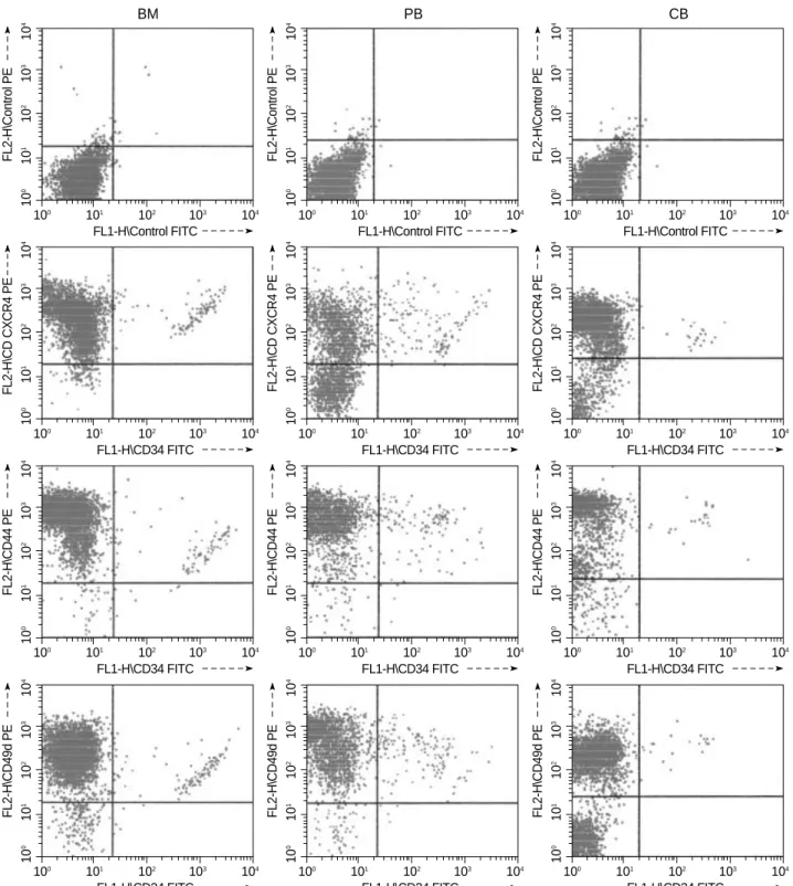
The proportions of CXCR4+ cells in the NCs were sig- nificantly higher in CB (82.76±5.89%) than in BM (62.31
±15.64%) or PB (76.35±23.7) (p=0.0493). However, the percentages of CD49d+ and CD44+ cells in CB were not statistically different from BM or PB. (Table 1, Fig. 1)
The percentage of CD34+CXCR4+, CD34+CD49d+, and CD34+CD44+ cells within the NC populations and within the CD34+ cell populations
Although the CD34+CXCR4+ and CD34+CD44+ cells within the NC populations in CB were not statistically dif- ferent from PB or BM, the CD34+CD49d+ cells were sig- nificantly lower in CB (0.91±0.49%) than in BM (2.38± 1.19%) or PB (4.23±3.14%) (p=0.0085) (Table 2). How- ever, in the CB CD34+ cell populations, the proportions of CD34+CXCR4+, CD34+CD49d+, and CD34+CD44+
cells were not significantly different from BM or PB (Table 3).
Values given represent a mean±the standard deviation. CB, cord blood;
BM, bone marrow; PB, peripheral blood; *: significant difference between CB and BM; �: significant difference between CB and PB.
CB (n=13) BM (n=8) PB (n=10) CXCR4+ cells (%) 82.76±5.89 62.31±15.64* 76.35±23.7� CD49d+ cells (%) 94.20±7.18 83.48±13.86 86.49±10.44 CD44+ cells (%) 98.61±1.3 98.65±1.14 98.51±1.73 Table 1.The proportions of CXCR4+, CD49d+, and CD44+ cells within the NC populations
Values given represent a mean±the standard deviation. CB, cord blood;
BM, bone marrow; PB, peripheral blood; *: significant difference between CB and BM; �: significant difference between CB and PB.
CB (n=13) BM (n=8) PB (n=10) CD34+CXCR4+ cells (%) 0.95±0.49 2.09±0.83 3.46±3.37 CD34+CD49d+ cells (%) 0.91±0.49 2.38±1.19* 4.23±3.14� CD34+CD44+ cells (%) 1.52±0.64 2.06±0.97 5.17±3.65 Table 2.The proportions of CD34+ cells expressing CXCR4, CD49d, and CD44 within the NC populations
Values given represent a mean±the standard deviation. CB, cord blood;
BM, bone marrow; PB, peripheral blood.
CB (n=13) BM (n=8) PB (n=10) CD34+CXCR4+ cells (%) 94.11±7.11 82.54±12.93 74.81±30.71 CD34+CD49d+ cells (%) 97.90±2.38 98.46±2.56 91.29±10.42 CD34+CD44+ cells (%) 98.24±1.51 99.00±1.54 98.66±1.66 Table 3.The proportions of CD34+ cells expressing CXCR4, CD49d, and CD44 within the CD34+ cell populations
Cell cycle status
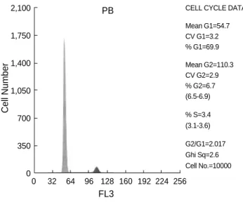
Most of NCs of CB (87.83±6.71%), BM (87.62±5.01%), and PB (88.38±32%) were in G0/G1 phase and they were not statistically different. The cell fractions in S/G2/M phase
showed no significant differences between those fractions in CB (12.17±6.75%) and BM (12.4±5.07%) or PB (11.6
±1.35%) (Fig. 2).
Fig. 1.Representative flow cytometric profile of CXCR4, CD44, CD49d and CD34 expression on the isolated nucleated cells from bone marrow (BM), mobilized peripheral blood (PB) and cord blood (CB).
FL2-H\Control PE
FL1-H\Control FITC 100101102 103104
100 101 102 103 104
FL2-H\Control PE
FL1-H\Control FITC 100101102 103104
100 101 102 103 104
FL2-H\Control PE
FL1-H\Control FITC 100101102 103104
100 101 102 103 104
FL2-H\CD CXCR4 PE
FL1-H\CD34 FITC 100101102 103104
100 101 102 103 104
FL2-H\CD CXCR4 PE
FL1-H\CD34 FITC 100101102 103104
100 101 102 103 104
FL2-H\CD CXCR4 PE
FL1-H\CD34 FITC 100101102 103104
100 101 102 103 104
FL2-H\CD44 PE
FL1-H\CD34 FITC 100101102 103104
100 101 102 103 104
FL2-H\CD44 PE
FL1-H\CD34 FITC 100101102 103104
100 101 102 103 104
FL2-H\CD44 PE
FL1-H\CD34 FITC 100101102 103104
100 101 102 103 104
FL2-H\CD49d PE
FL1-H\CD34 FITC 100101102 103104
100 101 102 103 104
FL2-H\CD49d PE
FL1-H\CD34 FITC 100101102 103104
100 101 102 103 104
FL2-H\CD49d PE
FL1-H\CD34 FITC 100101102 103104
100 101 102 103 104
BM PB CB
DISCUSSION
In hematopoietic stem cell transplantations, CB offers sub- stantial advantages when compared to BM or PB. There is the rapid availability of cells and the less stringent requirements for HLA identity between the donor and recipient because of the lower risk of acute and chronic graft versus host disease.
The major disadvantage of CBSCT is the delayed recovery of neutrophils and platelets, which could increase the risk of life-threatening infections and bleedings (4, 5). Although the engraftment following CBSCT is strongly correlated with infused nucleated cell numbers (2), very little is known about why the engraftment speed is slower in CBSCT than in BMT or PBSCT.
To rescue the patient’s hematopoietic systems after myeloab- lative chemo- or radiotherapy, the intravenously transplant- ed hematopoietic stem cells (HSCs) have to migrate to the bone marrow’s microenvironment for their ultimate prolif- eration and differentiation. In this homing process, various adhesion molecules present on both HSC and endothelial cells are involved (6, 7). The CD34+ cells exhibit other adhe-
sion receptors they are involved in the multistep process of HSCT: homing in to the BM, adhesion to the BM microen- vironment and finally, the stem cell differentiation. CXCR4 is a dominantly expressed chemokine receptor on primitive human blood cells within all stages of human development (21). Stromal cell-derived factor-1 (SDF-1), which is produced by stromal cells in various organs, is a powerful chemoattrac- tant for its receptor CXCR4. The SDF-1/CXCR-4 interac- tion is clinically relevant during embryonic development, hematopoiesis and the migration of CD34+ stem cells. We revealed that although the proportions of CD34+CXCR4+
cells within the NC or CD34+ cell populations were not statistically different in BM, PB and CB, the proportions of CXCR4+ cells in NC populations were significantly higher in CB than in BM or PB. These results suggest that NCs, except CD34+ cells expressing CXCR4, were more abun- dant in CB than in BM or PB.
The integrins, such as VLA-4 or VLA-5, they mediate the adhesion of HSC to extracellular matrix proteins. The mobi- lization of BM CD34+ cells into the PB is followed by a de- crease of VLA-4 and VLA-5 expression (22, 23), and the low frequency of CD49d+ on PB CD34+ cells has been also report- ed by many authors (24-27). In our study, we observed the same percentage of CD49d+ cells within the CD34+ cell pop- ulations in the BM, PB and CB. However, CD34+CD49d+
cells within the NC populations in the CB were significant- ly lower than in BM or PB.
In conclusion, our results suggest that the migrating poten- tial of CB would be enhanced with increased CXCR4 expres- sion on the NCs. However, the adhesion potential of CB CD34+ cells would be less than that of PB and BM because of the lower expression of CD34+CD49d+ cells within the NC populations. These findings may help explain why the
Fig. 2.Representative cell cycle analysis of the isolated nucleated cells from bone marrow (BM), mobilized peripheral blood (PB) and cord blood (CB).
927
772
618
463
309
154
0
CELL CYCLE DATA
Mean G1=65.4 CV G1=5.4
% G1=85.0
Mean G2=130.0 CV G2=4.6
% G2=6.5 (5.4-7.5)
% S=8.5 (7.3-9.8)
G2/G1=1.985 Ghi Sq=5.1 Cell No.=10000
0 32 64 96 128 160 192 224 256 FL3
BM
Cell Number
1,380
1,150
920
690
460
230
0
CELL CYCLE DATA
Mean G1=58.4 CV G1=3.8
% G1=88.2
Mean G2=117.3 CV G2=3.4
% G2=10.7 (10.4-11.0)
% S=1.0 (0.9-1.2)
G2/G1=2.008 Ghi Sq=5.6 Cell No.=10000
0 32 64 96 128 160 192 224 256 FL3 LIN
CB
Cell Number
2,100
1,750
1,400
1,050
700
350
0
CELL CYCLE DATA
Mean G1=54.7 CV G1=3.2
% G1=69.9
Mean G2=110.3 CV G2=2.9
% G2=6.7 (6.5-6.9)
% S=3.4 (3.1-3.6)
G2/G1=2.017 Ghi Sq=2.6 Cell No.=10000
0 32 64 96 128 160 192 224 256 FL3
PB
Cell Number
lower CB cell dose is required and engraftment is delayed in CBSCT.
REFERENCES
1. Gluckman E, Broxmeyer HA, Auerbach AD, Friedman HS, Douglas GW, Devergie A, Esperou H, Thierry D, Socie G, Lehn P, Cooper S, English D, Kurtzberg J, Bard J, Boyse EA. Hematopoietic recon- stitution in a patient with Fanconi’s anemia by means of umbilical cord blood from an HLA-identical sibling. N Engl J Med 1989; 321:
1174-8.
2. Kurtzberg J, Laughlin M, Graham ML, Smith C, Olson JF, Halperin EC, Ciocci G, Carrier C, Stevens CE, Rubinstein P. Placental blood as a source of hematopoietic stem cells for transplantation into unre- lated recipients. N Engl J Med 1996; 335: 157-66.
3. Wagner JE, Rosenthal J, Sweetman R, Shu XO, Davies SM, Ram- say NK, McGlave PB, Sender J, Cairo MS. Successful transplanta- tion of HLA-matched and HLA-mismatched umbilical cord blood from unrelated donors: Analysis of engraftment and acute graft ver- sus host disease. Blood 1996; 88: 795-802.
4. Rubinstein P, Carrier C, Scaradavou A, Kurtzberg J, Adamson J, Migliaccio AR, Berkowitz RL, Cabbad M, Dobrila NL, Taylor PE, Rosenfield RE, Stevens CE. Outcomes among 562 recipients of pla- cental blood transplants from unrelated donors. N Engl J Med 1998;
339: 1565-77.
5. Rocha V, Wagner JE Jr, Sobocinski KA, Klein JP, Zhang MJ, Horo- witz MM, Gluckman E. Graft-versus-host disease in children who have received a cord blood or bone marrow transplant from an HLA- identical sibling. Eurocord and International Bone Marrow Trans- plant Registry Working Committee on Alternative Donor and Stem Cell Sources. N Engl J Med 2000; 342: 1846-54.
6. Whetton AD, Graham GJ. Homing and mobilization in the stem cell niche. Trends Cell Biol 1999; 9: 233-8.
7. Kronenwett R, Martin S, Haas R. The role of cytokines and adhesion molecules for mobilization of peripheral blood stem cells. Stem Cells 2000; 18: 320-30.
8. Watanabe T, Dave B, Heiman DG, Lethaby E, Kessinger A, Tal- madge JE. GM-CSF-mobilized peripheral blood CD34+ cells differ from steady-state bone marrow CD34+ cells in adhesion molecule expression. Bone Marrow Transplant 1997; 19: 1175-81.
9. Barbosa IL, De Sausa ME, Godinho MI, Teixeira AM, Carvalhais A. Analysis of surface markers on CD34+ cells, isolated from cord blood and G-CSF primed peripheral blood. Bone Marrow Trans- plant 1998; 22: S56.
10. Kroger N, Zeller W, Hassan HT, Dierlamm J, Zander AR. Difference between expression of adhesion molecules on CD34+ cells from bone marrow and G-CSF-stimulated peripheral blood. Stem Cells 1998; 16: 49-53.
11. Bellucci R, De Propris MS, Buccisano F, Lisci A, Leone G, Tabilio A, De Fabritiis P. Modulation of VLA-4 and L-selectin expression on normal CD34+ cells during mobilization with G-CSF. Bone Mar- row Transplant 1999; 23: 1-8.
12. Voermans C, Van Hennik PB, Van der Schoot CE. Homing of human
hematopoietic stem and progenitor cells: New insights, New chal- lenges? J Hematother Stem Cell Res 2001; 10: 725-38.
13. Cavers M, Khoshkbijari BA, Macey M, McCarthy DA, Irshad S, Brown KA. Differential expression of 1 and 2 integrins and L- selectin on CD4+ and CD8+ T lymphocytes in human blood: com- parative analysis between isolated cells, whole blood samples and cryopreserved preparations. Clin Exp Immunol 2002; 127: 60-5.
14. Hemler ME. VLA proteins in the integrin family. Structures, functions and their roles on leukocytes. Annu Rev Immunol 1990; 8: 365-400.
15. Kishimoto TK, Larson RS, Corbi AL, Dustin ML, Staunton DE, Springer TA. The leukocyte integrins. Adv Immunol 1989; 46: 149- 82.
16. Jetmore A, Plett PA, Tong X, Wolber FM, Breese R, Abonour R, Orschell-Traycoff CM, Srour EF. Homing efficiency, cell cycle kinet- ics, and survival of quiescent and cycling human CD34+ cells trans- planted into conditioned NOD/SCID recipients. Blood 2002; 99:
1585-93.
17. Nilsson SK, Dooner MS, Quesenberry PJ. Synchronized cell-cycle induction of engrafting long-term repopulating stem cells. Blood 1997; 90: 4646-50.
18. Gothot A, van der Loo JC, Clapp DW, Srour EF. Cell cycle-related changes in repopulating capacity of human mobilized peripheral blood CD34+ cells in non-obese diabetic/severe combined immune- deficient mice. Blood 1998; 92: 2641-9.
19. Barker JN, Davies SM, DeFor T, Ramsay NK, Weisdorf DJ, Wagn- er JE. Survival after transplantation of unrelated donor umbilical cord blood is comparable to that of human leukocyte antigen-matched unrelated donor bone marrow: results of a matched-pair analysis.
Blood 2001; 97: 2957-61.
20. Ooi J, Iseki T, Takahashi S, Tomonari A, Nagayama H, Ishii K, Ito K, Sato H, Takahashi T, Shindo M, Sekine R, Ohno N, Uchimaru K, Nagamura F, Shirafuji N, Tojo A, Tani K, Asano S. A clinical comparison of unrelated cord blood transplantation and unrelated bone marrow transplantation for adult patients with acute leukemia in complete remission. Br J Haematol 2002; 118: 140-3.
21. Rosu-Myles M, Khandaker M, Wu DM, Keeney M, Foley SR, How- son-Jan K, Yee IC, Fellows F, Kelvin D, Bhatia M. Characteriza- tion of chemokine receptors expressed in primitive blood cells dur- ing human hematopoietic ontogeny. Stem cells 2000; 18: 374-81.
22. To LB, Haylock DN, Simmons PJ, Juttner CA. The biology and clini- cal uses of blood stem cells. Blood 1997; 89: 2233-58.
23. Prosper F, Stroncek D, McCarthy JB, Verfaillie CM. Mobilization and homing of peripheral blood progenitors is related to reversible downregulation of 4 1 integrin expression and function. J Clin Invest 1998; 101: 2456-67.
24. Mohle R, Moore MA, Nachman RL, Rafii S. Transendothelial migra- tion of CD34+ and mature hematopoietic cells: an in vitro study using a human bone marrow endothelial cell line. Blood 1997; 89:
72-80.
25. Dercksen MW, Gerritsen WR, Rodenhuis S, Dirkson MK, Slaper- Cortenbach IC, Schaasberg WP, Pinedo HM, von dem Borne A, Van der Schoot CE. Expression of adhesion molecules on CD34+ cells:
CD34+ L-selectin+ cells predict a rapid platelet recovery after peri- pheral blood stem cell transplantation. Blood 1995; 85: 3313-9.
26. Watanabe T, Dave B, Heimann DG, Lethaby E, Kessinger A, Tal- madge JE. GM-CSF-mobilized peripheral blood CD34+ cells differ from steady state bone marrow CD34+ cells in adhesion molecule expression. Bone Marrow Transplant 1997; 19: 1175-81.
27. Bellucci R, De Propris MS, Buccisano F, Lisci A, Leone G, Tabilio A, de Fabritiis P. Modulation of VLA-4 and L-selectin expression on normal CD34+ cells during mobilization with G-CSF. Bone Marrow Transplant 1999; 23: 1-8.