- 115 - 고신대학교 의과대학 학술지 제23권 제2호
Kosin Medical Journal
Vol. 23. No. 2, pp. 115∼118, 2008
Multiple Spinal Intradural Schwannomas
Combined with Sequestered Lumbar Disc Herniation
- A Case Report -
Dae-Yong Kim, Jin-Wook Kim, Ju-Ho Jeong, Jae-Hoon Cho Department of Neurosurgery, Kosin University College of Medicine, Busan, Korea
――― Abstract ――――――――――――――――――――――――――――――――――――――――
In this report, we describe a case of multiple intradural schwannomas in the spine combined with a upward sequestered disc herniation at L4-L5. It is often difficult to differentiate this tumor from lumbar disc herniation, especially a sequestered hernia. Gadolinium-enhanced MR images were a useful preoperative examination modality for differentiating these lesions from other diseases. Microscopically, the intradural tumors were successfully removed. The dura maters of the L2 and S1 were opened microsurgically, allowing the nerve fibers involved in the tumor to be identified. The involved fibers were cut around the tumor, and the lesion was resected while the intact nerve fibers were preserved. An upward sequestered lumbar disc herniation at L4-L5 was also successfully removed. Based on histological examination of the resected specimen, the tumors were diagnosed as schwannomas. Microsurgery allowed the tumors to be removed with minimal impairment from cutting of nerve fibers in the each nerve root.
―――――――――――――――――――――――――――――――――――――――――――――――――Key words : Schwannoma, Sequestered herniation, Lumbar disc herniation
교신저자:정 주 호
주소 : 602-702, 부산광역시 서구 암남동 34번지 고신대학교 의과대학 신경외과학
TEL : 051-990-6465, FAX: 051-990-3042 E-mail : [email protected]
Introduction
Schwannomas, benign tumors of the nervous system originating in the neural sheath, most commonly manifest sporadically and only as a single neoplasm.1) However 3 to 4% of patients with a schwannoma present in the form of multiple lesions.2) It is often difficult to differentiate this tumor from lumbar disc herniation, especially a sequestered hernia.3) Surgical referral should be considered for the patient with a documented lumbar disk herniation that correlates precisely with clinical findings. In this report, we describe a case of multiple intradural schwannomas in the spine combined with an upward sequestered disc herniation at L4-L5.
Case Report
A 41-year-old woman was referred to our hospital
having severe pain in the lower back and both lower extremities and gaiting disturbance for two months. She had no history of trauma or previous lower back surgery. The primary medical facility where she underwent lumbar computed tomography could not improve the pain with conservative treatment. Therefore, that hospital referred her to our hospital. A radiologist in that hospital could not find any pathology in the compued tomography. The neurologic examination revealed weakness of the extensor hallucis longus and decreased ankle reflex in both lower extremitis. There are no apparent foot-drop in both lower extremities. Straight leg raise was positive at 30°
bilaterally. A sensory deficit over the saddle area was observed, but bladder and bowel function were normal.
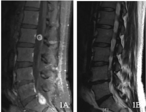
Magnetic resonance image (MRI) showed a mass-like lesion at the level of L2, L4 and S1. Gadolinium-enhanced MRI demonstrated two large enhanced masses at the level of L2 and S1(Fig. 1A) . It also revealed an upward sequestered lumbar disc herniation at L4-L5(Fig. 1B). To remove the masses, the patient underwent the L2 total laminectomy, the left L4-L5 facetectomy and the unroofing of the sacrum. Microscopically, the intradural tumors were
고신대학교 의과대학 학술지 제23권 2호, 2008
- 116 - Fig. 1 (A) Gadolinium-enhanced magnetic resonance image demonstrates two round-well enhanced masses at the level of L2 and S1.
(B) Spinal magnetic resonance image shows an upward sequestered lumbar disc herniation at L4-L5.
1A 1B
Fig. 2 Intraoperative photographs demonstrate the schwannomas at L2 (A) and S1 (B).
2A
2B
successfully removed. The dura maters of the L2 and S1 were opened microsurgically, allowing the nerve fibers involved in the tumor to be identified. The involved fibers were cut around the tumor, and the lesion was resected while the intact nerve fibers were preserved(Fig. 2A and 2B).
An upward sequestered lumbar disc herniation at L4-L5 was also successfully removed. Based on histological examination of the resected specimens at L2 and S1, the tumors were diagnosed as schwannomas. A frozed section comfirmed the presence of a disc fragment without neoplastic activity about upward sequestered disc
herniation. Microsurgery allowed the tumors to be removed with minimal impairment from cutting of nerve fibers in the each nerve root. After surgery, the pain in the the lower back and both lower extremities and gaiting disturbance were improved. She gained full recovery in 3 months.
Discussion
Acute low-back pain is one of the most common problems encountered by primary-care physicians.3) A few patients have severe neurologic impairment or evidence of cancer or other serious underlying systemic illness. For such patients, a broad differential diagnosis must be considered, and a prompt work-up and specialty consultation may be necessary. For most patients with acute low-back pain, extensive laboratory and imaging tests are unnecessary, and rapid improvement can be expected with only simple treatment measures. Physical therapy is useful in patients with refractory symptoms. Magnetic resonance imaging and other sophisticated spinal imaging should usually be reserved for patients who are being considered for an operation. Surgical referral should be considered for the patient with a documented lumbar disk herniation that correlates precisely with clinical findings.
Careful analysis of this case reveals that the original primary-care physician missed the correct diagnosis not only because of failure to see the abnormalities, but also failure to recognize them-because of lack or lapse of knowledge. The primary-care physician failed to recognize that the initial axial views of computed tomography did not match the neurologic symptom. In the case of mismatch between the neurologic examination and neuroradiolgic study, the physician should be consider further evalution such as MRI, electromyography, myelography and so on.
Furthermore, the physician should not confirm the diagnosis without full adequate studies.4)
Extraspinal causes of radiculopathy are rare, but they should be considered, especially in the differential diagnosis of patients who have dysesthesia in a lower extremity and who are more than fifty years old and have pain at night, a history of malignant tumor, or a radiculopathy of the third or fourth lumbar-nerve root. In dealing with elderly
Multiple Spinal Intradural Schwannomas Combined with Sequestered Lumbar Disc Herniation
- 117 - patients who have radiculopathy, one should be suspicious that the cause is outside the spine.5)
Typically, schwannoma presents frequently as a single benign neoplasm of the nervous system originating in the neural sheath. Occasionally it presents in multiple form or simultaneously arises from several points along the peripheral nervous system, including cranial nerves, spinal roots, the brachial and lumbosacral plexuses, or major peripheral nerves.1)
Several syndromes, including the well-known neurofibromatoses, are associated with an increased frequency of schwannomas.6) The defining feature of neurofibromatosis 2 (NF2) is the presence of bilateral vestibular schwannomas. The case presented in this article is multiple spinal intradural scwannoma without neurofibromatosis. This kind of multiple schwannomas is now considered as schwannomatosis. Recent reports have suggested that some patients develop multiple schwannomas without any other associated stigmata of neurofibromatosis and that schwannomatosis may thus be regarded as a distinct clinical entity.7~9)
Schwannomatosis is a recently recognized third major form of neurofibromatosis (NF) that causes multiple schwannomas without vestibular tumors diagnostic of NF2.7) Patients with schwannomatosis represent 2.4 to 5%
of all patients requiring schwannoma resection, and approximately one third of patients with schwannomatosis have anatomically localized disease with tumors limited to a single limb or segment of spine. Epidemiologic studies suggest that schwannomatosis is as common as NF2, but that familial occurrence is inexplicably rare. Patients with schwannomatosis overwhelmingly present with pain, which remains the primary clinical problem and indication for surgery. Diagnostic criteria for schwannomatosis are needed for both clinicians and researchers, but final diagnostic certainty will await the identification of the schwannomatosis locus itself.7)
Baser, et al have modified the schwannomatosis diagnostic criteria so that all patients with definite or possible schwannomatosis must not fulfill any of the existing sets of diagnostic criteria for NF2 and have no evidence of vestibular schwannoma on high-quality MRI
scan, no first-degree relative with NF2, and no known constitutional NF2 mutation.8)
T here are a few strategic points in differentiating sequestered disc herniations from schwannoma. The former may extend down root sleeve or into neuroforamen, but do not erode bone. Generally in a case of the former, gadolinium-enhanced MR images shows no enhancement and nerve root may be identified as a separate structure10). Conclusion
It is often difficult to differentiate schwannoma from lumbar disc herniation, especially a sequestered hernia.
Gadolinium-enhanced MR images were a useful preoperative examination modality for differentiating this lesion from other diseases. In the case of mismatch between the neurologic examination and neuroradiolgic study, the physician should be consider further evalution such as MRI, electromyography, myelography, CT of abdominopelvis and so on. Furthermore, the physician should not confirm the diagnosis without full adequate studies.
References
1) Ahn JY, Kwon SO, Choi EW, Lee BH : Multiple Schwannomas. case report. J Korean Neurosurg Soc 33 : 299-301, 2003
2) Purcell S, Dixon S : Schwannomatosis-an unusual variant of neurofibromatosis or a distinct clinical entity? Arch Dermatol 125 : 390-393, 1989
3) Deen HG Jr. : Diagnosis and management of lumbar disk disease. Mayo Clin Proc 71 : 283-7, 1996
4) Berlin L : Malpractice issues in radiology. Possessing ordinary knowledge. AJR Am J Roentgenol 166 : 1027-9, 1996 5) Kleiner JB, Donaldson WF 3rd, Curd JG, Thorne RP :
Extraspinal causes of lumbosacral radiculopathy. J Bone Joint Surg Am 73 : 817-21, 1991
6) Ghyra A, Israel J, Santander C, Acuna D : Schwannoma of the brachial plexus with intrathoracic extension. Thorax 35 : 703-704, 1980
7) MacCollin M, Chiocca EA, Evans DG, Friedman JM, Horvitz R, Jaramillo D, et al : Diagnostic criteria for schwannomatosis.
Neurology 64 : 1838-45, 2005
8) Baser ME, Friedman JM, Evans DG : Increasing the specificity of diagnostic criteria for schwannomatosis. Neurology 66 : 730-732, 2006
9) Shishiba T, Niimura M, Ohtsuka F, Tsuru N : Multiple cutaneous neurilemmomas as a skin manifestation of
고신대학교 의과대학 학술지 제23권 2호, 2008
- 118 - neurilemmomatosis. J Am Acad Dermatol 10 : 744-754, 1984 10) Lee JS, Suh KT : Intradural disc herniation at L5-S1 mimicking an intradural extramedullary spinal tumor: a case report. J Korean Med Sci 21 : 778-780, 2006