https://doi.org/10.5624/isd.2018.48.3.201
Introduction
Pulp stones(denticles or nodules) are discrete calcified masses that can be seen in the pulp chamber of any decid- uous or permanent tooth.1-3 They can be found in healthy, diseased, and even unerupted or impacted teeth of all age groups.2-4 The most commonly affected tooth is the first molar on both jaws, followed by the second molar. The least commonly affected teeth are the incisors and ca- nines.3,5 The presence of pulp stones does not affect the threshold of electric pulp testing6 and should not be con-
sidered as a disorder requiring endodontic treatment.5 The formation of pulp stone is not understood, although some etiological factors have been proposed, such as aging, pulp degeneration, inductive interactions between the epi- thelium and pulp tissue, genetic predisposition, long-stand- ing irritants(caries, deep fillings, chronic inflammation, and abrasion), orthodontic tooth movement, trauma, peri- odontal disease, drugs, anemia, arteriosclerosis, acromeg- aly, and Marfan syndrome. Additionally, a high prevalence of pulp stones has been reported in patients with cardiovas- cular disease and kidney, gall, and salivary gland stones.4
In the literature, panoramic,7,8 periapical,3,9,10 bite-wing radiographs,3,9,11 and cone-beam computed tomography (CBCT)2,12,13 have been used to evaluate the presence of pulp stones. The prevalence of pulp stones varies widely from 8% to 95%, depending on the population, study de-
A comparative study of cone-beam computed tomography and digital panoramic radiography for detecting pulp stones
Melek Tassoker1,*, Guldane Magat1, Sevgi Sener1
1Department of Oral and Maxillofacial Radiology, Necmettin Erbakan University, Faculty of Dentistry, Konya, Turkey
ABSTRACT
Purpose: The aim of this study was to compare cone-beam computed tomography(CBCT) and digital panoramic radiography(DPR) for the detection of pulp stones.
Materials and Methods: DPR and CBCT images of 202 patients were randomly selected from the database of our department. All teeth were evaluated in sagittal, axial, and coronal sections in CBCT images. The systemic condition of patients, the presence of pulp stones, the location of the tooth, the group of teeth, and the presence and depth of caries and restorations were recorded. The presence of pulp stones in molar teeth was compared between DPR and CBCT images.
Results: Pulp stones were identified in 105(52.0%) of the 202 subjects and in 434(7.7%) of the 5,656 teeth examined.
The prevalence of pulp stones was similar between the sexes and across various tooth locations and groups of teeth (P>.05). A positive correlation was observed between age and the number of pulp stones(ρ=0.277, P<.01). Pulp stones were found significantly more often in restored or carious teeth(P<.001). CBCT and DPR showed a significant difference in the detection of pulp stones(P<.001), which were seen more often on DPR than on CBCT.
Conclusion: DPR, as a 2D imaging system, has inherent limitations leading to the misinterpretation of pulp stones.
Restored and carious teeth should be carefully examined for the presence of pulp stones. CBCT imaging is recom- mended for a definitive assessment in cases where there is a suspicion of a pulp stone on DPR.(Imaging Sci Dent 2018; 48: 201-12)
KEY WORDS: Dental Pulp Calcification; Cone-Beam Computed Tomography; Radiography; Panoramic
Copyright ⓒ 2018 by Korean Academy of Oral and Maxillofacial Radiology
This is an Open Access article distributed under the terms of the Creative Commons Attribution Non-Commercial License(http://creativecommons.org/licenses/by-nc/3.0) which permits unrestricted non-commercial use, distribution, and reproduction in any medium, provided the original work is properly cited.
Imaging Science in Dentistry·pISSN 2233-7822 eISSN 2233-7830 Received July 5, 2018; Revised July 24, 2018; Accepted August 6, 2018
*Correspondence to : Dr. Melek Tassoker
Department of Oral and Maxillofacial Radiology, Necmettin Erbakan University, Faculty of Dentistry, Karacigan mh Ankara cd nm: 74/A Karatay, 42050, Konya, Turkey
Tel) 90-332-220-0025-1236, Fax) 90-332-220-00-45, E-mail) [email protected]
sign, and radiographic method employed.3,4 Histology- based studies have reported higher prevalence rates than radiographic studies because calcified masses smaller than 200μm cannot be seen on radiographs.5,14,15 Panoramic radiography has benefits regarding the examination of all teeth at the same time with a single exposure, and it uses minimal ionizing radiation.7 It is an integral part of dental check-ups, and previous studies have revealed that pulp stones can be detected well on panoramic radiography, as well as on bite-wing and periapical radiography.7,8 How- ever, 2-dimensional(2D) radiographic techniques may tend to underreport such calcifications.2 CBCT is a superior 3- dimensional(3D) imaging modality that is often used in endodontic practice to overcome the limitations of 2D tech- niques. It helps to manage complex endodontic problems by facilitating the separate examination of all teeth and root canals and the localization of calcified canals.2 The objective of this study was to compare CBCT and digital panoramic radiography(DPR) for the detection of pulp stones and to identify any potential correlation between the presence of pulp stones and factors such as age, sex, the tooth involved, restoration, and caries.
Materials and Methods
Patients
This retrospective study was approved by the Necmettin Erbakan University Faculty of Dentistry Research Ethics Committee and complied with the guidelines laid out in the Declaration of Helsinki(decision No. 2017.11). Sub- jects were referred to our radiology department if they re- quired CBCT analysis as part of their oral examination, diagnosis, and/or treatment planning.
DPR and CBCT scans of patients at least 18 years of age in whom the roots of all permanent teeth were completed were included in the study. The exclusion criteria were as follows: 1) teeth with root resorption, 2) teeth with metal crowns, 3) teeth with previous root canal treatment, and 4) poor-quality DPR or CBCT images. Patients’ characteris- tics(age, sex, and systemic disease) were obtained from their medical records. A total of 5,656 teeth(including all types of teeth, except for third molars) from 202 individu- als were analyzed.
Image acquisition
In all cases, DPR was performed using a panoramic unit (Morita Veraviewepocs 3D R100-P, J Morita MFG Corp., Kyoto, Japan) at 70kVp, 10mA, and 10s, according to the manufacturer’s recommended protocol.
CBCT images were acquired in a sitting position using a Morita 3D Accuitomo 170 device(J Morita MFG Corp., Kyoto, Japan), which was operated at 90kVp and 5mA, with a 17.5-s rotation time, a voxel size of 250μm, and a 100-×100-mm field of view, according to the manufac- turer’s recommended protocol.
Image analysis for pulp stones
The DPR images were exported as TIFF files and eval- uated by a blinded investigator with 6 years of oral and maxillofacial radiology experience in a darkened room. To
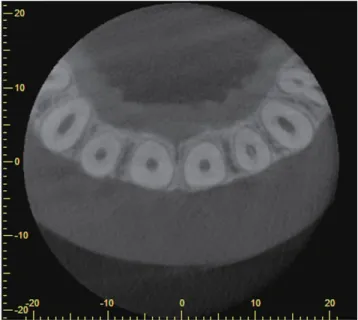
Fig. 1. A cropped panoramic radiograph represents pulp stones in the upper and lower molars.
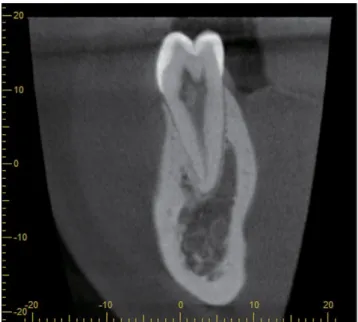
Fig. 2. A pulp stone is found in a left lateral incisor on an axial cone-beam computed tomographic slice.
view the raw data set, a 2.66 GHz Intel Xeon PC with 3.25 GB of RAM and Windows XPTM Professional operating system and a 27ʺ Dell U2711HTM monitor(U2711HTM;
Dell, Round Rock, TX, USA) with a resolution of 2,560×
1,600 pixels were used. All CBCT images were evaluated using i-Dixel software(J Morita MFG Corp., Kyoto, Japan) in all 3 planes(sagittal, axial, and coronal) on a flat-screen monitor by the same examiner(Figs. 1-4). For optimal visu- alization, the contrast and brightness of images were ad-
justed using the image-processing tool of the software.
Teeth were scored as having a pulp stone when a definite radiopaque mass was observed in the pulp space. The pre- sence of a pulp stone was also analyzed according to the presence of caries and restoration, the location of the tooth, the group of teeth, and the depth of restoration and caries (superficial: up to one-third of the dentin affected, medium:
one-third to two-thirds of the dentin affected, deep: two- thirds to all of the dentin affected but without pulp expo- sure).
For reasons related to image quality, only the DPR and CBCT images of posterior teeth(1,616 molar teeth) were compared in terms of pulp stone occurrence.
Statistical analysis
All observations and comparisons of the DPR and the CBCT imaging modalities in terms of pulp stone identifi- cation were evaluated using the Pearson chi-square test, odds ratios, and the Cohen kappa. To test intra-examiner reproducibility, 25% of the sample was re-examined in the same manner after an interval of 30 days. The relationship between age and the presence of pulp stones was deter- mined by the Spearman correlation test. All statistical anal- yses were performed using SPSS version 21.0(IBM Corp, Armonk, NY, USA) with a significance level of 5%.
Results
Of the 202 individuals, 75(37.1%) were male(mean age, 20.65±5.00 years) and 127(62.9%) were female(mean age, 21.42±6.15 years). The mean age of the patients in the sample was 21.13±5.75 years(range, 18-53 years).
The kappa values were excellent(between 0.90 and 0.97) for all observations. The variables evaluated with regard to the presence or absence of pulp stones are described in Table 1. The prevalence of pulp stones was not significant- ly different by sex(P>.05). Pulp stones were detected in 105 subjects(52%) and in 434(7.7%) of the 5,656 teeth examined. The prevalence of pulp stones in the maxillary and mandibular arches was found to be almost equal(P>
.05). The occurrence of pulp stones was highest in the max- illary canines(9.7%), followed by the mandibular incisors (9.2%), and mandibular premolars(8.3%)(P>.05). The mandibular canines were the least affected group of teeth (6.4%). Teeth with restorations and caries showed a signi- ficantly higher prevalence(73.5% and 62.1%, respective- ly) of pulp stones than teeth without restorations or caries (P<.05).
Of the 105 patients with pulp stones, 96.2% had no sys-
Fig. 3. A pulp stone is seen in a lower premolar on a sagittal cone- beam computed tomographic slice.
Fig. 4. Pulpal calcifications are found in the lower molars on a cor- onal cone-beam computed tomographic slice.
temic disease, while 3.8% had a systemic(cardiovascular, endocrine, or respiratory) disease. A positive correlation (Spearman correlation test) was observed between age and the number of pulp stones(ρ=0.277, P<.05).
Tables 2 and 3 summarize the distribution of teeth with or without pulp stones according to tooth location, type of tooth, and depth of restoration and caries.
Tables 4 and 5 show the risk of the presence of these cal- cifications, both in all teeth and stratified between the max- illary and mandibular arches, according to the presence and type of restorations and caries. The presence of restorations and caries was associated with a higher likelihood of pulp stones by 5.33 and 9.23 times in all teeth examined, respec- tively(P<.05). As the depth of the restoration increased, the likelihood of pulp stone presence increased(Table 4).
However, pulp stones were more frequently found in teeth with superficial caries in both the mandibular and maxil- lary arches(Table 5).
Table 6 presents the likelihood of pulp stone identifica- tion in each group of teeth in the presence of restorations
and caries. Restored and maxillary canines with caries demonstrated the highest risk of occurrence of pulp stones (OR=9.10; 15.34, respectively, P<.05) compared with other tooth categories.
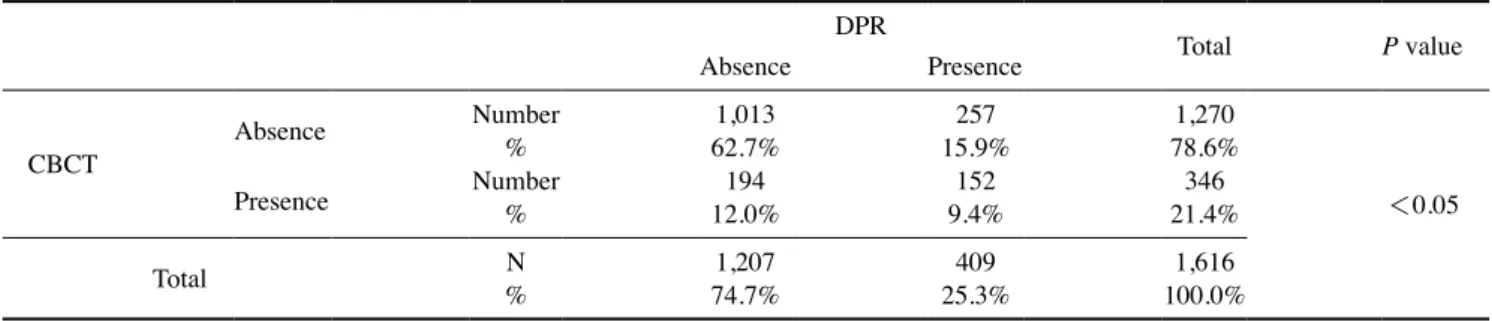
Overall, 9.4% of the teeth(152 of 1,616) were found to have a pulp stone by both DPR and CBCT, whereas 15.9%
of the teeth(252 of 1,616) had a pulp stone that was only detected by DPR. More teeth with pulp stones were found using DPR than using CBCT(P<.05)(Table 7).
Discussion
This study was performed to compare CBCT and DPR for the detection of pulp stones and to identify any poten- tial correlation between the presence of pulp stones and age, sex, the tooth involved, restoration, and caries. Pulp stones were identified in 7.7%(434 of 5,656) of the teeth examined and 52%(105 of 202) of all individuals evalu- ated. This is consistent with the findings of Patil et al.,12 who used CBCT and reported a prevalence of 50.93%, and
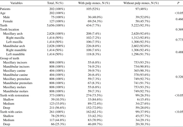
Table 1. Number(N) and prevalence(%) of pulp stones according to demographic data and tooth-related variables
Variables Total, N(%) With pulp stones, N(%) Without pulp stones, N(%) P
Patients 202(100%) 105(52%) 97(48%)
Age 202(100%) <0.05
Male 75(100%) 36(48.0%) 39(52.0%) 0.466
Female 127(100%) 69(54.3%) 58(45.7%)
Teeth 5,656(100%) 434(7.7%) 5,222(92.3%)
Tooth location 0.369
Maxillary arch 2,828(100%) 208(7.4%) 2,620(92.6%)
Right maxilla 1,414(50%) 102(7.2%) 1,312(92.8%) 0.773
Left maxilla 1,414(50%) 106(7.5%) 1,308(92.5%)
Mandibular arch 2,828(100%) 226(8.0%) 2,602(92.0%)
Right mandible 1,414(50%) 108(7.6%) 1,306(92.4%) 0.488
Left mandible 1,414(50%) 118(8.3%) 1,296(91.7%)
Group of teeth
Maxillary incisors 808(100%) 55(6.8%) 753(93.2%)
0.326
Mandibular incisors 808(100%) 74(9.2%) 734(90.8%)
Maxillary canine 404(100%) 39(9.7%) 365(90.3%)
Mandibular canine 404(100%) 26(6.4%) 378(93.6%)
Maxillary premolars 808(100%) 59(7.3%) 749(92.7%)
Mandibular premolars 808(100%) 67(8.3%) 741(91.7%)
Maxillary molars 808(100%) 55(6.8%) 753(93.2%)
Mandibular molars 808(100%) 59(7.3%) 749(92.7%)
Teeth with restoration 373(100%) 274(73.5%) 99(26.5%) <0.05
Shallow 39(10.5%) 33(84.6%) 6(15.4%)
Medium 123(33.0%) 89(72.4%) 34(27.6%)
Deep 211(56.6%) 152(72.0%) 59(28.0%)
Teeth with caries 261(100%) 162(62.1%) 99(37.9%) <0.05
Shallow 78(29.9%) 33(42.3%) 45(57.7%)
Medium 117(44.8%) 83(70.9%) 34(29.1%)
Deep 66(25.3%) 46(69.7%) 20(30.3%)
Table 2. Associations between the presence or absence of pulp stones and the type of tooth and restoration LocationToothTotal teeth N(%)
Teeth with pulp stones(WPS)Teeth without pulp stone(WtPS) Without restoration N(%)
Shallow N(%)
Medium N(%)
Total of Deep NWPS N(%) (%)
Without restoration N(%)
Shallow N(%)
Medium N(%)
Total of Deep NWtPS N(%) (%) 1418862117911111202Central incisorRight maxilla 88.6%93.1%3.0%1.0%0.5%0.5%6.9%0.5%0.5%5.4%100.0% 174187652615-18202Lateral incisor 86.1%3.0%2.5%1.0%92.6%7.4%0.5%4.0%100.0%3.0% 1524118018721-12202Canine 92.6%1.0%2.0%0.5%7.4%89.1%1.0%0.5%5.9%100.0% 171183741First premolar1931-15202 90.6%3.5%2.0%0.5%84.7%9.4%1.5%0.5%7.4%100.0% --218935179Second premolar11220213 1.0%93.6%1.5%2.5%88.6%6.4%0.5%5.9%100.0% ---176819111First molar1720210 87.1%4.0%3.5%94.6%5.4%5.0%100.0%0.5% -182187212315111Second molar202 0.5%90.1%1.0%92.6%1.0%0.5%7.4%100.0%1.5%5.4% Left maxilla18722-183152-11202Central incisor2 90.6%92.6%1.0%1.0%7.4%1.0%5.4%100.0%1.0% 1119152184-2--9202Lateral incisor 100.0%4.5%1.0%5.4%91.1%1.0%2.5%94.6% 224617816648-14202Canine2 1.0%88.1%3.0%2.0%11.9%82.2%4.0%1.0%6.9%100.0% -17462218418115202First premolar2 1.0%91.1%3.0%1.0%86.1%8.9%1.0%0.5%7.4%100.0% -177193754Second premolar9117202 2.0%95.5%3.5%2.5%4.5%87.6%0.5%0.5%3.5%100.0% -18042419012129202First molar 94.1%2.0%1.0%2.0%5.9%89.1%0.5%1.0%4.5%100.0% 175185811Second molar171--16202 91.6%4.0%0.5%0.5%86.6%8.4%0.5%7.9%100.0% 1833619-174171-202Central incisorRight mandible1 86.1%90.6%1.5%3.0%9.4%0.5%0.5%8.4%100.0% 206421701824-14202Lateral incisor2 1.0%90.1%3.0%2.0%84.2%9.9%2.0%1.0%6.9%100.0% --319511Canine190716202 96.5%0.5%1.5%0.5%0.5%94.1%3.0%100.0%3.5% -17818973113139202First premolar 88.1%93.6%1.5%0.5%3.5%6.4%0.5%1.5%4.5%100.0% -202Second premolar153118217820311 100.0%7.4%0.5%0.5%1.5%9.9%88.1%0.5%1.5%90.1%
Table 2. Associations between the presence or absence of pulp stones and the type of tooth and restoration LocationToothTotal teeth N(%)
Teeth with pulp stones(WPS)Teeth without pulp stone(WtPS) Without restoration N(%)
Shallow N(%)
Medium N(%)
Total of Deep NWPS N(%) (%)
Without restoration N(%)
Shallow N(%)
Medium N(%)
Total of Deep NWtPS N(%) (%) --16914418715429202First molar 92.6%6.9%2.0%83.7%7.4%2.0%1.0%4.5%100.0% -181188331202141112Second molar 93.1%1.5%1.5%0.5%89.6%6.9%0.5%0.5%5.9%100.0% 141881431170Left mandible41-9202Central incisor 93.1%6.9%1.5%0.5%6.9%84.2%2.0%0.5%4.5%100.0% 181111178-21-120202Lateral incisor 89.6%0.5%0.5%0.5%88.1%10.4%0.5%9.9%100.0% 1839119173-2-16202Canine1 85.6%90.6%4.5%0.5%9.4%1.0%0.5%7.9%100.0% ----1913First premolar18811110202 93.1%94.6%1.5%100.0%5.4%0.5%5.0% 1798421652334-16202arolmond preSec 88.6%1.0%4.0%2.0%7.9%81.7%1.5%2.0%100.0%11.4% 18118825--14--14202First molar 2.5%3.0%93.1%89.6%6.9%100.0%6.9% --173186671648202Second molar4 85.6%92.1%3.0%3.5%2.0%7.9%2.0%4.0%100.0% N: number, %: prevalence
Table 2. Continued
Table 3. Associations between the presence or absence of pulp stones and the type of tooth and caries LocationToothTotal teeth N(%)
Teeth with pulp stones(WPS)Teeth without pulp stone(WtPS) Without restoration N(%)
Shallow N(%)
Medium N(%)
Total of Deep NWPS N(%) (%)
Without restoration N(%)
Shallow N(%)
Medium N(%)
Deep N(%)
Total of WtPS N(%) --Right maxilla1Central incisor1882218314311202 0.5%93.1%1.0%1.0%100.0%90.6%6.9%1.5%5.4% 17818751321513Lateral incisor9202 92.6%2.5%0.5%1.5%88.1%7.4%0.5%1.5%1.0%4.5%100.0% 184187-1220215-4-11Canine 92.6%0.5%1.0%7.4%91.1%2.0%5.4%100.0% 183232176191312202First premolar3 87.1%90.6%1.0%1.5%1.0%0.5%9.4%1.5%1.5%5.9%100.0% 1341184189-Second premolar1-202111 0.5%93.6%2.0%91.1%6.4%0.5%0.5%5.4%100.0% 18619123-20211--29First molar 1.5%94.6%1.0%100.0%92.1%5.4%1.0%4.5% -187242179152112202Second molar 92.6%1.0%2.0%88.6%1.0%1.0%100.0%7.4%5.9%0.5% ---2318218715213202Central incisorLeft maxilla 92.6%1.0%1.5%7.4%90.1%1.0%6.4%100.0% 1911813612021110-Lateral incisor-1 100.0%5.0%0.5%5.4%0.5%3.0%1.5%94.6%89.6% 24178-4-174-419202Canine1 86.1%88.1%2.0%0.5%11.9%2.0%9.4%100.0% 18184--184-1114First premolar2022 8.9%91.1%91.1%1.0%0.5%0.5%6.9%100.0% 186193331Second premolar9---9202 95.5%1.5%1.5%4.5%0.5%4.5%100.0%92.1% ----121218720212190First molar 0.5%1.0%94.1%92.6%5.9%100.0%5.9% -185221783171214202Second molar 100.0%6.9%1.0%0.5%8.4%88.1%1.0%1.5%1.0%91.6% -Central incisor1441Right mandible19174342183202 1.5%90.6%1.0%2.0%6.9%86.1%9.4%0.5%2.0%100.0% 1781823-11520212Lateral incisor202 90.1%1.5%0.5%88.1%1.0%9.9%0.5%1.0%7.4%100.0% 1952711875-25202Canine- 0.5%96.5%1.0%2.5%92.6%3.5%1.0%2.5%100.0% 181189-53First premolar13-229202 93.6%89.6%2.5%1.5%4.5%6.4%1.0%100.0%1.0% --18Second premolar111182180202021 100.0%8.9%0.5%0.5%9.9%89.1%0.5%0.5%90.1%
Table 3. Associations between the presence or absence of pulp stones and the type of tooth and caries LocationToothTotal teeth N(%)
Teeth with pulp stones(WPS)Teeth without pulp stone(WtPS) Without restoration N(%)
Shallow N(%)
Medium N(%)
Total of Deep NWPS N(%) (%)
Without restoration N(%)
Shallow N(%)
Medium N(%)
Deep N(%)
Total of WtPS N(%) First molar102121518214-187202 90.1%92.6%2.0%0.5%5.0%7.4%1.0%0.5%1.0%100.0% 17818836131412Second molar8202 93.1%1.5%3.0%0.5%88.1%6.9%0.5%1.0%1.5%4.0%100.0% 188114411822024Left mandible1Central incisor81 0.5%100.0%4.0%0.5%0.5%2.0%6.9%90.1%2.0%0.5%93.1% 123-181177211-18202Lateral incisor 89.6%1.5%0.5%87.6%10.4%0.5%1.0%8.9%100.0% -18332177119202Canine3115 100.0%7.4%0.5%1.5%9.4%87.6%0.5%1.0%1.5%90.6% ---220211187139191First premolar 92.6%1.5%0.5%94.6%5.4%1.0%100.0%4.5% 2317914-Second premolar1742-219202 88.6%0.5%2.0%86.1%1.0%11.4%9.4%100.0%1.0% 14212183188-2-12202First molar 93.1%1.0%0.5%1.0%90.6%6.9%1.0%5.9%100.0% 178186332Second molar163292022 88.1%92.1%1.5%1.5%1.0%1.5%7.9%1.0%1.0%4.5%100.0% N: number, %: prevalence
Table 3. Continued
higher than the findings of da Silva et al.,2 who detected pulp stones in 31.9% of patients. Previous studies reported wide variation in the prevalence of pulp stones(from 8%
to 95%) depending on the population, study design, and radiographic method employed.3,4 A comparison of previ- ous studies that were conducted using various methods is presented in Table 8.
Consistent with previous research,2,10,14 the results of this study showed that the prevalence of pulp stones did not have a statistically significant relationship with sex. Other studies contradicting our findings3,15 stated that more pulp stones were seen in women, which was associated with a higher prevalence of bruxism in women, which can lead to long-term irritation of the teeth.3
The current study revealed a positive correlation between age and the number of pulp stones. Additionally, it was
found that the presence of restorations and caries increased the likelihood of pulp stones by 5.33 and 9.23 times in all teeth examined, respectively. Teeth with restorations and caries showed a significantly higher prevalence(73.5% and 62.1%, respectively) of pulp stones than teeth without res- torations or caries. Chronic irritations such as restoration and tooth caries, as well as parafunctions, have been sug- gested to be predisposing factors for pulp stone formation.3 Other studies in the literature3,16,17 have reported that pulp stones were not related to age. However, the duration, in- tensity, and frequency of those irritations may also be cru- cial. Therefore, there is a need for longitudinal studies in which radiographic follow-up is performed in patients in order to resolve this issue.3
The results of this study indicated that as the depth of restoration increased, the risk of pulp stone occurrence increased, in accordance with da Silva et al.,2 who used CBCT for pulp stone evaluation in their study. This may be explained through the hypothesis that chronic pulpal irritation leads the pulpodentinal complex to form pulp stones as a defense reaction.2,3,14 Deeper restorations may cause greater irritation closer to the pulp and more vascu- lar wall injuries.2 However, it was found that pulp stones were more likely to be detected in teeth with superficial caries in both the mandibular and maxillary arches. This can be explained in terms of the design of this study. This was a cross-sectional study and we had no information about how long the teeth had been exposed to the caries.
This can be considered a limitation of this study. Even if the caries were superficial, the duration of the irritants may
Table 4. Odds ratios for the presence of pulp stones based on the presence and depth of restorations according to tooth location(maxillary or mandibular arch)
Variables Odds ratio
Maxillary arch Mandibular arch Total
Teeth with restorations 5.30(P<0.05) 5.39(P<0.05) 5.33(P<0.05)
Shallow 1.14(P>0.05) 4.23(P<0.05) 2.20(P>0.05)
Medium 4.02(P<0.05) 5.76(P<0.05) 4.90(P<0.05)
Deep 6.41(P<0.05) 4.32(P<0.05) 5.24(P<0.05)
Table 5. Odds ratios for the presence of pulp stones based on the presence and depth of caries according to tooth location(maxillary or mandibular arch)
Variables Odds ratio
Maxillary arch Mandibular arch Total
Teeth with caries 8.22(P<0.05) 10.08(P<0.05) 9.23(P<0.05)
Shallow 20.49(P<0.05) 16.22(P<0.05) 18.19(P<0.05)
Medium 6.03(P<0.05) 4.61(P<0.05) 5.26(P<0.05)
Deep 1.51(P>0.05) 9.97(P<0.05) 5.43(P<0.05)
Table 6. Odds ratios for the presence of pulp stones according to the presence of restorations, caries, and group of teeth
Variables Odds ratio
Teeth with
restorations Teeth with caries Group of teeth
Maxillary incisors 8.95(P<0.05) 6.96(P<0.05) Mandibular incisors 3.84(P<0.05) 10.67(P<0.05) Maxillary canine 9.10(P<0.05) 15.34(P<0.05) Mandibular canine 4.40(P<0.05) 7.80(P<0.05) Maxillary premolars 2.98(P<0.05) 10.85(P<0.05) Mandibular premolars 7.53(P<0.05) 8.29(P<0.05) Maxillary molars 3.48(P<0.05) 5.40(P<0.05) Mandibular molars 5.82(P<0.05) 13.20(P<0.05)
have played a crucial role.3 Furthermore, our study sample was relatively limited, and further prospective studies are needed to confirm this result.
The present study found that the prevalence of pulp stones in the maxillary and mandibular arches was almost equal. This finding is consistent with some studies,2,12,14 but not with others.11,16,18 The distribution of pulp stones according to the group of teeth did not show any statisti- cally significant trends. The prevalence of pulp stones was highest in the maxillary canines(9.7%), followed by the mandibular incisors(9.2%) and mandibular premolars (8.3%), and lowest in the maxillary incisors and maxillary molars(6.8%). This may be attributed to the relatively lim- ited sample size, and further investigations are required.
Contrary to our findings, previous studies1,11-13,16 reported that pulp stones were mostly observed in molar teeth. Most studies were conducted with only posterior teeth, which could lead to an overestimation of the actual prevalence.3 Furthermore, the radiological method is an important con- sideration. Both anterior and posterior teeth should be ex- amined to determine the prevalence of pulp stones. CBCT is a relatively new imaging method in endodontic practice that allows all teeth to be examined separately in different
views, which is useful for localizing calcified canals.19 This method overcomes the superimposition of structures and has been found to be the best technique for the examina- tion of pulp stones.2 Although histological analysis pro- vides reliable results, it is only useful as an ex vivo meth- od.20 Since pulp stones may obscure or change the root canal anatomy and lead to a poorer outcome of root canal treatment, using CBCT to determine whether pulp stones are present has been argued to be justified.2
The present study indicated that the number of teeth with pulp stones was higher on DPR than on CBCT, as 9.4%
of teeth with pulp stones(152 of 1,616) were detected on both DPR and CBCT, whereas 15.9%(252 of 1,616) were detected only on DPR. DPR, as a projectional form of ra- diography, may overestimate or underestimate the preva- lence of pulp stones8 due to image distortion and super- imposition. The spatial resolution of an imaging method is defin ed as its capability to resolve fine details.21 Because of its low spatial resolution and 2D nature,22 DPR might not able to define pulp stones as well as CBCT. CBCT overcomes the superimposition of structures and has been found to be the best technique for the examination of pulp stones.2
Table 7. Distribution of pulp stone findings on digital panoramic radiography(DPR) and cone-beam computed tomography(CBCT)
DPR Total P value
Absence Presence
CBCT Absence Number 1,013 257 1,270
<0.05
% 62.7% 15.9% 78.6%
Presence Number 194 152 346
% 12.0% 9.4% 21.4%
Total N 1,207 409 1,616
% 74.7% 25.3% 100.0%
Table 8. Pulp stone studies in the literature
Authors Year Population Methodology Prevalence
(% of teeth)
Yaacob and Hamid25 1986 Malaysian Histology 6.7
Baghdady et al.1 1988 Iraqi Bite-wing radiographs 14.8
Arys et al.17 1993 Belgian Microradiography and light microscopy 95
Al-Hadi Hamasha and Darwazeh16 1998 Jordanian Periapical and bite-wing radiographs 22.4
Ranjitkar et al.11 2002 Australian Bite-wing radiographs 10.1
Turkal et al.15 2013 Turkish Panoramic radiographs 12.7
Kannan et al.14 2015 Malaysian Periapical radiographs 15.7
da Silva et al.2 2016 Brazilian CBCT 9.5
Hsieh et al.13 2017 Taiwanese CBCT 31.3
Present study 2018 Turkish CBCT and panoramic radiography 7.7
CBCT: cone-beam computed tomography
As DPR is an integral part of dental check-ups, and pre- vious studies have revealed that pulp stones can be detect- ed well on panoramic radiography, as well as on bite-wing and periapical radiography, and in light of the fact that the patient radiation dose is lower for DPR, this study compar- ed DPR and CBCT for pulp stone detection. Patients were not subjected to any additional radiation exposure as part of this study, since data were collected from patients who had both DPR and CBCT records. In a recent study,23 it was reported that DPR showed minimal to no distortion.
However, the highest distortion was found in the anterior region. Therefore, in this study, DPR and CBCT images of only the posterior teeth(1,616 molar teeth) were com- pared in terms of pulp stone detection.
Most previous studies have used conventional radiogra- phy or histological examinations to diagnose pulp stones.
The prevalence of pulp stones has been found to be higher in histological studies than in conventional radiography studies24 because calcified masses smaller than 200μm cannot be seen on radiographs.5,14,15 This is probably due to the inherent shortcomings of 2D imaging systems. It is difficult to distinguish such small calcifications from the results of overlapping with other tissues. CBCT is a supe- rior method for detecting pulp stones compared to conven- tional radiography, as it has greater specificity and accu- racy and overcomes the overlapping limitation.13
In conclusion, within the limitations of this study, the presence of pulp stones was not found to be related with sex, the location of the tooth, or the group of teeth, but pulp stones were found more often in teeth with restorations and caries. It can be concluded that chronic pulp irritation might lead to pulp stone formation. This was a cross-sec- tional study, and there is a need for longitudinal studies with larger samples including radiographic follow-up ex- aminations in order to more precisely characterize the ef- fect of the depth of caries and restorations on pulp stones.
DPR, as a 2D imaging system, has inherent limitations that lead to the misinterpretation of pulp stones. CBCT imaging is advised for the examination of pulp stones once there is a suspicion of a pulp stone on DPR.
References
1. Baghdady VS, Ghose LJ, Nahoom HY. Prevalence of pulp stones in a teenage Iraqi group. J Endod 1988; 14: 309-11.
2. da Silva E, Prado MC, Queiroz PM, Nejaim Y, Brasil DM, Groppo FC, et al. Assessing pulp stones by cone-beam com- puted tomography. Clin Oral Investig 2017; 21: 2327-33.
3. Sener S, Cobankara FK, Akgünlü F. Calcifications of the pulp chamber: prevalence and implicated factors. Clin Oral Inves-
tig 2009; 13: 209-15.
4. Nayak M, Kumar J, Prasad LK. A radiographic correlation bet- ween systemic disorders and pulp stones. Indian J Dent Res 2010; 21: 369-73.
5. Goga R, Chandler NP, Oginni AO. Pulp stones: a review. Int Endod J 2008; 41: 457-68.
6. Moody AB, Browne RM, Robinson PP. A comparison of mono- polar and bipolar electrical stimuli and thermal stimuli in deter- mining the vitality of human teeth. Arch Oral Biol 1989; 34:
701-5.
7. Horsley SH, Beckstrom B, Clark SJ, Scheetz JP, Khan Z, Far- man AG. Prevalence of carotid and pulp calcifications: a corre- lation using digital panoramic radiographs. Int J Comput Assist Radiol Surg 2009; 4: 169-73.
8. Movahhedian N, Haghnegahdar A, Owji F. How the prevalence of pulp stone in a population predicts the tisk for kidney stone.
Iran Endod J 2018; 13: 246-50.
9. Ravanshad S, Khayat S, Freidonpour N. The prevalence of pulp stones in adult patients of Shiraz Dental School, a radiographic assessment. J Dent(Shiraz) 2015; 16: 356-61.
10. Gulsahi A, Cebeci AI, Ozden S. A radiographic assessment of the prevalence of pulp stones in a group of Turkish dental pati- ents. Int Endod J 2009; 42: 735-9.
11. Ranjitkar S, Taylor JA, Townsend GC. A radiographic assess- ment of the prevalence of pulp stones in Australians. Aust Dent J 2002; 47: 36-40.
12. Patil SR, Ghani HA, Almuhaiza M, Al-Zoubi IA, Anil KN, Misra N, et al. Prevalence of pulp stones in a Saudi Arabian sub- population: a cone-beam computed tomography study. Saudi Endod J 2018; 8: 93-8.
13. Hsieh CY, Wu YC, Su CC, Chung MP, Huang RY, Ting PY, et al. The prevalence and distribution of radiopaque, calcified pulp stones: a cone-beam computed tomography study in a northern Taiwanese population. J Dent Sci 2018; 13: 138-44.
14. Kannan S, Kannepady SK, Muthu K, Jeevan MB, Thapasum A.
Radiographic assessment of the prevalence of pulp stones in Malaysians. J Endod 2015; 41: 333-7.
15. Turkal M, Tan E, Uzgur R, Hamidi MM, Colak H, Uzgur Z.
Incidence and distribution of pulp stones found in radiographic dental examination of adult Turkish dental patients. Ann Med Health Sci Res 2013; 3: 572-6.
16. al-Hadi Hamasha A, Darwazeh A. Prevalence of pulp stones in Jordanian adults. Oral Surg Oral Med Oral Pathol Oral Radiol Endod 1998; 86: 730-2.
17. Arys A, Philippart C, Dourov N. Microradiography and light microscopy of mineralization in the pulp of undemineralized human primary molars. J Oral Pathol Med 1993; 22: 49-53.
18. Tamse A, Kaffe I, Littner MM, Shani R. Statistical evaluation of radiologic survey of pulp stones. J Endod 1982; 8: 455-8.
19. Patel S, Durack C, Abella F, Shemesh H, Roig M, Lemberg K.
Cone beam computed tomography in Endodontics-a review.
Int Endod J 2015; 48: 3-15.
20. Moss-Salentijn L, Hendricks-Klyvert M. Calcified structures in human dental pulps. J Endod 1988; 14: 184-9.
21. Brullmann D, Schulze RK. Spatial resolution in CBCT ma- chines for dental/maxillofacial applications-what do we know today? Dentomaxillofac Radiol 2015; 44: 20140204.
22. Shahbazian M, Vandewoude C, Wyatt J, Jacobs R. Compara-
tive assessment of panoramic radiography and CBCT imaging for radiodiagnostics in the posterior maxilla. Clin Oral Investig 2014; 18: 293-300.
23. Kayal RA. Distortion of digital panoramic radiographs used for implant site assessment. J Orthod Sci 2016; 5: 117-20.
24. Bevelander G, Johnson PL. Histogenesis and histochemistry of pulpal calcification. J Dent Res 1956; 35: 714-22.
25. Yaacob HB, Hamid JA. Pulpal calcifications in primary teeth:
a light microscope study. J Pedod 1986; 10: 254-64.