Spontaneous Perforation of Common Bile Duct: Abscess Formation
Presenting as a Choledochal Cyst
INTRODUCTION
Spontaneous perforation of the bile duct without traumatic or iatrogenic injury is extremely rare. It is very difficult to diagnose, before surgical exploration (1). We report a case of abscess formation related to spontaneous perforation of the common bile duct (CBD) by gallstones, which presented as a choledochal cyst on computed tomography (CT). A diagnosis of spontaneous CBD perforation and gallstone spillage related abscess was made based on magnetic resonance cholangiopancreatography (MRCP) before surgical treatment.
CASE REPORT
A 48-year-old female presented with a 2 day history of epigastric pain. She had no relevant medical history. On clinical examination, epigastric area tenderness was observed. The patient’s white blood cell count was 9.3 - 109/L with 82.4% neutrophils.
Aspartate aminotransferase was mildly increased to 90 IU/L. Other laboratory results were within normal range. Portal phase contrast-enhanced abdominal CT was performed and revealed a cystic lesion with thickened walls and internal calcified gallstones in the portocaval space. The cystic lesion communicated with the CBD and a small calcified gallstone was noted in the distal CBD (Fig. 1). We suspected that the cystic structure was a choledochal cyst, Todani classification type II (biliary diverticulum), with inflammation (2). Small calcified gallstone at distal CBD suspiciously extracted from choledochal cyst. For further evaluation, MRCP was performed. MRI was
This is an Open Access article distributed under the terms of the Creative Commons Attribution Non-Commercial License (http://creativecommons.org/licenses/
by-nc/3.0/) which permits unrestricted non-commercial use, distribution, and reproduction in any medium, provided the original work is properly cited.
Received: October 7, 2016 Revised: November 16, 2016 Accepted: November 30, 2016 Correspondence to:
Dae Jung Kim, M.D.
Department of Radiology, CHA Bundang Medical Center, CHA University, 351 Yatap- dong, Bundang-gu, Seongnam-si, Gyeonggi-do 13496, Korea.
Tel. +82-31-780-5425 Fax. +82-31-780-5381 Email: choikim75@gmail.com
Copyright © 2016 Korean Society of Magnetic Resonance in Medicine (KSMRM)
Case Report
Spontaneous perforation of the bile duct without any traumatic or iatrogenic injury is extremely rare. We report a case of abscess formation related to spontaneous perforation of the common bile duct by a gallstone, mimicked a cholecochal cyst.Keywords: Bile duct; Perforation; Gallstone;
Magnetic resonance cholangiopancreatography
Cho Hee Kim1, Dae Jung Kim1, Kyoung Ah Kim1, Sung Hoon Choi2, Chang-Il Kwon3
1Department of Radiology, CHA Bundang Medical Center, CHA University, Gyeonggi-do, Korea
2Department of Surgery, CHA Bundang Medical Center, CHA University, Gyeonggi-do, Korea
3Department of Internal Medicine, CHA Bundang Medical Center, CHA University, Gyeonggi-do, Korea
performed using a 3 Tesla system. Three dimensional heavily T2-weighted MRCP including an automatic maximum intensity projection reconstruction was performed in the coronal orientation. Breath hold axial and coronal T2- weighted single shot turbo spine echo images and breath hold axial T1-weighted dual echo gradient images were obtained. Diffuse weighted image (DWI) was obtained by applying two different b factors of 400 and 800 s/mm2. After
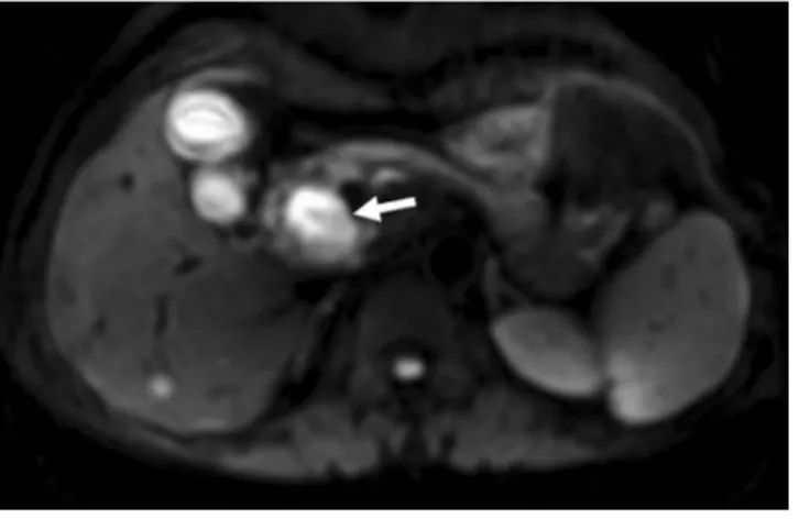
intravenous injection of 20 mL gadobenate dimeglumine (MultiHance; Bracco, Princeton, NJ, USA), dynamic contrast- enhanced imaging was obtained. Hepatobiliary phase images were obtained at 90 minute. Three dimensional heavily T2 weighted MRCP showed no visualization of the cystic lesion at CT, and revealed diffuse narrowing of the CBD by mass effect. The cystic lesion at CT had high signal intensity on DWI and low signal intensity on Fig. 1. (a) Axial portal phase contrast enhanced CT shows a cystic lesion with a thick wall in the portocaval space (arrows). An asterisk denotes the common bile duct (CBD).
(b) Axial portal phase contrast enhanced CT shows a cystic lesion with a thick wall and internal calcified gallstones (arrowhead) in the portocaval space. An asterisk denotes the CBD. (c, d) Axial and coronal portal phase contrast enhanced CT shows communication between the cystic lesion and the CBD (curved arrows). An asterisk denotes the CBD. (e) Axial portal phase contrast enhanced CT shows a small calcified gallstone in the distal CBD (arrowhead).
a
d b
e c
apparent diffusion coefficient (ADC) map (Fig. 2). We thus suspected that the cystic structure was an abscess related to gallstone spillage due to spontaneous CBD perforation.
After the gallstone was removed by endoscopic retrograde cholangiopancreatography (ERCP), elective laparoscopy was performed with the presumptive diagnosis of abscess with gallstone spillage related to spontaneous perforation.
Intraoperatively, the CBD, common hepatic artery and portal vein were difficult to isolate due to severe inflammation and adhesion. An abscess was seen in the posterior aspect of the CBD and a focal defect of the CBD was observed (Fig. 3). Laparoscopic CBD resection with Roux-en-Y hepaticojejunostomy, abscess excision and cholecystectomy was performed. The patient improved and was discharged eight days postoperatively.
DISCUSSION
Spontaneous perforation of the bile duct without any traumatic or iatrogenic injury was first described by Freeland in 1882 (3). It is relatively more common in infants and children than adults and is typically related to congenital malformation in those populations. Spontaneous perforation of the bile duct in adults has been reported in 70 cases in the English literature and the etiology has not been established, but intramural infection, intramural ischemia Fig. 2. (a) Three dimensional MRCP shows mild displacement of the CBD (arrows). The right posterior intrahepatic duct (arrowhead) drains at the mid CBD and the cystic duct (curved arrow) drains at the right posterior intrahepatic duct. (b) A cystic lesion in the portocaval space demonstrates high signal intensity on diffusion weighted MRI (b factor = 800 s/mm2) (arrow). (c) A cystic lesion in the portocaval space demonstrates low signal intensity on the apparent diffusion coefficient map (arrow).
a b
c
Fig. 3. The surgical specimen shows a mural defect in the CBD (arrow).
or increased intraductal pressure due to obstruction by gallstones are commonly considered and a combination of these factors is likely responsible for bile duct perforation (1, 4, 5). Bile duct perforation is uncommonly associated with tumor and the resulting gradual increase in intraductal pressure. The sudden increase in pressure caused by gallstone obstruction is the more common cause; 70% of the reported cases are associated with gallstone obstruction (1, 4, 6). When spontaneous perforation of the bile duct in adults is categorized by perforation site, extrahepatic bile duct perforations are common, but the most common site is the CBD (4). In this case, the perforation site was the CBD and the cause was obstruction by gallstones.
Spontaneous perforation of the bile duct in adults presents as an acute abdominal condition and is difficult to diagnose prior to surgical intervention (1). It usually presents as bile peritonitis or intraabdominal abscess, among other conditions. Sometimes, ultrasonography, CT, or MRCP are useful for detecting pathologic lesions or fluid collection, but a definite diagnosis of spontaneous perforation of the bile duct is made based on intraoperative detection of bile ascites or abscess with gallstones (5).
In the case of bile peritonitis, scintigraphy, ERCP and MRCP with a hepatocyte-selective contrast agent enable localization of bile leaks (7). In this case, we first diagnosed biliary diverticulum and CBD obstruction by gallstones on CT. Biliary diverticulum, or Todani type II choledochal cyst, is a rare disorder representing between 0.8% and 5% of all reported choledochal cyst cases. It presents with variable symptoms and, in some cases, coexistent gallstones (8).
MRCP was performed for evaluation of biliary diverticulum and we diagnosed abscess-related gallstone spillage due to spontaneous CBD perforation. On DWI, high signal intensity and low ADC values indicate diffusion restriction, such as high viscosity of the cystic lesion, malignant tumor, etc. (9) (Fig. 2).
If spontaneous perforation of the bile duct is diagnosed in adults, the treatment is based on the patient’s condition.
If the patient’s condition is stable, nonsurgical treatment, which consists of percutaneous drainage of bile collection and decompression of the biliary tree, is considered. ERCP with biliary drainage is first considered for internal drainage and gallstone extraction from the CBD to stop bile leakage, preventing or improving cholangitis (7). A percutaneous drainage procedure is a useful option for intraperitoneal bile and abscess drainage. General surgical treatment via laparoscopy or open procedure in cases of spontaneous CBD perforation consists of drainage of intraperitoneal bile
or abscess, CBD exploration with primary repair, T-tube replacement, and cholecystectomy (1, 5). The most common perforation site in the case of spontaneous perforation is the anterior wall of the CBD, but primary closure is more difficult at other sites and a more invasive operation should be considered. Sometimes intraoperative choledochoscopy was needed to locate the pathologic lesions and rule out distal obstruction (10). In this case, primary repair of the perforation site could not be performed because of severe inflammation, posterior wall perforation and normal variation of the bile duct - the right posterior intrahepatic duct drains at the mid CBD and the cystic duct drains at the right posterior intrahepatic duct (Fig. 2). Laparoscopic CBD resection with Roux-en-Y hepaticojejunostomy, abscess excision and cholecystectomy were performed.
In conclusion, spontaneous perforation of the bile duct in adults is a very rare condition and DWI is helpful for diagnosis of abscess formation related to spontaneous perforation of the common bile duct by a gallstone in this case.
REFERENCES
1. Kang SB, Han HS, Min SK, Lee HK. Nontraumatic perforation of the bile duct in adults. Arch Surg 2004;139:1083-1087
2. Wiseman K, Buczkowski AK, Chung SW, Francoeur J, Schaeffer D, Scudamore CH. Epidemiology, presentation, diagnosis, and outcomes of choledochal cysts in adults in an urban environment. Am J Surg 2005;189:527-531;
discussion 531
3. Freeland J. Rupture of the hepatic duct. Lancet 1882;1:731- 732
4. Mizutani S, Yagi A, Watanabe M, et al. T tube drainage for spontaneous perforation of the extrahepatic bile duct. Med Sci Monit 2011;17:CS8-11
5. Lee HK, Han HS, Lee JH, Min SK. Nontraumatic perforation of the bile duct treated with laparoscopic surgery. J Laparoendosc Adv Surg Tech A 2005;15:329-332
6. Piotrowski JJ, Van Stiegmann G, Liechty RD. Spontaneous bile duct rupture in pregnancy. HPB Surg 1990;2:205-209 7. Thompson CM, Saad NE, Quazi RR, Darcy MD, Picus DD,
Menias CO. Management of iatrogenic bile duct injuries:
role of the interventional radiologist. Radiographics 2013;33:117-134
8. Ouaissi M, Kianmanesh R, Belghiti J, et al. Todani Type II congenital bile duct cyst: European Multicenter Study of the French Surgical Association and literature review. Ann Surg 2015;262:130-138
9. Qayyum A. Diffusion-weighted imaging in the abdomen and pelvis: concepts and applications. Radiographics 2009;29:1797-1810
10. Megison SM, Votteler TP. Management of common bile duct obstruction associated with spontaneous perforation of the biliary tree. Surgery 1992;111:237-239
