60
INTRODUCTION
Granular cell tumors (GCTs) can develop in different parts of the body, but usually occur in the skin or the subcutaneous soft tissue of the head and neck. GCTs of the neurohypophy- sis or pituitary stalk are very rare [1-8]. They account for less than 0.1% of all primary brain tumors, and approximately 1–
1.5% of adult brain tumors [9]. In most cases reported to date, GCTs have been found in the posterior pituitary gland. GCT of the neurohypophysis is difficult to diagnose preoperatively, owing to the lack of specific imaging features [10]. In this pa- per, we report the clinical, radiological, anatomical, and path- ological findings of a patient with GCT of the pituitary stalk, along with a literature review.
CASE REPORT
A 60-year-old man presented to our clinic complaining of intermittent headache and dizziness for 3 months. The patient had no specific neurological or hormonal symptoms. Magnet- ic resonance imaging (MRI) showed iso-signal intensity in the
Granular Cell Tumor in the Pituitary Stalk: A Case Report
Soo Jeong Park, Youn Hyuk Chang, Na-Rae Yang, Eui Kyo Seo
Department of Neurosurgery, Ewha Womans University Mokdong Hospital, Seoul, Korea
Received December 14, 2014 Revised February 16, 2015 Accepted March 24, 2015 Correspondence Eui Kyo Seo
Department of Neurosurgery, Ewha Womans University Mokdong Hospital,
1071 Anyangcheon-ro, Yangcheon-gu, Seoul 158-710, Korea
Tel: +82-2-2650-2651 Fax: +82-2-2650-5052 E-mail: [email protected]
Granular cell tumors (GCTs) have been reported in various tissues, especially the skin and subcutane- ous soft tissue of the head and neck. We report a 60-year-old man who presented with intermittent head- ache and dizziness for 3 months, but no other neurological symptoms. Magnetic resonance imaging (MRI) showed the presence of a mass in the pituitary stalk, and contrast-enhanced MRI showed nodular en- hancement in this region. The lesion was completely excised microscopically via a frontotemporal (pteri- onal) approach. On pathological examination, a final diagnosis of a typical GCT was made.
Key Words Granular cell tumor; Pituitary neoplasms.
pituitary stalk on T1-weighted imaging (T1WI), and contrast- enhanced MRI showed nodular enhancement in this region (Fig. 1). However, these findings were not conclusive, and the differential diagnoses included metastasis, lymphoma, and gli- oma. A pituitary function test revealed high levels of thyroid- stimulating hormone (22.02 μlU/mL). Other laboratory find- ings for hormones and infection markers were normal. The patient was diagnosed with hypothyroidism, and he was pre- operatively administered 50 µg of levothyroxine sodium. Sur- gery was performed for pathologic confirmation.
The tumor adjacent to the pituitary stalk was completely ex- cised via a frontotemporal (pterional) approach. Thinning of the pituitary stalk had been caused by the tumor (Fig. 2). Mac- roscopically, the tumor was a light gray, round, mass-like lesion, 1×1 cm in size. It was relatively solid compared to other pitu- itary gland adenomas. Histopathologic examination revealed a fragment of brain parenchyma, with dense fibrocollagenous tissue admixed with granular cell nests, and multifocal lym- phocytic infiltration. The tumor cells had abundant granular cytoplasm, showed diffuse weak positivity for CD68, and dif- fuse, weak to strong positivity for S-100 (Fig. 3). These results were conclusive for the diagnosis of a typical GCT.
Transient diabetes insipidus occurred immediately post-sur- gery. However, the patient recovered, without the use of hor- mone replacement therapy, within 1 month of surgery. In addi- tion, postoperative MRI revealed an intact pituitary stalk (Fig. 4).
CASE REPORT Brain Tumor Res Treat 2015;3(1):60-63 / pISSN 2288-2405 / eISSN 2288-2413 http://dx.doi.org/10.14791/btrt.2015.3.1.60
This is an Open Access article distributed under the terms of the Creative Commons Attribution Non-Commercial License (http://creativecommons.org/licenses/by-nc/3.0) which permits unrestricted non-commercial use, distribution, and reproduction in any medium, provided the original work is properly cited.
Copyright © 2015 The Korean Brain Tumor Society, The Korean Society for Neuro- Oncology, and The Korean Society for Pediatric Neuro-Oncology
SJ Park et al.
61
DISCUSSION
Anatomically, the neurohypophysis consists of the posterior pituitary gland, pituitary stalk, infundibulum, and median emi- nence. The cellular elements include pituicytes, microglias, and the distal parts of nerve cells from anastomosed blood vessels and the hypothalamus. Pituicytes are considered to be modi- fied neuroglial cells, and show positive staining for glial fibril- lary acidic protein; they have been classified into five different types on the basis of their ultrastructural characteristics: ma- jor, dark, ependymal, oncocytic, and granular [11]. Granular pi- tuicytes contain many granums. GCTs, the most common pri-
mary tumors that develop in the pituitary gland, have similar granums, and some studies have suggested that these tumors originate from granular pituicytes [3,12,13]. Primary tumors that develop in the neurohypophysis are rare, and are known by many different terms such as pituicytoma, infundibuloma, granular cell myoblastoma, choristoma, and GCT [14]. Of these, GCTs, granular cell myoblastomas, and choristomas are syn- onymous, and are composed of polygonal cells with finely gran- ular, eosinophilic, strong periodic acid Schiff-positive cyto- plasm. The cells show little nuclear pleomorphism and no mitotic figures. Tumor cells are reactive for S-100 and CD68 on immu- nohistochemistry, as observed in the present case [15]. These
A B C D
Fig. 1. Preoperative magnetic resonance imaging findings. A: A preoperative T1-weighted gadolinium-enhanced axial image shows a homog- enous enhanced round mass (white arrow). B: A T1-weighted gadolinium-enhanced coronal image shows the pituitary stalk (white arrow). C:
An anterior view of image B shows a round mass (white arrowhead). D: A T2-weighted axial image shows a mass (arrow) with low signal intensity.
A B
Fig. 2. Surgical findings. A: The tumor (asterisk) is round, and adjacent to the pituitary stalk. B: After tumor excision, the pituitary stalk (arrow) ap- pears thinned, but remains intact.
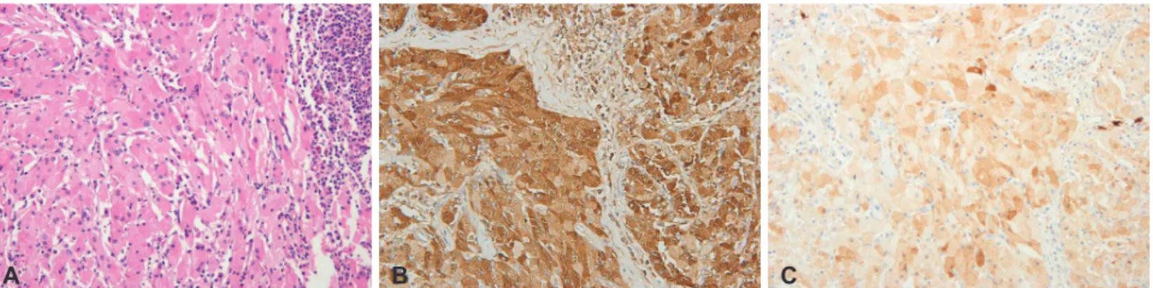
A B C
Fig. 3. Histopathologic findings. A: The tumor consists of large polygonal cells with ample granular cytoplasm and small, oval, eccentric nuclei (he- matoxylin and eosin; original magnification ×100). B: The tumor shows dense fibrocollagenous tissue admixed with granular cell nests. Multifocal lymphocytic infiltration can also be observed (original magnification ×100). C: Immunostaining for S-100 shows diffuse weak to strong positivity (original magnification ×100).
62 Brain Tumor Res Treat 2015;3(1):60-63 GCT of the Pituitary Stalk
tumors can be differentiated from pituitary adenomas by us- ing endocrine markers such as chromogranin A, growth hor- mone, and adrenocorticotropic hormone [16].
A previous study reported that the number of cases of GCT involving the posterior pituitary gland was small, and 50 of these cases were symptomatic GCTs. Furthermore, all these cases were clinically benign.
GCTs of the neurohypophysis are difficult to diagnose pre- operatively, owing to the lack of specific imaging features [10].
However, despite this, computed tomography (CT) and MRI are useful for visualizing tumor expansion to the sellar or su- prasellar region. CT usually shows hyperdense tumors with con- trast enhancement. MRI shows high signal intensity on T1WI for normal posterior pituitary gland, while gadolinium dieth- ylenetri-aminepentaacetic acid-enhanced MRI shows iso-in- tense tumors with uneven contrast enhancement on T1WI, in- dicating fat components or neural hormones, as observed in this case. The radiologic findings in this case suggested the pos- sibility of tumor, inflammatory pseudotumor, infection, or au- toimmune disease. Infection, inflammatory pseudotumor, and autoimmune disease were excluded on the basis of the labora- tory findings, and it remained only to distinguish between dif- ferent tumor entities using pathological studies. As the imaging findings could not differentiate between metastases, lympho- ma, and glioma, pathologic confirmation using a surgical speci- men was necessary. It is rarely possibility to diagnose metastatic tumors on the basis of negative tumor marker results, and patho- logic confirmation is indispensable for establishing treatment plans for lymphomas. Pathological examination to determine the degree of cell differentiation and malignancy is also neces- sary to distinguish between high- and low-grade gliomas.
The clinical features of GCTs in the posterior pituitary gland are mainly non-specific. The tumors are usually small, and have
no space-occupying effects. Furthermore, they are usually as- ymptomatic; granular cell nests or pinhead-sized GCTs are ob- served in 6.4–17% of autopsy cases. Their frequency is similar to that found in the pituitary stalk and the posterior hypothal- amus [1]. In rare cases, the tumors can be large, and the large size can result in headaches, visual defects, and endocrine prob- lems such as hypocortisolism and acromegaly [2,17]. The cur- rent case was not associated with hormonal or ophthalmolog- ic symptoms because of the small tumor size. Therefore, complete tumor resection was possible before the tumor became symp- tomatic and began to destroy the pituitary stalk.
Little is known about the natural progression of GCTs, and significant research using large cohorts of patients with GCT has not been performed. Most GCTs are benign, slow-grow- ing tumors. However, they are occasionally associated with in- vasion or recurrence, and therefore, complete surgical resec- tion is the treatment of choice. However, because of the benign, indolent nature of the tumor, partial resection is recommend- ed when there is risk to major blood vessels or vital structures.
In the present case, the tumor was located in the pituitary stalk, and in order to minimize stalk damage, a biopsy was performed for the differential diagnosis. Biopsy findings revealed a solid tumor, and safe surgical resection was deemed possible. In cas- es where stalk injury is likely, further resection would not be performed, and the tumor would remain untreated, as radia- tion therapy is not effective for GCTs.
Here, we report a rare case of GCT of the pituitary stalk, which is difficult to diagnose without pathologic confirmation. The GCT was completely resected, and the patient recovered fully from transient post-operative diabetes insipidus. Long-term follow-up is necessary in cases such as this, as the natural his- tory of GCTs is poorly understood.
Conflicts of Interest
The authors have no financial conflicts of interest.
REFERENCES
1. Cohen-Gadol AA, Pichelmann MA, Link MJ, et al. Granular cell tumor of the sellar and suprasellar region: clinicopathologic study of 11 cases and literature review. Mayo Clin Proc 2003;78:567-73.
2. Cone L, Srinivasan M, Romanul FC. Granular cell tumor (choristoma) of the neurohypophysis: two cases and a review of the literature. AJNR Am J Neuroradiol 1990;11:403-6.
3. Halbauer DJ, Mészáros I, Dóczi T, et al. Rare sellar region tumors. Pathol Oncol Res 2003;9:134-7.
4. Higuchi M, Tsuji M, Ikeda H. Symptomatic hypophyseal granular cell tumour: endocrinological and clinicopathological analysis. Br J Neuro- surg 1997;11:582-6.
5. Hurley TR, D’Angelo CM, Clasen RA, Wilkinson SB, Passavoy RD. Mag- netic resonance imaging and pathological analysis of a pituicytoma: case report. Neurosurgery 1994;35:314-7; discussion 317.
6. Rhee JS, Wackym PA, Hague K, Wolfe D, King WA. Granular cell tumor of the pituitary fossa. Ann Otol Rhinol Laryngol 2002;111:754-8.
7. Vogelgesang S, Junge MH, Pahnke J, Gaab MR, Warzok RW. August 2001:
Fig. 4. Postoperative magnetic resonance imaging findings. A post- operative T1-weighted gadolinium-enhanced coronal image shows resection of the tumor adjacent to the pituitary stalk. The pituitary stalk remains intact.
SJ Park et al.
63
Sellar/suprasellar mass in a 59-year-old woman. Brain Pathol 2002;12:
135-6, 139.
8. Shizukuishi T, Abe O, Haradome H, Fukushima T, Katayama Y, Sugi- tani M. Granular cell tumor of the neurohypophysis with optic tract edema. Jpn J Radiol 2014;32:179-82.
9. Wilkinson MD, Fulham MJ, Besser M. Neuroimaging findings in a su- prasellar granular cell tumor. J Comput Assist Tomogr 2003;27:26-9.
10. Iglesias A, Arias M, Brasa J, Páramo C, Conde C, Fernandez R. MR im- aging findings in granular cell tumor of the neurohypophysis: a difficult preoperative diagnosis. Eur Radiol 2000;10:1871-3.
11. Takei Y, Seyama S, Pearl GS, Tindall GT. Ultrastructural study of the hu- man neurohypophysis. II. Cellular elements of neural parenchyma, the pituicytes. Cell Tissue Res 1980;205:273-87.
12. Figarella-Branger D, Dufour H, Fernandez C, Bouvier-Labit C, Grisoli F, Pellissier JF. Pituicytomas, a mis-diagnosed benign tumor of the neuro-
hypophysis: report of three cases. Acta Neuropathol 2002;104:313-9.
13. Moriyama E, Matsumoto Y, Meguro T, Mano S. Suprasellar granular cell tumor. Neurol Med Chir (Tokyo) 1996;36:237-40.
14. Tomita T, Gates E. Pituitary adenomas and granular cell tumors. Inci- dence, cell type, and location of tumor in 100 pituitary glands at autop- sy. Am J Clin Pathol 1999;111:817-25.
15. Ellison D, Love S, Chimelli L, et al. Neuropathology: A Reference Text of CNS Pathology. 3rd ed. Philadelphia: Elsevier; 2013.
16. Nakamura T, Hirato J, Hotchi M, Kyoshima K, Nakamura Y. Astrocyto- ma with granular cell tumor-like changes. Report of a case with histo- chemical and ultrastructural characterization of granular cells. Acta Pathol Jpn 1990;40:206-11.
17. Losa M, Saeger W, Mortini P, et al. Acromegaly associated with a granu- lar cell tumor of the neurohypophysis: a clinical and histological study.
Case report. J Neurosurg 2000;93:121-6.