Received on February 21, 2013. Revised on March 4, 2013. Accepted on March 11, 2013.
CC This is an open access article distributed under the terms of the Creative Commons Attribution Non-Commercial License (http://creativecommons.org/licenses/by-nc/3.0) which permits unrestricted non-commercial use, distribu- tion, and reproduction in any medium, provided the original work is properly cited.
*Corresponding Author. Young Sang Kim, Department of Biochemistry, College of Natural Sciences, Chungnam National University, Daejeon, Korea. Tel: 82-42-821-5487; Fax: 82-42-822-7548; E-mail: [email protected]
Keywords: Membrane-bound form, Interleukin-12, p35 subunit, p40 subunit, MethA fibrosarcoma Abbreviations: TAA, tumor-associated autigen; CTL, cytotoxic T lymphocyte
Tumor Cell Clone Expressing the Membrane-bound Form of IL-12p35 Subunit Stimulates Antitumor Immune Responses Dominated by CD8 + T Cells
Hoyong Lim, Seon Ah Do, Sang Min Park and Young Sang Kim*
Department of Biochemistry, College of Natural Sciences, Chungnam National University, Daejeon 305-764, Korea
IL-12 is a secretory heterodimeric cytokine composed of p35 and p40 subunits. IL-12 p35 and p40 subunits are sometimes produced as monomers or homodimers. IL-12 is also pro- duced as a membrane-bound form in some cases. In this study, we hypothesized that the membrane-bound form of IL-12 subunits may function as a costimulatory signal for se- lective activation of TAA-specific CTL through direct priming without involving antigen presenting cells and helper T cells.
MethA fibrosarcoma cells were transfected with expression vectors of membrane-bound form of IL-12p35 (mbIL-12p35) or IL-12p40 subunit (mbIL-12p40) and were selected under G418-containing medium. The tumor cell clones were ana- lyzed for the expression of mbIL-12p35 or p40 subunit and for their stimulatory effects on macrophages. The respon- sible T-cell subpopulation for antitumor activity of mbIL- 12p35 expressing tumor clone was also analyzed in T cell subset-depleted mice. Expression of transfected membrane- bound form of IL-12 subunits was stable during more than 3 months of in vitro culture, and the chimeric molecules were not released into culture supernatants. Neither the mbIL- 12p35-expressing tumor clones nor mbIL-12p40-expressing tumor clones activated macrophages to secrete TNF-α.
Growth of mbIL-12p35-expressing tumor clones was more accelerated in the CD8+ T cell-depleted mice than in CD4+ T cell-depleted or normal mice. These results suggest that CD8+ T cells could be responsible for the rejection of mbIL-12p35-expressing tumor clone, which may bypass acti- vation of antigen presenting cells and CD4+ helper T cells.
[Immune Network 2013;13(2):63-69]
INTRODUCTION
Pro-inflammatory cytokine interleukin-12 (IL-12) was origi- nally identified as a NK cell stimulatory factor (1) and a cyto- toxic T lymphocyte (CTL) maturation factor (2,3). IL-12 in- duces Th1 responses (4,5) enhances the generation of CTL and lymphokine activated killer cells (6,7). IL-12 also stim- ulates T cells to produce cytokines including IFN-γ, GM-CSF and TNF-α (8).
IL-12 is a heterodimer formed by p40 and p35 subunits.
The p40 subunit is mainly produced in excess of p35 subunit in dendritic cells and macrophages, and these cells also pro- duce p40 homodimers (9). In addition, both monomers and homodimers of p40 are produced in transfected cells (10) and in mice (11). The biological function of p40 homodimer is controversial yet; acts as a receptor antagonist (12), induces production of TNF-α in macrophages (13,14), and promotes the migration of dendritic cells and macrophages (15,16).
IL-12 is also found as a cell surface-associated form in a hu- man monocytes and a mouse macrophage cell line, and is up-regulated in response to IFN-γ and LPS stimulation (17).
In addition, an alternative membrane-associated form of the p35 molecule may be produced (18). However, the biological
function of membrane-associated forms of the IL-12 and the p35 subunit has not been determined yet.
Administration of recombinant IL-12 exerts anti-tumor effect in experimental tumor models (19,20), but toxicities asso- ciated with systemic administration hamper clinical extension (21-23). To avoid the toxic problem of recombinant cyto- kines, the cytokine gene transfer method was adopted to ach- ieve local cytokine production. Alternatively, expression of cytokine as a membrane-bound form further confined the functional range of the cytokines and lowered toxicity of vari- ous cytokines (24); TNF-α (25,26), GM-CSF (12,27-29), IFN- γ (27), IL-2 (30-34), IL-4 (35,36), and IL-12 (32,33,35,37,38).
We hypothesized that a tumor cell vaccine may avoid such side effect if it stimulates selectively the TAA-specific CD8+ T cells, without involving CD4+ T helper cells and antigen presenting cells like dendritic cells (39).
We generated MethA tumor clones expressing membrane- bound form of IL-12p35 (mbIL-12p35) or IL-12p40 (mbIL- 12p40) and investigated if macrophages are activated by the mbIL-12 expressing tumor clones. Moreover, we examined the tumor growth of mbIL-12p35 expressing tumor clone in CD4+ or CD8+ T cell-depleted mice to understand which T cell subpopulation functions mainly in the immune responses to the tumor clone cells.
MATERIALS AND METHODS Cells and mice
The methylcholanthrene-induced fibrosarcoma MethA (BALB/c origin) was maintained in RPMI-1640 medium (Gibco-BRL, Rockville, MD), supplemented with 10% heat-inactivated fetal bovine serum (FBS; Gibco-BRL), 2 mM L-glutamine (Sigma, St. Louis, MO), 100 U/ml penicillin (Sigma, St. Louis, MO), 100μg/ml streptomycin (Sigma, St. Louis, MO), and at 37oC under 5% CO2. Female BALB/c mice were obtained from Daehan Biolink (Eumseong, Korea), and all mice were used between 6∼8 weeks of age. All animal procedures were ap- proved and guided by the committee for experimental animal care and use at Chungnam National University.
Antibodies and reagents
Control rat-IgG, PE-anti-IL-12p40, PE-anti-CD4, PE-anti-CD8, purified anti-Ld, PE-conjugated goat anti-rat IgG, PE-con- jugated rat anti-mouse IgG, and recombinant mouse IL-2 and IL-12 were purchased from BD PharMingen (San Diego, CA).
Purified anti-IL-12p35 mAb was purchased from Santa Cruz
Biotechnology (Santa Cruz, CA). Enzyme-linked immuno- sorbent assay (ELISA) kit for mouse TNF-α was purchased from R&D Systems (Menneapolis, MN). G418 were purchased from Sigma (St. Louis, MO).
Vector construction, transfection, and drug selection The construction of pcDNA3.1(+) based chimeric cDNAs ex- pression vectors of mbIL-12p35 and mbIL-12p40 was pre- viously described (38). The 240 bp TNF-α cDNA fragment encoding transmembrane region, cytoplasmic region, and part of the extracellular region was followed by the cDNA frag- ments of IL-12p35 (encoding 215 amino acids) or IL-12p40 (encoding 335 amino acids). MethA cells were transfected with mbIL-12p35, mbIL-12p40, or mock vector alone using the Lipofectamine 2000 (Invitrogen). After 24 hr, cells were plated in 96 well plates (0.5 cell/well) in G418 (1 mg/ml)- containing medium. Viable colonies were usually visible 2∼3 weeks after transfection.
Isolation of mouse peritoneal macrophages
Mice were injected with 0.8 ml of 4% thioglycollate (DIFCO, Detroit, USA) intraperitoneally, and 48 hr later, macrophages were obtained by peritoneal lavage with sterile RPMI-1640 medium containing 1% FBS. Cells were washed three times with RPMI-1640 at 4oC and were maintained in 5% CO2 at 37oC, and plated at a concentration of 1×10⁶ cells/ml (in a volume of 0.5 ml/well) in a 24 well plates. After 1 hr, non-ad- herent cells were removed by washing, and MMC (mito my- cine-C)-inactivated tumor clones (0.5 ml/well) were mixed for co-culture. As a control, LPS (2μg/ml) were added to the adherent cells.
FACS analysis
To analyze the expression of mbIL-12p35 or mbIL-12p40 on cell surface, mbIL-12p35 or mbIL-12p40 clones were in- cubated for 30 min at 4oC with fluorochrome-conjugated IL-12 p35 or p40-specific antibody appropriately diluted in staining buffer (1×PBS containing 0.02% sodium azide, and 2% FBS), respectively. Cells were then washed with staining buffer three times. Stained cells were analyzed on a FACSCalibur flow cytometer with CELLQuest (BD, San Jose, CA).
ELISA
To test of whether mbIL-12p40 and mbIL-12p35 proteins re- leased on tumor clones, 5×10⁵ cells of wild type, mock vector transfected, mbIL-12p35 and mbIL-12p40 clones were cultured
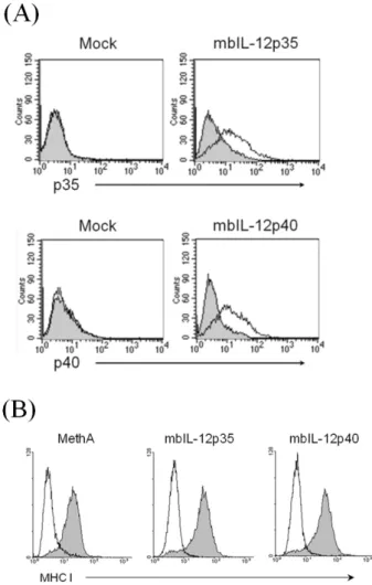
Figure 1. Expression of mbIL-12p35, mbIL-12p40, and MHC class I molecules on transfected MethA tumor cell clones. (A) The mock vector transfected, mbIL-12p35, and mbIL-12p40 expressing tumor clones were stained with anti-IL-12 antibody and analyzed by FACS, (B) Wild type MethA tumor cells, and mbIL-12p35 and mbIL-12 p40 expressing tumor clones were stained with anti-Ld antibody and analyzed by FACS.
for 48 hr in 2 ml of normal medium, respectively. To inves- tigate whether production of TNF-α on macrophages by mbIL-12p35 or mbIL-12p40 tumor clones, 1×10⁶ cells of mouse peritoneal macrophages were co-cultured with 5×10⁵ cells of MMC-treated wild type, mock vector transfected, mbIL-12p35 and mbIL-12p40 expressing tumor clones for 24 hr, respec- tively. All culture supernatants were harvested and measured for TNF-α and IL-12 levels by specific ELISA kit of correspond- ing cytokines following the manufacturer’s instructions. As a positive controls for IL-12 and TNF-α, culture supernatant from the LPS (2μg/ml)-treated mouse peritoneal macrophages was used.
Tumor growth in CD4+ T cell or CD8+ T cell-depleted mice
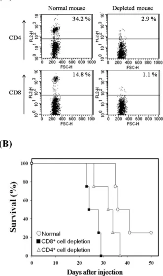
To deplete CD4+ T cells or CD8+ T cells in vivo, mice were injected intraperitoneally with antibodies specific to CD4 (GK1.5, 200μg/time) or CD8 (53-6.72, 200μg/time), re- spectively, on days -3, 0, 3, 7. Depletion of the target cells was confirmed by FACS analysis using CD4 and CD8-specific antibodies. After injection of mbIL-12p35 expressing tumor clone (5×10⁴ cells) on day 0, survival was monitored.
RESULTS
The mbIL-12 subunit molecules are stably expressed on tumor cell surface stably
The MethA cells were transfected with mbIL-12p35 or mbIL- 12p40 expression vectors based on pcDNA3.1(+) vector.
After transfection, cells were selection with G418-containing medium, and the drug-resistant clones were isolated and cul- tured in mass. The drug-resistant clones were then analyzed for the expression of mbIL-12p35 or mbIL-12p40. To analyze the effect of mbIL-12 on the expression of MHC class I mole- cules, the expression level of MHC class I (Ld) on the tumor clones was analyzed by FACS analysis. The expression of mbIL-12 p35 or p40 molecules was stable for more than three months in vitro culture (Fig. 1A), and they were expressed equivalent levels of MHC class I (Ld) (Fig. 1B), suggesting that the expression of the mbIL-12 subunits do not affect severely the expression of MHC class I.
The mbIL-12p35 and the mbIL-12p40 molecules are not released from the tumor clones
To analyze whether the mbIL-12p35 or the mbIL-12p40 mole- cules are cleaved and released from the mbIL-12p35 or the
mbIL-12p40 expressing tumor clones, culture supernatants of wild type MethA cells, mock vector transfected, mbIL-12p35 and mbIL-12p40 expressing clones were analyzed by ELISA for the presence of IL-12 or its subunits. As a positive control for IL-12, culture supernatant from LPS (2μg/ml)-treated peri- toneal macrophages was used (Fig. 2A). The purity of iso- lated macrophages was measured by FACS analysis using an- ti-CD11 antibody. After 48 hr, the culture supernatants were harvested and measured for IL-12 by ELISA. The mbIL-12p35 or mbIL-12p40 was not detected in any culture supernatant but LPS-activated macrophages (Fig. 2B). This result indicates
Figure 3. The mbIL-12p35 and the mbIL-12p40 expressing tumor clones do not induce TNF-α production on macrophages. Mouse peritoneal macrophages (1×10⁶ cells) were co-cultured with MMC- inactivated wild type MethA cells, mock vector transfected, mbIL- 12p35, or mbIL-12p40 transfected clones (5×10⁵ cells), respectively.
After 24 hr, culture supernatants were harvested and measured for TNF-α level by TNF-α specific ELISA. Culture supernatant from LPS (2μg/ml)-treated mouse peritoneal macrophages was used as a posi- tive control. ND, not detected.
Figure 2. The mbIL-12p35 and the mbIL-12p40 molecules on trans- fected tumor clones are not released. (A) The isolated mouse peri- toneal macrophages were stained with specific antibody to CD11b and analyzed by FACS. (B) Cells (5×10⁵ cells) of the wild type, mock vector transfected, mbIL-12p35, and mbIL-12p40 transfected clones were cultured for 48 hr in 2 ml normal medium, and then culture supernatants were analyzed for IL-12 by ELISA. As a positive control, culture supernatant from LPS (2μg/ml)-treated mouse peritoneal macrophages was used. ND, not detected.
that the membrane-bound form of p35 and p40 molecules are not released from the tumor clones, thus, only cells in phys- ical contact with the mbIL-12p35 or the mbIL-12p40 tumor clone would be affected.
The mbIL-12p35 or the mbIL-12p40 expressing tumor clones fails to induce TNF-α production in macro- phages
Soluble form of the IL-12p40 monomer or homodimer in- duces production of TNF-α on primary macrophages were reported (13). To investigate whether the mbIL-12p35 or the mbIL-12p40 clone induces production of TNF-α on macro- phages, isolated peritoneal macrophages from the normal mouse were co-cultured with MMC-inactivated wild type
MethA cells, mock vector transfectant clone, mbIL-12p35 or mbIL-12p40 expressing tumor clone, respectively. In contrast to the LPS-treated macrophage group, TNF-α levels of the other groups were similar to the group cultured macrophages only (Fig. 3), indicating that the mbIL-12p40 or the mbIL- 12p35 expressing tumor clones do not activate macrophages to produce TNF-α.
Depletion of CD8+ T cells accelerates IL-12p35 ex- pressing MethA tumor cell growth
In previous study, we evaluated tumorigenicity of mbIL-12 subunit expressing tumor clones, and found that the IL-12p35 expressing tumor clone was less tumorigenic than mbIL- 12p40 expressing tumor clone (38). The results suggest that the mbIL-12p35 expressing tumor clone is more effective than mbIL-12p40 expressing tumor clone to activate anti-tumor im- mune responses. To analyze which T cell subpopulation plays a critical role in the anti-tumor immune responses, mice were depleted CD4+ or CD8+ T cells by injecting specific monoclonal antibodies (Fig. 4A). The depleted mice were then injected with mbIL-12p35 expressing tumor clone sub- cutaneously, and survival was monitored. As shown in Fig.
4B, the group depleted with CD8+ T cell subpopulation was most susceptible to the mbIL-12p35 expressing tumor clone,
Figure 4. Survival of T cell subset-depleted mice injected with mbIL- 12p35 expressing tumor clone. To deplete CD4+ or CD8+ T cells in vivo, mice were injected with antibodies specific to CD4 (GK1.5, 200μg/time) or CD8 (53-6.72, 200μg/time) intraperitoneally on days -3, 0, 3, 7, respectively. The depletion of CD4+ or CD8+ T cells was confirmed by FACS analysis of peripheral blood cells from the mice (A). CD4+ or CD8+ T cell-depleted mice were injected with 5×10⁵ cells of mbIL-12p35 expressing tumor clone subcutaneously, and survival (B) were monitored.
suggesting that the CD8+ CTLs are responsible for the im- mune response to mbIL-12p35 expressing tumor clone.
Taken together, the expression of membrane-bound form of cytokine, p35 and p40 subunit of IL-12, was stable for ex- tended period of time, and the molecules were not released into culture supernatants. Moreover, both the mbIL-12p35 and the mbIL-12p40 expressing tumor clones did not activate
macrophages in vitro, but the mbIL-12p35 expressing tumor clone lost tumorigenicity by involving mainly CD8+ T cells.
DISCUSSION
We reported previously that the mbIL-12p35 expressing MethA tumor clone was highly immunogenic in vivo, so that the tu- mor clone failed to form tumor in BALB/c syngeneic mice (38). In this study we investigated more about the expression characteristics of membrane-bound form of IL-12 subunits and their effects on macrophages in vitro. The expression of membrane-bound form of IL-12 p35 or p40 subunits was sta- ble for more than 3 months, and the molecules were not re- leased into culture supernatants. The peritoneal macrophages were not activated by the mbIL-12p35 expressing tumor clones in vitro, but growth of the mbIL-12p35 expressing tu- mor clone in CD8+ T cell-depleted mice was accelerated than in CD4+ T cell-depleted mice and normal mice. These results suggest that the mbIL-12p35 expressing tumor clone cells may stimulate CD8+ T cells by direct priming, without involving antigen presenting cells or CD4+ T helper cells.
The main concern in the clinical application of IL-12 in tu- mor therapy is its systemic side effects. Various toxicities of recombinant IL-12 were recorded in mice and human; ele- vated transaminases, leukopenia, and liver dysfunction (21-23, 40). Tumor cells genetically engineered to express IL-12 also showed side effects (41,42). In our laboratory, IL-2 or IL-4 was expressed on MethA tumor cells and their anti-tumor effects were analyzed (30,34,36). As expected the membrane-bound form of cytokine chimeric molecules with TNF-α were stably expressed for a long time. The chimeric cytokine molecules were not detected in culture supernatant, suggesting that the membrane-bound form may not be shed so much.
With anti-tumor effect of tumor clone expressing the mbIL- 12p35 molecule, we have been interested in elucidating which cell populations are critical to display such anti-tumor effects. As an indirect way we analyzed stimulatory effect with the tumor clones expressing mbIL-12p35 or p40 on peri- toneal macrophages. Clearly the tumor clones were not effec- tive to stimulate the macrophages. These results do not rec- oncile with the positive effect of soluble p40 monomer or ho- modimer to induce TNF-α in macrophages (13). We spec- ulate that the membrane-bound form of p40 on tumor cells may require proper orientation to interact with specific IL-12 receptors. The membrane-bound form may have limited flexi- bility or totally different orientation. Interestingly, the mbIL-
12p35 expressing tumor clone showed anti-tumor effect (38), but the tumor clone failed to activate macrophages to pro- duce TNF-α in vitro. These results suggest that the antigen presenting cells may not be critical to induce anti-tumor effect by the mbIL-12p35 expressing tumor clone, at least in vitro condition. Further study is required to prove whether antigen presenting cells are dispensable for the anti-tumor effects.
Consistently, the growth in vivo of the mbIL-12p35 expressing tumor clone was accelerated in CD8+ T cell-depleted mice than CD4+ T cell-depleted mice as previously reported. We also reported previously that NK cells were not critical in the induction of anti-tumor immunity induced by mbIL-12p35 ex- pressing tumor clone (38).
To develop a tumor cell vaccine that stimulates TAA-specif- ic CD8+ T cells selectively, without involving CD4+ T helper cells and antigen presenting cells like dendritic cells or macro- phages, tumor cells should be equipped with the ability to provide signal 1 and costimulatory signals to fully activate CTLs. Practically, we could induce anti-tumor effect by genet- ically modifying tumor cells to express membrane-bound form of p35 subunit of IL-12.
ACKNOWLEDGEMENTS
This research was supported by Basic Science Research Pro- gram through the National Research Foundation of Korea (NRF) funded by the Ministry of Education, Science and Tech- nology (2010-0006746), and partly by the research fund of Chungnam National University (2012).
CONFLICTS OF INTEREST
The authors have no financial conflict of interest.
REFERENCES
1. Kobayashi, M., L. Fitz, M. Ryan, R. M. Hewick, S. C. Clark, S. Chan, R. Loudon, F. Sherman, B. Perussia, and G.
Trinchieri. 1989. Identification and purification of natural kill- er cell stimulatory factor (NKSF), a cytokine with multiple bi- ologic effects on human lymphocytes. J. Exp. Med. 170:
827-845.
2. Gately, M. K., D. E. Wilson, and H. L. Wong. 1986. Synergy between recombinant interleukin 2 (rIL 2) and IL 2-depleted lymphokine-containing supernatants in facilitating allogeneic human cytolytic T lymphocyte responses in vitro. J. Immu- nol. 136: 1274-1282.
3. Stern, A. S., F. J. Podlaski, J. D. Hulmes, Y. C. Pan, P. M.
Quinn, A. G. Wolitzky, P. C. Familletti, D. L. Stremlo, T.
Truitt, and R. Chizzonite, et al. 1990. Purification to homoge- neity and partial characterization of cytotoxic lymphocyte ma- turation factor from human B-lymphoblastoid cells. Proc.
Natl. Acad. Sci. U. S. A. 87: 6808-6812.
4. Hsieh, C. S., S. E. Macatonia, C. S. Tripp, S. F. Wolf, A.
O'Garra, and K. M. Murphy. 199. Development of TH1 CD4+
T cells through IL-12 produced by Listeria-induced macro- phages. Science 260: 547-549.
5. Manetti, R., P. Parronchi, M. G. Giudizi, M. P. Piccinni, E.
Maggi, G. Trinchieri, and S. Romagnani. 1993. Natural killer cell stimulatory factor (interleukin 12 [IL-12]) induces T helper type 1 (Th1)-specific immune responses and inhibits the de- velopment of IL-4-producing Th cells. J. Exp. Med. 177:
1199-1204.
6. Gately, M. K., B. B. Desai, A. G. Wolitzky, P. M. Quinn, C. M. Dwyer, F. J. Podlaski, P. C. Familletti, F. Sinigaglia, R. Chizonnite, and U. Gubler, et al. 1991. Regulation of hu- man lymphocyte proliferation by a heterodimeric cytokine, IL-12 (cytotoxic lymphocyte maturation factor). J. Immunol.
147: 874-882.
7. Trinchieri, G. 1998. Interleukin-12: a cytokine at the interface of inflammation and immunity. Adv. Immunol. 70: 83-243.
8. Weiss, J. M., J. J. Subleski, J. M. Wigginton, and R. H.
Wiltrout. 2007. Immunotherapy of cancer by IL-12-based cy- tokine combinations. Expert. Opin. Biol. Ther. 7: 1705-1721.
9. D'Andrea, A., M. Rengaraju, N. M. Valiante, J. Chehimi, M.
Kubin, M. Aste, S. H. Chan, M. Kobayashi, D. Young, and E. Nickbarg, et al. Production of natural killer cell stimulatory factor (interleukin 12) by peripheral blood mononuclear cells.
J. Exp. Med. 176: 1387-1398.
10. Gillessen, S., D. Carvajal, P. Ling, F. J. Podlaski, D. L.
Stremlo, P. C. Familletti, U. Gubler, D. H. Presky, A. S. Stern, and M. K. Gately. 1995. Mouse interleukin-12 (IL-12) p40 ho- modimer: a potent IL-12 antagonist. Eur. J. Immunol. 25:
200-206.
11. Heinzel, F. P., A. M. Hujer, F. N. Ahmed, and R. M. Rerko.
1997. In vivo production and function of IL-12 p40 homodimers. J. Immunol. 158: 4381-4388.
12. Ling, P., M. K. Gately, U. Gubler, A. S. Stern, P. Lin, K.
Hollfelder, C. Su, Y. C. Pan, and J. Hakimi. 1995. Human IL-12 p40 homodimer binds to the IL-12 receptor but does not mediate biologic activity. J. Immunol. 154: 116-127.
13. Jana, M., S. Dasgupta, R. N. Saha, X. Liu, and K. Pahan.
2003. Induction of tumor necrosis factor-alpha (TNF-alpha) by interleukin-12 p40 monomer and homodimer in microglia and macrophages. J. Neurochem. 86: 519-528.
14. Jana, M. and K. Pahan. 2009. IL-12 p40 homodimer, but not IL-12 p70, induces the expression of IL-16 in microglia and macrophages. Mol. Immunol. 46: 773-783.
15. Cooper, A. M. and S. A. Khader. 2007. IL-12p40: an in- herently agonistic cytokine. Trends. Immunol. 28: 33-38.
16. Khader, S. A., S. Partida-Sanchez, G. Bell, D. M. Jelley-Gibbs, S. Swain, J. E. Pearl, N. Ghilardi, F. J. Desauvage, F. E. Lund, and A. M. Cooper. 2006. Interleukin 12p40 is required for dendritic cell migration and T cell priming after Mycobacte- rium tuberculosis infection. J. Exp. Med. 203: 1805-1815.
17. Fan, X., V. Sibalic, E. Niederer, and R. P. Wüthrich. 1996.
The proinflammatory cytokine interleukin-12 occurs as a cell membrane-bound form on macrophages. Biochem. Biophys.
Res. Commun. 225: 1063-1067.
18. Wolf, S. F., P. A. Temple, M. Kobayashi, D. Young, M. Dicig, L. Lowe, R. Dzialo, L. Fitz, C. Ferenz, R. M. Hewick, et al.
1991. Cloning of cDNA for natural killer cell stimulatory fac- tor, a heterodimeric cytokine with multiple biologic effects on T and natural killer cells. J. Immunol. 146: 3074-3081.
19. Brunda, M. J., L. Luistro, R. R. Warrier, R. B. Wright, B. R.
Hubbard, M. Murphy, S. F. Wolf, and M. K. Gately. 1993.
Antitumor and antimetastatic activity of interleukin 12 against murine tumors. J. Exp. Med. 178: 1223-1230.
20. Nastala, C. L., H. D. Edington, T. G. McKinney, H. Tahara, M. A. Nalesnik, M. J. Brunda, M. K. Gately, S. F. Wolf, R.
D. Schreiber, W. J. Storkus, et al. 1994. Recombinant IL-12 administration induces tumor regression in association with IFN-gamma production. J. Immunol. 153: 1697-1706.
21. Atkins, M. B., M. J. Robertson, M. Gordon, M. T. Lotze, M.
DeCoste, J. S. DuBois, J. Ritz, A. B. Sandler, H. D. Edington, P. D. Garzone, J. W. Mier, C. M. Canning, L. Battiato, H.
Tahara, and M. L. Sherman. 1997. Phase I evaluation of intra- venous recombinant human interleukin 12 in patients with advanced malignancies. Clin. Cancer Res. 3: 409-417.
22. Car, B. D., V. M. Eng, J. M. Lipman, and T. D. Anderson.
1999. The toxicology of interleukin-12: a review. Toxicol.
Pathol. 27: 58-63.
23. Leonard, J. P., M. L. Sherman, G. L. Fisher, L. J. Buchanan, G. Larsen, M. B. Atkins, J. A. Sosman, J. P. Dutcher, N. J.
Vogelzang, and J. L. Ryan. 1997. Effects of single-dose inter- leukin-12 exposure on interleukin-12-associated toxicity and interferon-gamma production. Blood 90: 2541-2548.
24. Kim, Y. S. 2009. Tumor Therapy Applying Membrane-bound Form of Cytokines. Immune Netw. 9: 158-168.
25. Li, Q., L. Li, W. Shi, X. Jiang, Y. Xu, F. Gong, M. Zhou, C. K. 3rd Edwards, and Z. Li. 2006. Mechanism of action differences in the antitumor effects of transmembrane and se- cretory tumor necrosis factor-alpha in vitro and in vivo.
Cancer Immunol. Immunother. 55: 1470-1479.
26. Rieger, R., D. Whitacre, M. J. Cantwell, C. Prussak, and T.
J. Kipps. 2009. Chimeric form of tumor necrosis factor-alpha has enhanced surface expression and antitumor activity.
Cancer Gene Ther. 16: 53-64.
27. el-Shami, K. M., E. Tzehoval, E. Vadai, M. Feldman, and L.
Eisenbach. 1999. Induction of antitumor immunity with modi- fied autologous cells expressing membrane-bound murine cytokines. J. Interferon. Cytokine. Res. 19: 1391-1401.
28. Soo Hoo, W., K. A. Lundeen, J. R. Kohrumel, N. L. Pham, S. W. Brostoff, R. M. Bartholomew, and D. J. Carlo. 1999.
Tumor cell surface expression of granulocyte-macrophage colony-stimulating factor elicits antitumor immunity and pro- tects from tumor challenge in the P815 mouse mastocytoma tumor model. J. Immunol. 162: 7343-7349.
29. Yei, S., R. M. Bartholomew, P. Pezzoli, A. Gutierrez, E.
Gouveia, D. Bassett, W. Soo Hoo, and D. J. Carlo. 2002.
Novel membrane-bound GM-CSF vaccines for the treatment
of cancer: generation and evaluation of mbGM-CSF mouse B16F10 melanoma cell vaccine. Gene Ther. 9: 1302-1311.
30. Chang, M. R., W. H. Lee, J. W. Choi, S. O. Park, S. G. Paik, and Y. S. Kim. 2005. Antitumor immunity induced by tumor cells engineered to express a membrane-bound form of IL-2.
Exp. Mol. Med. 37: 240-249.
31. Choi, J. W., H. Y. Lim, M. R. Chang, J. Y. Cheon, and Y.
S. Kim. 2008. Anti-tumor immunity induced by tumor cells expresing a membrane-bound form of IL-2 and SDF-1. Animal Cells and Systems 12: 193-201.
32. Ji, J., J. Li, L. M. Holmes, K. E. Burgin, X. Yu, T. E. Wagner, and Y. Wei. 2002. Glycoinositol phospholipid-anchored inter- leukin 2 but not secreted interleukin 2 inhibits melanoma tu- mor growth in mice. Mol. Cancer Ther. 1: 1019-1024.
33. Ji, J., J. Li, L. M. Holmes, K. E. Burgin, X. Yu, T. E. Wagner, and Y. Wei. 2004. Synergistic anti-tumor effect of glyco- sylphosphatidylinositol-anchored IL-2 and IL-12. J. Gene Med. 6: 777-785.
34. Sonn, C. H., H. R. Yoon, I. O. Seong, M.-R. Chang, Y. C.
Kim, H. C. Kang, S. C. Suh, and Y. S. Kim. 2006. MethA Fibrosarcoma Cells Expressing Membrane-Bound Forms of IL-2 Enhance Antitumor Immunity. J. Microbiol. Biotech. 16:
1919-1927.
35. Chakrabarti, R., Y. Chang, K. Song, and G. J. Prud'homme.
2004. Plasmids encoding membrane-bound IL-4 or IL-12 strongly costimulate DNA vaccination against carcinoem- bryonic antigen (CEA). Vaccine 22: 1199-1205.
36. Kim, Y. S., C. H. Sonn, S. G. Paik, and A. L. Bothwell. 2000.
Tumor cells expressing membrane-bound form of IL-4 induce antitumor immunity. Gene Ther. 7: 837-843.
37. Cimino, A. M., P. Palaniswami, A. C. Kim, and P. Selvaraj.
2004. Cancer vaccine development: protein transfer of mem- brane-anchored cytokines and immunostimulatory molecules.
Immunol. Res. 29: 231-240.
38. Lim, H. Y., H. Y. Ju, H. Y. Chung, and Y. S. Kim. 2010.
Antitumor effects of a tumor cell vaccine expressing a mem- brane-bound form of the IL-12 p35 subunit. Cancer Biol.
Ther. 10: 336-343.
39. Baek, S., S. J. Lee, M. J. Kim, and H. Lee. 2012. Dendritic Cell (DC) Vaccine in Mouse Lung Cancer Minimal Residual Model; Comparison of Monocyte-derived DC vs. Hemato- poietic Stem Cell Derived-DC. Immune Netw. 12: 269-276.
40. Cohen, J. 1995. IL-12 deaths: explanation and a puzzle.
Science 270: 908.
41. Okada, Y., N. Okada, H. Mizuguchi, K. Takahashi, T.
Hayakawa, T. Mayumi, and N. Mizuno. 2004. Optimization of antitumor efficacy and safety of in vivo cytokine gene ther- apy using RGD fiber-mutant adenovirus vector for preexisting murine melanoma. Biochim. Biophys. Acta 1670: 172-180.
42. Sun, Y., K. Jurgovsky, P. Möller, S. Alijagic, T. Dorbic, J.
Georgieva, B. Wittig, and D. Schadendorf. 1998. Vaccination with IL-12 gene-modified autologous melanoma cells: pre- clinical results and a first clinical phase I study. Gene Ther.
5: 481-490.