Received on November 27, 2015. Revised on January 22, 2016. Accepted on January 29, 2016.
CC This is an open access article distributed under the terms of the Creative Commons Attribution Non-Commercial License (http://creativecommons.org/licenses/by-nc/4.0) which permits unrestricted non-commercial use, distribution, and reproduction in any me- dium, provided the original work is properly cited.
*Corresponding Author. Hyunah Lee, R&D Center, Pharmicell Co. Ltd., Seongnam 13229, Korea. Tel: 82-2-3496-0130; Fax: 82-2-3496-0159;
E-mail: [email protected]
#These authors contributed equally to this work.
Abbreviations: CRT, calreticulin; LLC, Lewis Lung Carcinoma; HSPs, Heat Shock Proteins
Immunogenic Cell Death Induced by Ginsenoside Rg3:
Significance in Dendritic Cell-based Anti-tumor Immunotherapy
Keum-joo Son1#, Ki ryung Choi1#, Seog Jae Lee2 and Hyunah Lee1*
1R&D Center, Pharmicell Co. Ltd., Seongnam 13229, 2Department of Thoracic and Cardiovascular Surgery, Jeju National University School of Medicine, Jeju 63241, Korea
Cancer is one of the leading causes of morbidity and mor- tality worldwide; therefore there is a need to discover new therapeutic modules with improved efficacy and safety.
Immune-(cell) therapy is a promising therapeutic strategy for the treatment of intractable cancers. The effectiveness of certain chemotherapeutics in inducing immunogenic tu- mor cell death thus promoting cancer eradication has been reported. Ginsenoside Rg3 is a ginseng saponin that has antitumor and immunomodulatory activity. In this study, we treated tumor cells with Rg3 to verify the significance of inducing immunogenic tumor cell death in antitumor therapy, especially in DC-based immunotherapy. Rg3 kil- led the both immunogenic (B16F10 melanoma cells) and non-immunogenic (LLC: Lewis Lung Carcinoma cells) tu- mor cells by inducing apoptosis. Surface expression of im- munogenic death markers including calreticulin and heat shock proteins and the transcription of relevant genes were increased in the Rg3-dying tumor. Increased calreticulin expression was directly related to the uptake of dying tu- mor cells by dendritic cells (DCs): the proportion of CRT+CD11c+cells was increased in the Rg3-treated group. Interestingly, tumor cells dying by immunogenic cell death secreted IFN-γ, an effector molecule for anti- tumor activity in T cells. Along with the Rg3-induced sup- pression of pro-angiogenic (TNF-α) and immunosup-
pressive cytokine (TGF-β) secretion, IFN-γ production from the Rg3-treated tumor cells may also indicate Rg3 as an effective anticancer immunotherapeutic strategy. The data clearly suggests that Rg3-induced immunogenic tu- mor cell death due its cytotoxic effect and its ability to in- duce DC function. This indicates that Rg3 may be an effec- tive immunotherapeutic strategy.
[Immune Network 2016;16(1):75-84]
Keywords: Ginsenoside Rg3, DC, Immunogenic cell death, CRT, HSPs
INTRODUCTION
Cancer is one of the leading causes of morbidity and mor- tality worldwide. Although, cancer progression is mainly driven by the expansion of tumor cells, tumor micro- environment and/or anti-tumor immunity may also play important roles (1). The main treatment options for cancer are surgery, chemotherapy, and radiotherapy however, these methods have serious side effects including toxicity to normal cell and tissue (2). It is thought that cancer cells show immunogenic or non-immunogenic characteristics in their growth e.g. B16F10, a mouse melanoma cell line and
LLC, a mouse lung cancer cell line, were shown to have immunogenic and non-immunogenic characteristics, re- spectively. Immunogenic tumors may respond more sensi- tively to immunotherapy.
Recent studies have shown the induction of immuno- genic tumor cell death by certain chemotherapeutics as promising strategy for cancer therapy. Immunogenic cell death is characterized by the early cell surface exposure of chaperone proteins calreticulin (CRT) and HSPs, which affect DC maturation and the uptake and presentation of tumor antigens by DCs (3-7). Thus, inducing immunogenic tumor cell death may enhance the effectiveness of DC-based antitumor therapeutic modules.
Among the saponins originating from plant, ginsenoside is derived from Ginseng, which was originally used in an- cient times for the treatment of diseases (8,9). One of the Ginsenosides, Rg3, has been known to kill tumor cells as well as modulate the immune system (10-12). Numerous studies have demonstrated that Rg3 suppresses tumor growth by inhibiting the invasive and metastatic ability of various tumors including lung (13-17) and ovarian carcino- ma cells (18) and/or by promoting the apoptosis of mela- noma cells (19). Very few studies have been done into whether the immunomodulatory activity of Rg3 has any anticancer effects, particularly the role of Rg3 in inducing immunogenic tumor cell death.
In this study, we treated tumor cells with ginsenoside Rg3 with the aim of verifying the significance of inducing immunogenic tumor cell death in antitumor therapy, espe- cially in DC-based immunotherapy.
MATERIALS AND METHODS
Animals
Pathogen-free female C57BL/6 mice, at 5∼6 weeks old, were purchased from the Orient Bio (Seongnam, South Korea). The mice were provided with water and food ad libitum and quarantined under a 12 h light: 12 h dark pho- toperiod in the animal care facility of the Animal Resource Center at the Asan Institute for Life Science and Technolo- gy, Asan Medical Center, Seoul, Korea. Animal care was performed following the ILAR guideline. The mice were acclimated for at least one week before any experiments were conducted.
Reagents
Ginsenoside Rg3 was supplied by Dr. Sung Ho Son (VitroSys Inc., Yeongju, Korea). Doxorubicin hydro- chloride was purchased from Sigma-Aldrich (St. Louis, MO, USA). Dulbecco’s modified Eagle’s medium (DMEM) and gentamicin were obtained from GIBCO laboratories (Grand Island, NY, USA) and fetal bovine serum (FBS) was obtained HyClone Laboratories (Logan, UT, USA).
The following antibodies for flow cytometric phenotyping were purchased from eBioscience (SanDiego, CA, USA);
fluorescence labeled-monoclonal Abs against CRT, HSP60, HSP70, HSP90, and Annexin V/PI. ELISA kits for cyto- kines including IFN-γ, IL-6, TGF-β1, and TNF-α were purchased from eBioscience (SanDiego, CA, USA).
Cell lines
C57BL/6 syngeneic Lewis Lung Carcinoma (LLC) and B16F10 (melanoma) cell lines were purchased from the American Type Culture Collection (ATCC) (Rockville, MD, USA). All cell lines were maintained in Dulbecco’s modified Eagle’s medium (DMEM) supplemented with 10% heat-inactivated fetal bovine serum (FBS) and 10 mg/ml gentamicin.
Cell viability assay
The Cell Counting Kit-8 (CCK-8, Dojindo Labratories, Kumamoto, Japan) was used to measure the cytotoxicity of Rg3 on LLC and B16F10 cells. The cells (2×10³ cells/well) were cultured in the presence of Rg3 (100, 200, 300, 400, 500 μg/ml) for 48 h in 96-well plates at 37oC in a 5% CO2 incubator. After 10 μl CCK-8 solution was added to each well, the plate was re-incubated for 3 h at 37oC and the absorbance at 450 nm was detected using a microplate reader (μ Quant MQX200; Bio-Tek, Winoo- ski, Vermont, USA).
Immunophenotypic analysis of cultured cells by flow cytometry
The phenotype and ability to induce immunogenicity of Rg3-treated cells (LLC, B16F10) were analyzed by direct immunofluorescence staining of cell surface antigens using fluorescein isothiocyanate (FITC)-or phycoerythrin (PE) conjugated antibodies against HSP60, HSP70, HSP90, and CRT. Single cells were incubated with fluorescence-la- beled surface antibodies in PBS with 0.1% sodium azide and 1% FBS (PBS-CS) for 40 min at 4oC. No more than
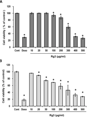
Figure 1. The effect of ginsenoside Rg3 treatment on the viability of LLC and B16F10 cells. The LLC (A) and B16F10 (B) cells (2×10³ cells/well) were seeded in 96-well plates and treated with different Rg3 concentrations (10∼500 μg/ml) and Do- xorubicin (0.01 μg/ml) for 48 h. Each value is the mean±SE of three experiments. Each experiment was done in triplicate.
Significant effects compared to control are indicated with asterisks (*p<0.05 or **p<0.01).
1 h after antibody labeling, cells in the 300 μl PBS-CS analyzed with CytoFLEX (Beckman-Coulter, Pasadena, CA, USA). Data was analyzed using Flowjo Software (LLC, Ashland, OR, USA).
Apoptosis induction and detection
Cell death was assessed by Annexin V-fluorescein iso- thiocyanate staining. Cells were collected, washed in PBS, and resuspended in an incubation buffer containing Annexin V-fluorescein isothiocyanate antibodies. The samples were kept in the dark and incubated for 15 minutes before the addition of 0.1% propidium iodide (PI). They were then analyzed on a CytoFLEX (Beckman- Coulter, Pasadena, CA, USA). Data were analyzed using Flowjo Software (LLC, Ashland, OR, USA).
ELISA assay of cytokine secretion
The concentration of cytokines secreted into the Rg3- treated tumor cell culture supernatants, were measured us- ing commercial ELISA kits for IFN-γ, IL-6, TGF-β1, and TNF-α (eBioscience, SanDiego, CA, USA).
Microarray protocol
Total RNA was isolated, labeled, and prepared for hybrid- ization to an 11K mouse oligonucleotide microarray gene chip (Macrogen Inc., Seoul, Korea) following the manu- facturer’s instructions. Hybridization was then conducted overnight using 15 μg of labeled RNA product, after which the arrays were scanned using Affymetrix scanners.
The gene expression profile of the cells were created using the Affymetrix system (Beyond Bioinformatics ISTECH AATC Gyeonggi, South Korea) in conjunction with the Mouse Genome 430A 2.0 Array, which contains approx- imately 54675 probes. Pre-treatment was conducted using the GCOS global scaling in GenPlex software (Istech Corp., Korea). Differences in the distribution of data were confirmed by comparing an MA plot of the control array to a plot of the experimental array. Data were considered significant when gene expression changed by at least two-fold at three consecutive time-points when compared to the expression of the control (0 h). Increased gene ex- pression also had to include at least one present call (Affymetrix algorithm) or both control points needed to be present when gene expression increased or decreased.
Statistical analysis
The experiments with same protocol were repeated at least 3 times. Data was expressed as the mean±standard error (SE). Statistical significance was determined by a two- tailed Student’s t-Test. P values less than 0.05 or 0.001 were interpreted as statistically significant.
RESULTS
Effect of Ginsenoside Rg3 on cell viability in LLC and B16F10
To evaluate the cytotoxicity of Rg3 against the LLC, non-immunogenic tumor cells and B16F10, immunogenic tumor cells, CCK-8 assay was performed using different
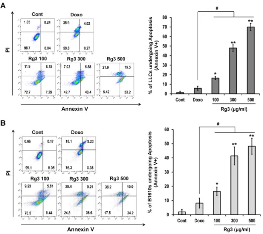
Figure 2. Apoptotic cell death was observed in LLC (A) and B16F10 (B) cell lines. Cells were treated for 18 h with the indicated Rg3 (100, 300, 500 μg/ml) and Doxorubicin (0.01 μg/ml) con- centrations. The percentage of apoptotic cells were determined by measuring Annexin V/PI ex- pression using flow cytometry.
Each experiment was done in triplicate. Significant effects ver- sus control are indicated with asterisks (*p<0.05, **p<0.01);
# indicates the significant chan- ges (#p<0.05) compared to Doxorubicin (0.01μg/ml).
concentrations of Rg3 (10, 20, 50, 100, 200, 300, 400, 500 μg/ml) for 48 h. As shown in Fig. 1A and B, Rg3 kill both LLC and B16F10 cells in a dose-dependent manner.
B16F10 cells were more sensitive to Rg3-induced cell death than LLC. Although much weaker than doxorubicin, a well-known chemotherapeutic agent, this data confirms the direct tumor cell cytotoxicity of Rg3.
Rg3 induced immunogenic death of LLC and B16F10 Assays were employed to investigate the type of cell death induced by Rg3. Rg3 induced apoptosis rather than necrosis. Rg3 induced the early (Annexin V+/PI−) and late (Annexin V+/PI+) stages of apoptosis in LLC and B16F10 tumor cells in a dose-dependent manner (Fig. 2). At a dose that evoked similar cell death (Fig. 1 Doxo 0.01 μg/ml, Rg3 500 μg/ml), this phenomenon was differentiated from Doxorubicin induced death, which was mainly ne- crotic (Annexin V−/PI+).
Expression of immunogenic cell death markers HSP60, HSP70, HSP90 and Calreticulin (CRT) in LLC and B16F10 cell lines
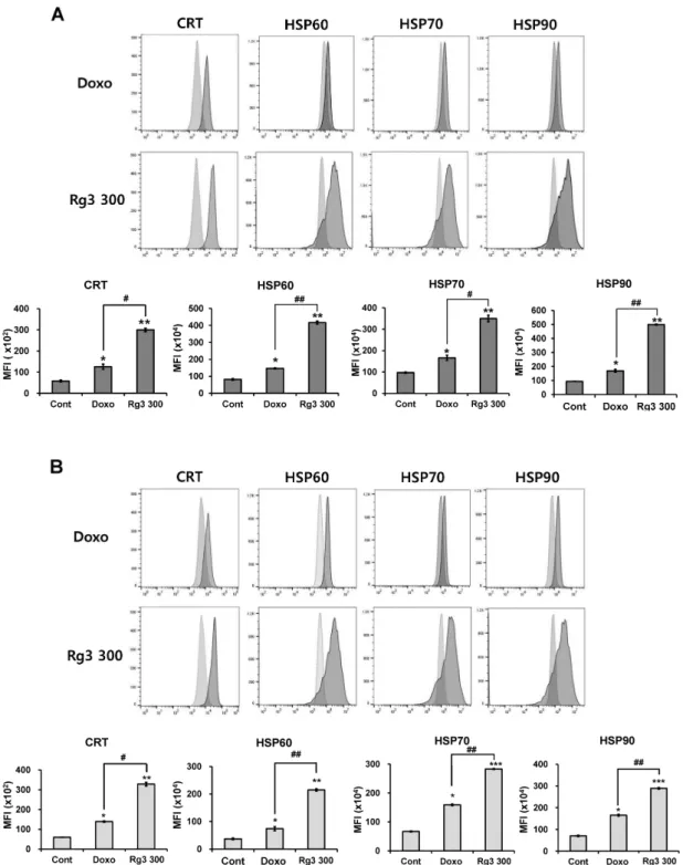
Surface expression of immunogenicity-inducing mole- cules on the tumor cells killed by Rg3: To observe the ability of Rg3 to induce immunogenic cell death, tumor cell surface molecular expressions were analyzed by flow cytometry. Significant expression of HSP60, HSP70, HSP90, and CRT on LLC and B16F10 cells was detected 18 hours after treatment with a 300 μg/ml dose (∼IC50) of Rg3 (Fig. 3A and B). These findings suggest that Rg3-treated tumor cells may be more easily taken up and presented to the immune system by DC, when compared to control cells. After Rg3 treatment, CRT expression in- creased more in B16F10 cells than in LLC cells. On the other hand, in LLC cells, higher levels of HSP were ex- pressed when compared to B16F10 cells.
Cytokine secretion from the Rg3 treated LLC and B16F10 cells: The most significant finding was that the secretion of IFN-γ from both tumor cell lines was induced
Figure 3. Rg3 induced the expression of CRT and HSPs on LLC (A) and B16F10 (B) cell lines. Representative histograms of one of the experiments and mean±SE of three experiments showing the expression of CRT, HSP60, HSP70, and HSP90 after 18 h of Rg3 treatment on tumor cells are shown. Each experiment was done in triplicate. Significant effects versus control are indicated with asterisks (*p
<0.05, **p<0.01, ***p<0.001); #,##indicates the significant changes (#p<0.05 or ##p<0.01) compared to Doxorubicin (0.01 μg/ml).
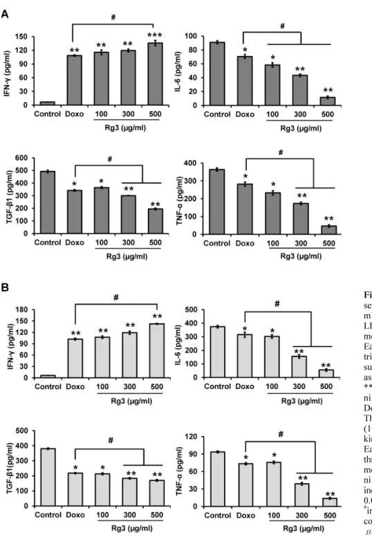
Figure 4. (A) The effect of gin- senoside Rg3 (100, 300, 500 μg/
ml) on cytokine secretion from LLC cells. Each value is the mean±SE of three experiments.
Each experiment was done in triplicate. Significant effects ver- sus control are indicated with asterisks (*p<0.05, **p<0.01,
***p<0.001); #indicate the sig- nificant changes compared to Doxorubicin (0.01 μg/ml). (B) The effect of ginsenoside Rg3 (100, 300, 500 μg/ml) on cyto- kine secretion from B16F10 cells.
Each value is the mean±SE of three experiments. Each experi- ment was done in triplicate. Sig- nificant effects versus control are indicated with asterisks (*p< 0.05, **p<0.01, ***p<0.001);
#indicates the significant changes compared to Doxorubicin (0.01 μg/ml).
by Rg3 treatment in dose-dependent manner (Fig. 4A and B). The secretion of inflammatory cytokines IL-6, TNF-α, and TGF-β1 from the Rg3-treated tumor cells were sig- nificantly reduced.
Increased DC uptake of Rg3 treated tumor cells Splenic DCs (CD11c+) were co-cultured with tumor cells pre-labeled with CRT for 18 h and, using FACS, we ana- lyzed the uptake of tumor cells by DCs (Fig. 5A and B).
Both LLC and B16F10 tumor cells expressed HSPs and
Figure 5. (A) LLC was incubated overnight with or without doxorubicin or Rg3 at 37oC. LLCs were then stained with CRT-FITC. DCs were labeled with CD11c-PerCP5.5 then co-cultured with CRT-FITC labeled LLC for 6 h. FITC and PerCP5.5 double stained cells were considered as DCs taken tumor cells. Each experiment was done in triplicate. (B) B16F10 was incubated overnight with or without doxorubicin or Rg3 at 37oC. B16F10s were then stained with CRT-FITC. DCs were labeled with CD11c-PerCP5.5 then co-cultured with CRT-FITC labeled B16F10 for 6 h. FITC and PerCP5.5 double stained cells were considered as DCs taken tumor cells. Each experiment was done in triplicate. Significant effects versus control are indicated with asterisks (*p<0.05); # indicates the significant changes (#p< 0.05) compared to Doxorubicin (0.01μg/ml).
CRT on their surface after Rg3 treatment (Fig. 3A and B).
To confirm the role of CRT as an ‘eat-me’ signal, DC up- take of Rg3-treated tumor cells expressing CRT was observed. The proportion of CRT+CD11c+ double pos- itive cells in the co-culture of Rg3-treated tumor cell and CD11c+ DCs was measured. Rg3 treatment clearly in- duced DC uptake of LLC (15.3% vs. 39% with 0 or 300 μg/ml Rg3 respectively) and B16F10 (7.72% vs. 52.8%
with 0 or 300 μg/ml Rg3 respectively) tumor cells.
Effect of Rg3 on immunogenic death-related gene ex- pression (microassay)
To observe the differential gene expression related to Rg3-induced immunogenic cell death, mRNA microarray analysis was performed (Fig. 6A and B). The gene tran- scription of CRT (CALR), Hsp60, and HMGB1 which are related to the immunogenicity induction of dead cells, was upregulated in the Rg3-treated tumor cells. On the other hand, VEGF (VEGFA) and TNF (TNFRSF9) receptor gene levels were downregulated by Rg3 treatment. As was expected, the transcription of apoptosis related genes
Figure 6. (A) Clustering of gene expression in LLC after treatment with Doxorubicin or Rg3. 11 genes were selected based on the fold change in expression (log2). Then, these genes were divided into control. The graph produced showed clusters using the Genplex software and the expression levels of genes are shown as graphs. (B) Clustering of gene expression in B16F10 after treatment with Doxorubicin or Rg3. 11 genes were selected based on the fold change in expression (log2). Then, these genes were divided into control.
The graph produced showed clusters using the Genplex software and the expression levels of genes are shown as graphs.
(ATG5, BCL2L13) were upregulated by Rg3 treatment.
DISCUSSION
Ginsenoside Rg3, one of the ginseng saponins, has been reported to possess a potent antitumor and immunomodu- latory activity. Both LLC lung cancer cells and B16F10 melanoma cells were killed by high doses of Rg3 (over 100 μg/ml) in a dose-dependent manner. Melanoma cells were more sensitive to Rg3-induced cytotoxicity than lung cancer cells. Nonetheless, the direct cytotoxicity of Rg3 to tumor cells is not strong enough to be compared to known chemotherapeutics like doxorubicin.
Along with its direct cytotoxicity, the immunomodu- latory activity of Rg3 has been observed as a mechanism of antitumor effect. Thus, the possibility of immunogenic tumor cell death induced by Rg3 has been studied. Immu- nogenic cell death is characterized by the early cell surface exposure of chaperone proteins including CRT and HSPs, which affect DC maturation and the uptake and pre- sentation of tumor antigens. Thus, the induction of im- munogenic tumor cell death may increase the efficacy of antitumor immunotherapy including DC-based therapeutic
cancer vaccines. Immunogenic cell death is initiated by the induction of apoptosis. Translocation of CRT or HSPs onto the dying cell surface is considered as a molecular mecha- nism underlying the increased immunogenicity of apop- totic tumor cells. In this study, Rg3-mediated induction of apoptotic tumor cell death and a subsequent increase in the expression of CRT and HSPs on the surface of dying lung cancer (LLC) and melanoma cells (B16F10) were ob- served. Induction of apoptosis and increased surface pro- tein expression of CRT or Hsp60 on the Rg3-induced dy- ing tumor cells were supported by an increase in the tran- scription of related apoptotic genes (BCL2L13, an apopto- sis facilitator / CALR or HSP60 for CRT or Hsp60, re- spectively) as observed using mRNA microarray analysis.
Another known marker indicating immunogenic cell death, HMGB1, showed increased gene transcription (2∼4 times) in the Rg3-induced dying tumor cells of both LLC and B16F10, when compared to the control. The role of CRT as an ‘eat-me’ signal, presented to DCs by dying tumor cells to signal their uptake, was confirmed by the co-cul- ture of Rg3-treated tumor cell with DCs. DCs took up the Rg3-treated tumor cells more readily than control cells.
This was observed by the increased proportion of CD11c
(DC marker) and CRT double positive cells using flow cytometry.
An interesting phenomenon related to the Rg3-induced immunogenic tumor cell death was the secretion of IFN-γ from Rg3-treated tumor cells. IFN-γ is known to be an antitumor effector molecule produced by T cells not by tu- mor cells. However, our previous study (20) reported that MC38 colon cancer cells killed by chemically inducing im- munogenic cell death secreted IFN-γ. In addition, over- expression of IFN-γ in B16F10 cells inhibited tumor growth and induced apoptotic cell death (21). Along with the Rg3-induced suppression of pro-angiogenic (TNF-α) or immune-suppressive cytokine (TGF-β) secretion, IFN- γ production from the Rg3-treated tumor cells also in- dicates that Rg3 may be an effective anticancer immuno- therapeutic module.
Although Rg3 induced the immunogenic cell death of both LLC, Lewis Lung Carcinoma and B16F10, melanoma cells, each parameter for cell death, surface molecule in- cluding CRT expression and cytokine secretion was differ- entially modulated. B16F10 melanoma cells are known to be immunogenic and were very sensitive to Rg3-induced cell death. However, compared to LLC, the cell death in- duced by Rg3 in B16F10 was more like necrosis. Surface expression of CRT was similar in the two tumor cell lines, but the expression of HSPs was higher in LLC, a typical non-immunogenic tumor cells. There were also a greater proportion of Rg3-treated melanoma cells taken up by DC in comparison to lung cancer cells.
Our data suggests that Rg3 induced immunogenic tumor cell death in not only immunogenic tumor like melanoma but also in non-immunogenic tumor like lung cancer cells.
This ability of Rg3 confers an advantage in using it as an immunotherapeutic module. Most importantly, Rg3-induced cell death can result in the conversion of non-immuno- genic to immunogenic tumor cells. Thus, normally intract- able non-immunogenic tumor cells like lung cancer cells could respond sensitively to antitumor immunity such as DC uptake.
ACKNOWLEDGEMENTS
The authors sincerely thank to Dr. Sung Ho Son at the Vitrosys Inc., Yeongju, Korea for providing ginsenoside Rg3. This study was supported by the grant from the National Research Foundation of Korea (#2014-055-842).
CONFLICTS OF INTEREST
The authors have no financial conflict of interest.
REFERENCES
1. Kim, J. W., S. Y. Jung, Y. H. Kwon, J. H. Lee, Y. M. Lee, B.
Y. Lee, and S. M. Kwon. 2012. Ginsenoside Rg3 attenuates tumor angiogenesis via inhibiting bioactivities of endothelial progenitor cells. Cancer Biol. Ther. 13: 504-515.
2. Nam, K. Y., J. E. Choi, S. C. Hong, K. M. Pyo, and J. D. Park.
2014. Recent Progress in Research on Anticancer Activities of Ginsenoside-Rg3. Kor. J. Pharm. 45: 1-10.
3. Cirone, M., R. L. Di, L. V. Lotti, V. Conte, P. Trivedi, R. Santarelli, R. Gonnella, L. Frati, and A. Faggioni. 2012. Activation of den- dritic cells by tumor cell death. Oncoimmunology 1: 1218-1219.
4. Fucikova, J., P. Kralikova, A. Fialova, T. Brtnicky, L. Rob, J.
Bartunkova, and R. Spisek. 2011. Human tumor cells killed by anthracyclines induce a tumor-specific immune response. Cancer Res. 71: 4821-4833.
5. Zitvogel, L., O. Kepp, L. Senovilla, L. Menger, N. Chaput, and G. Kroemer. 2010. Immunogenic tumor cell death for optimal an- ticancer therapy: the calreticulin exposure pathway. Clin. Cancer Res. 16: 3100-3104.
6. Panaretakis, T., O. Kepp, U. Brockmeier, A. Tesniere, A. C.
Bjorklund, D. C. Chapman, M. Durchschlag, N. Joza, G. Pierron, E. P. van, J. Yuan, L. Zitvogel, F. Madeo, D. B. Williams, and G. Kroemer. 2009. Mechanisms of pre-apoptotic calreticulin ex- posure in immunogenic cell death. EMBO J. 28: 578-590.
7. Obeid, M., A. Tesniere, F. Ghiringhelli, G. M. Fimia, L. Apetoh, J. L. Perfettini, M. Castedo, G. Mignot, T. Panaretakis, N. Casares, D. Metivier, N. Larochette, E. P. van, F. Ciccosanti, M. Piacentini, L. Zitvogel, and G. Kroemer. 2007. Calreticulin exposure dictates the immunogenicity of cancer cell death. Nat. Med. 13: 54-61.
8. Cragg, G. M., D. J. Newman, and K. M. Snader. 1997. Natural products in drug discovery and development. J. Nat. Prod. 60:
52-60.
9. Joo, E. J., Y. W. Ha, H. Shin, S. H. Son, and Y. S. Kim. 2009.
Generation and characterization of monoclonal antibody to ginse- noside rg3. Biol. Pharm. Bull. 32: 548-552.
10. Gao, J. L., G. Y. Lv, B. C. He, B. Q. Zhang, H. Zhang, N. Wang, C. Z. Wang, W. Du, C. S. Yuan, and T. C. He. 2013. Ginseng saponin metabolite 20(S)-protopanaxadiol inhibits tumor growth by targeting multiple cancer signaling pathways. Oncol. Rep. 30:
292-298.
11. Wang, C. Z., H. H. Aung, M. Ni, J. A. Wu, R. Tong, S. Wicks, T. C. He, and C. S. Yuan. 2007. Red American ginseng: ginseno- side constituents and antiproliferative activities of heat-processed Panax quinquefolius roots. Planta Med. 73: 669-674.
12. Liu, T. G., Y. Huang, D. D. Cui, X. B. Huang, S. H. Mao, L.
L. Ji, H. B. Song, and C. Yi. 2009. Inhibitory effect of ginsenoside Rg3 combined with gemcitabine on angiogenesis and growth of lung cancer in mice. BMC Cancer 9: 250.
13. Zhang, Q., X. Kang, and W. Zhao. 2006. Antiangiogenic effect of low-dose cyclophosphamide combined with ginsenoside Rg3 on Lewis lung carcinoma. Biochem. Biophys. Res. Commun. 342:
824-828.
14. Yi, C., X. B. Huang, and M. Hou. 2005. [Experimental study on effect of chemotherapy combined ginsengnoside Rg3 in treating pulmonary carcinoma]. Zhongguo Zhong. Xi. Yi. Jie. He. Za Zhi.
25: 58-59.
15. Hu, S. S., L. K. Zhou, Y. Ba, H. I. Li, and C. H. Zhu. 2011.
A meta-analysis of Ginsenoside Rg3 for non- small cell lung cancer. Clin. Oncol. Cancer Res. 8: 175-180.
16. Liu, J. W., J. X. Chen, L. H. Yu, Y. X. Tian, X. Y. Cui, Q. Yan, and L. Fu. 2004. [Inhibitory effect of ginsenoside-Rg3 on lung metastasis of mouse melanoma transfected with ribonuclease in- hibitor]. Zhonghua Zhong. Liu Za Zhi. 26: 722-725.
17. Xu, T. M., M. H. Cui, Y. Xin, L. P. Gu, X. Jiang, M. M. Su, D. D. Wang, and W. J. Wang. 2008. Inhibitory effect of ginseno- side Rg3 on ovarian cancer metastasis. Chin. Med. J. (Engl.) 121:
1394-1397.
18. Kim, Y. J., W. I. Choi, B. N. Jeon, K. C. Choi, K. Kim, T. J.
Kim, J. Ham, H. J. Jang, K. S. Kang, and H. Ko. 2014.
Stereospecific effects of ginsenoside 20-Rg3 inhibits TGF-beta1-in- duced epithelial-mesenchymal transition and suppresses lung cancer migration, invasion and anoikis resistance. Toxicology 322: 23-33.
19. Lee, S. G., B. S. Kim, and J. O. Nam. 2014. Ginsenoside Rg3 induces apoptosis in B16F10 melonoma cells. J. Life Sci. 24:
1001-1005.
20. Oh, S. J., C. K. Ryu, S. Y. Baek, and H. Lee. 2011. Cellular mech- anism of newly synthesized indoledione derivative-induced im- munological death of tumor cell. Immune Netw. 11: 383-389.
21. Park, D., D. K. Bae, J. H. Jeon, J. Lee, N. Oh, G. Yang, Y. H.
Yang, T. K. Kim, J. Song, S. H. Lee, B. S. Song, T. H. Jeon, S. J. Kang, S. S. Joo, S. U. Kim, and Y. B. Kim. 2011. Immuno- potentiation and antitumor effects of a ginsenoside Rg(3)-fortified red ginseng preparation in mice bearing H460 lung cancer cells.
Environ. Toxicol. Pharmacol. 31: 397-405.