Biomedical Science Letters 2018, 24(4): 349~356 https://doi.org/10.15616/BSL.2018.24.4.349 eISSN : 2288-7415
The Inhibitory Effect of Gooseberry on DNCB-induced Atopic Dermatitis in vivo and in vitro
Su-Jin Kim†,*
Department of Biotechnology and Convergence, Daegu Haany University, Kyungsan 38578, Korea
Generally, berry fruits have various pharmacological activities such as anti-inflammation, anti-oxidation and anti-cancer effects. The effects of gooseberry, a berry fruits, on atopic dermatitis (AD) have not been widely examined. The aim of this present study is to investigate whether gooseberry modulates AD. We examined the pharmacological effects of gooseberry on 2, 4-dinitrochlorobenzene (DNCB)-induced AD symptoms in mice. To determine the anti-atopic mechanism of gooseberry, we investigated its effects on the production of inflammatory cytokines and activation of nuclear factor-κB in PMA + ionophore -stimulated human mast cells (HMC-1). The results demonstrated that gooseberry attenuated AD clinical symptoms such as erythema, edema and dryness as well as histamine and IgE serum levels in DNCB-induced AD model mice. Additionally, gooseberry suppressed the expression of inflammatory cytokines and activation of nuclear factor-κB in stimulated HMC-1. These findings demonstrate that gooseberry is potential agent for treating AD and allergic inflammation.
Key Words: Gooseberry, Atopic dermatitis, Inflammatory cytokines, Nuclear factor-κB
INTRODUCTION
AD is a common chronic inflammatory skin disease that induces intense itching, edema, erythema, thickening, severe pruritus, and eczematous lesions of the skin (Leung and Bieber, 2003). Genetic factos, environmental factors and immune responses were reported to be associated with the pathogenesis and progression of AD (Bieber, 2008). Gener- ally, AD is typically treated with corticosteroids (Berke et al., 2012), but long-term treatments can cause serious side effects such as immunosuppression and epidermal barrier dysfunction (Shiohara et al., 2004). Thus, anti-atopic agents with fewer side effects are needed.
Mast cells contribute to allergic inflammation such as AD (Modena et al., 2016). Mast cells are important effector cells of IgE-mediated allergic inflammatory reactions and IgE levels are related to AD severity (Siraganian, 2003). It was previously reported that mast cells are present in larger numbers in AD lesional skin. In response to various stimuli, mast cells generate a variety of cytokines that contribute to the infiltration of immune cells to sites of inflammation in the skin (Trefzer et al., 2003). Therefore, the suppression of cytokine production is a useful therapeutic strategy for AD and allergic inflammation.
Nuclear factor-kappa B (NF-κB) performs a crucial func- tion by affecting the expression of various genes involved in inflammatory responses (Tegeder et al., 2001). In the nucleus,
Original Article
Received: November 9, 2018 / Revised: December 12, 2018 / Accepted: December 17, 2018
*Professor.
†Corresponding author: Su-Jin Kim. Department of Biotechnology and Convergence, Daegu Hanny University, Kyungsan 38578, Korea.
Tel: +82-53-819-1389, Fax: +82-53-819-1389, e-mail: [email protected]
○CThe Korean Society for Biomedical Laboratory Sciences. All rights reserved.
○CCThis is an Open Access article distributed under the terms of the Creative Commons Attribution Non-Commercial License (http://creativecommons.org/licenses/by-nc/3.0/) which permits unrestricted non-commercial use, distribution, and reproduction in any medium, provided the original work is properly cited.
NF-κB activates gene transcription; thus, NF-κB is important in the regulation of inflammatory responses, by controlling the transcription of inflammatory cytokine genes (Gadaleta et al., 2011). Increased NF-κB activity associated with the secretion of high levels of interleukin (IL)-6 and tumor necrosis factor (TNF)-α was shown to be involved in skin inflammation (Gilmore and Garbati, 2011). The results of previous studies demonstrated that NF-κB activation and the subsequent increase in inflammatory cytokine expression are important in AD pathology.
An increasing number of studies have shown that berry fruits have several pharmacological activities such as anti- inflammation, anti-oxidation and anti-cancer effects (Wan et al., 2012; Xu et al., 2016). Therefore, they are widely used as health care products worldwide. The precise bioactivities of gooseberry, Grossulariaceae family, are unknown. In the present study, we investigated the pharmacological effects of gooseberry on 2, 4-dinitrochlrobenzene (DNCB)-induced AD symptoms in mice. Additionally, we evaluated the effects of gooseberry on inflammatory cytokines production and NF-κB activation in stimulated-human mast cells (HMC-1).
MATERIALS AND METHODS Reagents
DNCB, PMA, calcium Ionophore A23187, avidin peroxi- dase (AP), dimethyl sulfoxide (DMSO) and other reagents were purchased from Sigma-Aldrich (St. Louis, MO, USA).
ELISA kit for human TNF-α/IL-6 was obtained from BD Biosciences. NF-κB antibodies (Abs) were obtained from Santa Cru Biotechnology (Santa Crzu CA, USA).
Animals
BALB/c mice (6 weeks, 19~20 g) were purchased from the Hyochang Science (Daegu, Korea). Animals were housed 6~7 heads per cage in pathogenfree environment to allow them to adapt the environmental changes.
Induction of AD-Like Skin Lesions and gooseberry treat- ment
DNCB (Sigma-Aldrich, St. Louis, MO, USA) was dis- solved in vehicle (3:1 acetone olive oil) and used as a sen-
sitizer for inducing AD-like skin lesions in mice (Yoon et al., 2015). The dorsal skin of BALB/c mice was shaved with depilatory and gauzed a day before sensitization. Mice were randomized divided into 4 groups (n=6/ group): vehicle, DNCB, and DNCB plus treatment of gooseberry (10 mg/kg) or gooseberry (100 mg/kg). Exposed skin was treated with vehicle or 200 μL of a 1% DNCB. On day 4 after sensiti- zation, the dorsal skin was challenged with a 0.5% DNCB (200 μL) solution three times per week. This procedure was repeated for 4 weeks and gooseberry was orally administrated every day for 2 weeks.
Evaluation of skin dermatitis severity
The severity of dermatitis was assessed according to the Eczema Area and Severity Index scoring system: 0, no symptoms; 1, mild symptoms; 2, moderate symptoms; and 3, severe symptoms. The severity of dermatitis was evaluated by the naked eye of three blind examiners. The sum of the individual scores was defined as the dermatitis score for erythema/haemorrhage, edema, excoriation/erosionand sca- ling/dryness (Hanifin et al., 2001).
Cell culture
HMC-1 was maintained in IMDM containing with 100 IU /mL penicillin, 100 μg/mL streptomycin, and 10% FBS at 37℃ in 5% CO2 atmosphere at 95% humidity. HMC-1 was stimulated with 50 nM of PMA plus1 μg/mL A23187 (PMACI).
MTT assay
To investigate the cell viability by gooseberry, the MTT colorimetric assay was performed. Briefly, cells were incu- bated with gooseberry (0.01~1 mg/mL) for 8 h and 50 μL of MTT solution was subsequently added and was incubated for 4 h. After then, the crystallized MTT (formazan) was dissolved in 1 mL of dimethyl sulfoxide and read the absor- bance of plate at 540 nm.
Cytokine assay
TNF-α and IL-6 secretion were measured by modification of an enzyme-linked immunosorbent assay (ELISA) as pre- viously described (Kim et al., 2010). Briefly, 96-well plates
were coated with anti-human monoclonal Abs and incubated overnight at 4℃. After additional washes, sample or standard solution of TNF-α and IL-6 were added and incubated for 2 h. Plates were exposed to biotinylated anti-human Abs was added and incubated for 2 h. After washing plates, AP and ABTS substrate containing H2O2 was sequentially added.
Finally, the optical density of plate was evaluated at 405 nm by a microplate reader.
Histamine assay
The mice were anesthetized with ether following an over- night fast and serum was obtained immediately after blood sampling by centrifugation. Concentrations of histamine in serum were measured with a specific ELISA kit according to the manufacturer's instructions (Neogen, Lexington, USA).
Western blot analysis
To isolate the nuclear extracts, cells were rinsed with PBS and nuclear extracts were prepared by Nuclear Extraction Reagents (Pierce Thermo Scientific, Rockford, USA). After bicinchoninic acid protein quantification, the supernatant was mixed with a sample buffer, separated by gel electro- phoresis, and transferred to membranes. After then, the mem- branes were blocked with 5% skim milk and subsequently reacted with primary Abs. After washing, membranes were then incubated with secondary Abs for 1 h. After washing with 0.1% PBST, protein bands were visualized using an ECL detection system purchased from Pierce Thermo Scien- tific (Rockford, IL, USA).
Statistical analysis
The experiments were shown a summary of the data from at least-three experiments and presented as the mean ± S.D.
Statistical evaluation of the results was performed by inde- pendent t-test. A value of P < 0.05 was considered statisti- cally significant.
RESULTS
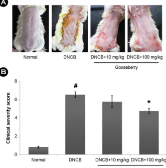
Effect of gooseberry on DNCB-induced AD in mice To characterize the contribution of gooseberry to AD symptoms in mice, DNCB was applied to BALB/c mice.
When the mice were treated for 2 weeks with gooseberry, DNCB-induced the AD symptoms such as erythema, edema and dryness were significantly improved (Fig. 1A). We con- firmed that the skin severity scores in the gooseberry group were significantly lowered compared to those in the DNCB- treated group (Fig. 1B).
Effect of gooseberry on IgE and histamine serum levels in AD mice
An important feature of AD is the pathological secretion of IgE and histamine (Saeki et al., 2009). Thus, we evaluated the effect of gooseberry on IgE and histamine levels in serum using ELISA. As shown in Fig. 2A and B, application of DNCB to mice resulted in increased release of IgE and his- tamine in the serum. In contrast, the gooseberry-treated group showed a considerable reduction in IgE and histamine levels in the serum. The inhibition rates of IgE and histamine by gooseberry (100 mg/kg) were approximately 40.2% and 39.3%, respectively (P<0.05).
Fig. 1. Effect of gooseberry on DNCB-induced AD in mice.
(A) Clinical feature of AD-like skin lesions. (B) The score of skin severity is represented. The results are presented as mean ± SD.
(#P<0.05; significantly different from vehicle control group, *P<
0.05; significantly different from DNCB-treated group).
A
B
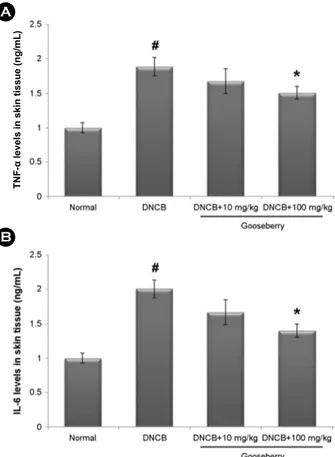
Effect of gooseberry on TNF-α and IL-6 levels in AD- like skin lesion
Increased levels of inflammatory cytokines are associated with the initiation of the inflammatory response in AD. We investigated the effect of gooseberry on TNF-α and IL-6 levels in AD-like skin lesions. At the end of the experiment, skin tissues were homogenized and subjected to ELISA.
TNF-α and IL-6 levels were significantly higher in the skin tissues of DNCB-treated mice than in those of controls.
However, administration of gooseberry reduced the effects induced by DNCB (Fig. 3). The inhibition rates of TNF-α and IL-6 levels by gooseberry (100 mg/kg) were approxi- mately 41.2% and 38.4%, respectively (P<0.05).
Effect of gooseberry on inflammatory cytokine release in PMACI-stimulated HMC-1 cells
We determined the anti-inflammatory mechanism of goo-
seberry in the human mast cell line. Cells were pretreated with gooseberry (0.01~1 mg/mL) and then incubated with PMACI for 8 h. First, the cytotoxic effect of gooseberry was measured via MTT assay. We observed that gooseberry did not affect cell viability (Fig. 4A). We determined whether gooseberry regulates the PMACI-induced production of TNF-α and IL-6 via ELISA. The results showed that goose- berry significantly suppressed the increases in TNF-α and IL-6 induced by PMACI in a dose-dependent manner. The maximum rates of TNF-α and IL-6 suppression by goose- berry (1 mg/mL) were approximately 45.7%, and 40.24%, respectively.
A
B
Fig. 3. The effect of gooseberry and TNF-α and IL-6 levels in AD-like skin lesion. (A and B) At the end of experiment, the skin tissues were cut out and homogenized. The level of TNF-α and IL-6 in the indicated groups was measured via ELISA. The results are pre- sented as mean ± SD. (#P<0.05; significantly different from vehicle control group, *P<0.05; significantly different from DNCB-treated group).
Fig. 2. Effect of gooseberry on the IgE and histamine serum levels. (A and B) Blood samples in DNCB-induced AD mice were collected and then levels of serum IgE and histamine were measured using ELISA method. The results are presented as mean ± SD. (#P<
0.05; significantly different from vehicle control group, *P<0.05;
significantly different from DNCB-treated group).
A
B
TNF-α levels in skin tissue (ng/mL)
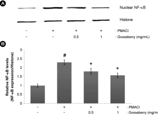
Effect of gooseberry on NF-κB activation in PMACI- stimulated HMC-1 cells
As the NF-κB activation is associated with the inflam- matory response, we predicted that the effects of gooseberry are mediated via suppression of NF-κB activation. As acti- vation of NF-κB requires the translocation of the NF-κB into nucleus, we evaluated the effects of gooseberry on the nuclear pool of NF-κB via western blot analysis. In PMACI- stimulated cells, the levels of NF-κB in the nucleus were increased, but gooseberry reduced these enhanced nuclear levels of NF-κB (Fig. 5A). The relative levels of NF-κB (in the nucleus) are shown in Fig. 5B.
DISCUSSION
Berry fruits are rich in flavonoids, polyphenols, and phe- nolic acids and berry fruits have been reported to have numerous pharmacological activities. Therefore, there are good dietary sources of substances with health benefits. Ho- wever, the precise mechanisms of the effects of gooseberry on AD remain unclear. In this study, we demonstrated the anti-atopic effects of gooseberry in in vivo and in vitro.
AD is known to be result from immune system dysregu- lation, ultimately resulting in allergic inflammation (Gold and Kemp, 2005). IgE dysregulation has been implicated in the pathogenesis of AD and it was reported that the serum IgE concentration is elevated in patients with AD (Allam and Novak, 2006; Brenninkmeijer et al., 2008). Steroid therapy is a crucial factor in the treatment of AD because of its re- markable anti-inflammatory activity. However, steroids can- not be administered long-term because of their deleterious side effects (Das and Panda, 2017). Therefore, natural pro- ducts have gained attention for treating AD (Shiohara et al., 2004). In this study, we found that gooseberry significantly reduced AD symptoms such as erythema, edema and dry- ness in mice. Additionally, we observed that administration of gooseberry suppressed DNCB-induced IgE levels in the serum. In pathological skin conditions, histamine is involved in inducing itching and edema (Minami and Kamei, 2004).
We showed that gooseberry attenuated DNCB-induced his- ta mine levels in the serum. Thus, gooseberry possibly may have therapeutic activity that attenuates the clinical symptoms of AD.
Accumulated experimental evidence shows that inflam- matory cytokines are related to the development of AD. It was also reported that TNF-α and IL-6 levels are elevated in patients with AD and plays an integral role in AD patho- genesis (Wong et al., 2001; Fedenko et al., 2011). These results indicate that new biological therapies for AD should focus on inhibiting inflammatory cytokines. In the present study, we confirmed that the levels of TNF-α and IL-6 were increased in AD-like skin lesions compared to those in con- trols and that treatment with gooseberry reduced these in- creased TNF-α and IL-6 levels. Additionally, we examined Fig. 4. Effects of gooseberry on the production of inflammatory
cytokines in PMACI-stimulated HMC-1 cells. Cells were pre reated with gooseberry (1~100 mg/mL) for 1 h and then stimulated with PMA (50 nM) plus A23187 (1 μg/mL) for 8 h. The TNF-α and IL-6 levels in cell supernatant were evaluated using ELISA.
The results are presented as mean ± SD. (#P<0.05; significantly different from control group, *P<0.05; significantly different from PMACI-treated group).
TNF-α production (ng/mL)
A
B
C
the regulatory effect of gooseberry on intracellular signaling molecules involved in PMACI signaling pathways in HMC-1.
The HMC-1 cell line is a useful for studying cytokine activa- tion pathways (Choi et al., 2011). Mast cells play differential roles in inflammation by initiating and regulating immune responses by releasing various cytokines and chemokines via differential intracellular signal transduction pathways (Harvima and Nilsson, 2011). In response to different stimuli, mast cells release an array of cytokines with the potential to cause inflammation (Galli et al., 2005). Cyclosporin A has been employed previously for treating AD, because it suppresses the cytokine production observed in cases of severe pediatric AD (Bunikowski et al., 2001). In this study, we demonstrated that gooseberry attenuated the release of TNF-α and IL-6 in PMACI-simulated HMC-1 cells. The inhibition rates of TNF-α and IL-6 by gooseberry (1 mg/
mL) were approximately 41.2% and 38.4%, respectively.
These results suggest that gooseberry exerts an anti-atopic effect by suppressing of inflammatory cytokine production.
The production of these cytokines is associated with
activation of the transcription regulator NF-κB (Gilmore and Garbati, 2011). In inactive state, complexes of NF-κB/
inhibitor of κB (IκB) are sequestered in the cytoplasm. In the inflammatory process, IκB kinase (IKK) complex phos- phorylate and degrade the IκB protein and NF-κB is trans- locate into the nucleus where it can combine the promoter of target genes and activate gene expression. Based on these results, suppression of NF-κB activation was identified as an anti-inflammatory strategy. Therefore, we examined whether the anti-atopic effect of gooseberry occurs through the regu- lation of NF-κB activation. The results demonstrate that gooseberry inhibited NF-κB translocation into the nucleus in stimulated HMC-1 cells. We hypothesized that gooseberry exerts anti-atopic effects via the regulation of NF-κB activa- tion. Although gooseberry attenuated NF-κB activation, the effects of gooseberry on pathways involving NF-κB (phos- phorylation of IκB-α and IKK activation) were not deter- mined. Therefore, further studies are necessary to clarify more precisely the role of gooseberry on the NF-κB pathway in mast-cell mediated inflammation.
Fig. 5. Effect of gooseberry on the NF-κB activation of PMACI-stimulated HMC-1 cells. Cells were pre-treated with gooseberry for 1 h and then stimulated with PMA (50 nM) plus A23187 (1 μg/mL) for 2 h. (A) Nuclear extracts were prepared and The NF-κB levels in nucleus measured via Western blot analysis. (B) The relative levels of NF-κB were repre- sented. The results are presented as mean ± SD.
(#P<0.05; significantly different from control group, *P<0.05; significantly different from PMACI-treated group).
Relative NF-κB levels (NF-κB expression/Histone)
A
B
In conclusion, gooseberry can regulate AD clinical sym- ptoms and and IgE and histamine serum levels in a DNCB- induced AD model. Additionally, we demonstrated that the anti-atopic activities of gooseberry can attributed to the re- gulation of inflammatory cytokine expression and NF-κB activation. These results demonstrate that gooseberry may be useful for treating AD and inflammatory skin diseases.
ACKNOWLEDGEMENT
This research was supported by Basic Science Research Program through the National Research Foundation of Korea (NRF) funded by the Ministry of Education (NRF- 2017R1D1A1B03031186).
CONFLICT OF INTEREST
No potential conflict of interest relevant to this article was reported.
REFERENCES
Allam JP, Novak N. The pathophysiology of atopic eczema. Clinical and Experimental Dermatology. 2006. 31: 89-93.
Berke R, Singh A, Guralnick M. Atopic dermatitis: an overview.
American Family Physician. 2012. 86: 35-42.
Bieber T. Atopic dermatitis. The New England Journal of Medicine.
2008. 358: 1483-1494.
Brenninkmeijer EE, Spuls PI, Legierse CM, Lindeboom R, Smitt JH, Bos JD. Clinical differences between atopic and atopiform dermatitis. Journal of the American Academy of Dermatology.
2008. 58: 407-414.
Bunikowski R, Gerhold K, Bräutigam M, Hamelmann E, Renz H, Wahn U. Effect of low-dose cyclosporin a microemulsion on disease severity, interleukin-6, interleukin-8 and tumor necrosis factor alpha production in severe pediatric atopic dermatitis.
International Archives of Allergy and Immunology. 2001. 125:
344-348.
Choi IY, Kim SJ, Kim MC, Kim HL, Shin HJ, Kang TH, Jeong HJ, Shim JS, Kim JH, Yang DC, Hong SH, Kim HM, Um JY. In- hibitory effects of the transgenic Panax ginsengs on phorbol ester plus A23187-induced IL-6 production and cyclooxygenase- 2 via suppression of NF-κB and MAPKs in HMC-1. Immuno- pharmacology And Immunotoxicology. 2011. 33: 205-210.
Das A, Panda S. Use of Topical Corticosteroids in Dermatology: An
Evidence-based Approach. Indian Journal of Dermatology.
2017. 62: 237-250.
Fedenko ES, Elisyutina OG, Filimonova TM, Boldyreva MN, Burmenskaya OV, Rebrova OY, Yarilin AA, Khaitov RM.
Cytokine gene expression in the skin and peripheral blood of atopic dermatitis patients and healthy individuals. Self Nonself.
2011. 2: 120-124.
Gadaleta RM, Oldenburg B, Willemsen EC, Spit M, Murzilli S, Salvatore L, Klomp LW, Siersema PD, van Erpecum KJ, van Mil SW. Activation of bile salt nuclear receptor FXR is repres- sed by pro-inflammatory cytokines activating NF-κB signaling in the intestine. Biochimica et Biophysica Acta. 2011. 1812:
851-858.
Galli SJ, Kalesnikoff J, Grimbaldeston MA, Piliponsky AM, Williams CM, Tsai M. Mast cells as "tunable" effector and immunoregulatory cells: recent advances. Annual Review of Immunology. 2005. 23: 749-786.
Gilmore TD, Garbati MR. Inhibition of NF-κB signaling as a strategy in disease therapy. Current Topics in Microbiology and Immunology. 2011. 349: 245-263.
Gold MS, Kemp AS. Atopic disease in childhood. The Medical Journal of Australia. 2005. 182: 298-304.
Hanifin JM, Thurston M, Omoto M, Cherill R, Tofte SJ, Graeber M. The eczema area and severity index (EASI): assessment of reliability in atopic dermatitis. EASI Evaluator Group. Experi- mental Dermatology. 2001. 10: 11-18.
Harvima IT, Nilsson G. Mast cells as regulators of skin inflam- mation and immunity. Acta Dermato Venereologica. 2011. 91:
644-650.
Kim SJ, Kim MC, Um JY, Hong SH. The beneficial effect of vanillic acid on ulcerative colitis. Molecules. 2010. 15: 7208-7217.
Leung DY, Bieber T. Atopic dermatitis. Lancet. 2003. 361: 151-160.
Minami K, Kamei CA. chronic model for evaluating the itching associated with allergic conjunctivitis in rats. International Immunopharmacology. 2004. 4: 101-108.
Modena BD, Dazy K, White AA. Emerging concepts: mast cell involvement in allergic diseases. Translational Reserch. 2016.
174: 98-121.
Mukaida N. Interleukin-8: an expanding universe beyond neutro- phil chemotaxis and activation. International Journal of Hema- tology. 2000. 72: 391-398.
Saeki H, Furue M, Furukawa F, Hide M, Ohtsuki M, Katayama I, Sasaki R, Suto H, Takehara K. Guidelines for management of atopic dermatitis. Journal of Dermatology. 2009. 36: 563-577.
Shiohara T, Hayakawa J, Mizukawa Y. Animal models for atopic
dermatitis: are they relevant to human disease. Journal of Dermatological Science. 2004. 36: 1-9.
Siraganian RP. Mast cell signal transduction from the high-affinity IgE receptor.Current Opinion in Immunology. 2003. 15: 639 -646.
Tegeder I, Pfeilschifter J, Geisslinger G. Cyclooxygenase-independent actions of cyclooxygenase inhibitors. FASEB Jornal. 2001. 15:
2057-2072.
Trefzer U, Hofmann M, Sterry W, Asadullah K. Cytokine and anti- cytokine therapy in dermatology. Expert Opinion on Biological Therapy. 2003. 3: 733-743.
Wan C, Yuan T, Cirello AL, Seeram NP. Antioxidant and α- glucosidase inhibitory phenolics isolated from highbush blue- berry flowers. Food Chemistry. 2012. 135: 1929-1937.
Wong CK, Ho CY, Ko FW, Chan CH, Ho AS, Hui DS, Lam CW.
Proinflammatory cytokines (IL-17, IL-6, IL-18 and IL-12) and Th cytokines (IFN-gamma, IL-4, IL-10 and IL-13) in patients with allergic asthma. Clinical and Experimental Immunology.
2001. 125: 177-183.
Xu W, Zhou Q, Yao Y, Li X, Zhang JL, Su GH, Deng AP. In- hibitory effect of Gardenblue blueberry (Vaccinium ashei Reade) anthocyanin extracts on lipopolysaccharide-stimulated inflammatory response in RAW 264.7 cells. Journal of Zhejiang University Science B. 2016. 17: 425-436.
Yoon HJ, Jang MS, Kim HW, Song DU, Nam KI, Bae CS, Kim SJ, Lee SR, Ku CS, Jang DI, Ahn BW. Protective effect of diet supplemented with rice prolamin extract against DNCB- induced atopic dermatitis in BALB/c mice. BMC Comple- mentary and Alternative Medicine. 2015. 15: 353.
https://doi.org/10.15616/BSL.2018.24.4.349
Cite this article as: Kim SJ. The Inhibitory Effect of Gooseberry on DNCB-induced Atopic Dermatitis in vivo and in vitro. Biomedical Science Letters. 2018. 24: 349 -356.