119
유제품 발효에서 유산균의 단백질 가수분해 시스템
장운기․설국환․김민경․한기성․정석근․오미화․박범영․함준상*
농촌진흥청 국립축산과학원
Proteolytic Systems of Lactic Acid Bacteria in Milk Fermentation
Oun-Ki Chang, Kuk-Hwan Seol, Min-Kyung Kim, Gi-Sung Han, Seok-Geun Jeong, Mi-Hwa Oh, Beom-Young Park and Jun-Sang Ham*
National Institute of Animal Science, RDA, Suwon 441-350, Korea
Abstract
Lactic acid bacteria (LAB) have been used as starter cultures in the manufacturing processes of fermented dairy products such as cheese and yogurt. LAB have a proteolytic system to use the nitrogen source from milk for their growth. The proteolytic system involved in casein utilization provides cells with essential amino acids during growth in milk and is also of industrial importance, because of its contribution to the development of the organoleptic properties such as flavor of fermented milk products. In the most extensively studied LAB, Lactococcus lactis, the main features of the proteolytic system comprise 3 groups. The first is proteinase, which initially cleaves the milk protein to peptides. The second group consists of transport systems for the internalization of oligopeptides, which are involved in the cellular uptake of small peptides and amino acids. The third group, peptidases in the cell, cleaves peptides into smaller peptides and amino acids. This review is to provide the information about the proteolytic system of LAB.
Keywords: LAB, proteolytic system, milk, proteinase, fermented milk
* Corresponding author: Jun-Sang Ham, National Institute of Animal Science, RDA, Suwon 441-350, Korea. Tel.: +82-31-290-1692, Fax:
+82-31-290-1697, E-mail: [email protected]
서 론
많은 종류의Lactobacillus, Lactococcus, Leuconostoc, Pedi- ococcus, 그리고 Streptococcus의 종을 포함한 유산균(lactic acid bacteria, LAB)은 그람양성균으로 비포자 및 catalase-negative, 조건혐기성으로 정의될 수 있다(Axelsson, 1998).
유산균들은 다양한 발효식품, 음료, 사료를 생산하는데 적 용되어져 왔는데(Leroy과 Devuyst, 2004), 어떤 균들은 특히 Lactobacillus 종 예를 들면 probiotics들은 건강을 증진시키는 요인으로 인해 시장성이 확대되어 가고 있다(Saxelin 등, 2005).
뿐만 아니라 어떠한 균들은 우유 단백질로부터 생리활성 peptide를 생산하기도 한다(Korhonen과 Pihlanto, 2003). 게다
가 유산균의 잠재된 가능성은 살아있는 운반체로서 유용한 molecule(백신, 약)을 장으로 운반하는 역할을 담당한다(Nouaille 등, 2003).
유산균의 가장 중요한 적용은 의심할 여지없이 다양한 발효유제품 제조에서starter로서의 사용이라 할 수 있다. 특 히Streptococcus thermophilus(S. thermophilus), Lactococcus lactis (L. lactis), Lactobacillus helveticus(L. helveticus), 그리고 Lacto- bacillus delbrueckii subsp. bulgaricus(L. bulgaricus)는 아주 광 범위하게starter로서 사용되며, 그들은 중요한 경제적 가치 를 지닌다. 우유발효가공에 있어서, 유산균의 단백가수분해 시스템(proteolytic system)은 중요한 역할을 담당하는데, 그 것은 유산균들이 우유 내에서 성장하기 위한 수단이며, 그로
인해 성공적인 발효가 되기 때문이다. 유산균은 영양요구성
(auxotrophy)의 특징을 지니고 있어 우유 내 질소의 원료로
아미노산이나peptide가 충분하지 않기 때문에 외부로부터
이들을 공급받아야만 한다. 이러한 것들은 우유 단백질에 많이 존재하고, 아미노산의 주요 원료로 이용되는 casein의 가수분해에 의해 제공되어진다.
일반적으로 유산균에 의한casein의 이용은 단백분해효소
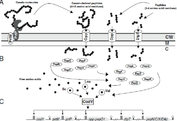
(cell envelope protease, CEP)를 이용하여 단백질을 oligopeptide 로 분해하는 것을 시작으로 진행되어진다. 그 다음 연속적 으로 유산균의 특수한 수송 시스템(transport system)에 의해 서 이 분해된 oligopeptide들을 더 작은 peptide로 분해하기 위하여 유산균 내부로 운반해 오는 과정 그리고 마지막으로 내부로 들어온 peptide들은 다양한 peptidase의 협동작용으 로 인해 peptide들이 di- 혹은 tri-peptide로 분해되거나 혹은 아미노산으로 분해되는 과정으로 이루어진다(Fig. 1)(Kunji 등, 1996; Christensen 등, 1999).
많은 유산균들이CEP를 함유하고 있지만, 이 균들 중 어
떠한 것들은nonstarter 유산균으로 CEP를 함유하지 않는 유
산균도 존재하므로 peptide와 아미노산 생산을 위해서는
starter 유산균의 선발이 중요하다고 볼 수 있다. 이것은 또 한 산업적으로 중요성을 지닌다. 왜냐하면 유산균의 성장뿐 아니라, peptide, 아미노산 그리고 그들이 생산한 또 다른 물
Fig. 1. Representation of the proteolytic system of lactococci in casein breakdown containing theire regulation (model of Doeven et al., 2005). CW: cell wall, M: membrane, C: cytoplasm, A: casein breakdown by cell envelope proteaseand transport and peptide internalization by transport system. B: intracelluar peptidase action on oligopeptide transported in cell. C: regulation of cell envelope protease by CodY
질들은 발효유의 조직과 향미 형성에 영향을 미치는 것은 잘 알려진 사실이기 때문이다. 그러므로 많은 수의 연구는 이렇게 산업적으로 관련이 있는 특성의 근본적인 경로를 규 명하는 것들이 진행되고 있으며, 지금까지 유산균 중에서 가장 광범위하게 사용되는dairy starter인 L. lactis에 대하여 진행되어왔다(Kok과 de Vos, 1994; Poolman 등, 1995; Kunji 등, 1996; Mierau 등, 1997; Christensen 등, 1999; Doeven 등, 2005). L. lactis의 단백질 가수분해 시스템은 casein 분해, 운 반, casein 유래 peptide 그리고 그것의 조절작용에 대한 완전 한 모델은 이미 연구가 많이 진행되어 왔다(Guedon 등, 2001a, b; den Hengst 등, 2005a, b).
현재까지 유산균starter로 사용되며, 유전체가 분석된 유 산균은 L. lactis(Bolotin 등, 2001), S. thermophilus(Bolotin 등, 2004; Delorme 등, 2011), Lactobacillus sakei(Chaillou 등, 2005)이며, 다른 프로바이오틱 유산균으로 B. longum supsp.
longum(Ham 등, 2011), L. plantarum(Kleerebezem 등, 2003), L. acidophilus(Altermann 등, 2005), 그리고 L. johnsonii(Pridmore 등, 2004) 등이 있다.
본 총론은 식품 발효에 연관이 있는 생화학적 및 유전학
적으로 특성화된 단백질 가수분해 시스템 특히 첫 번째 단 계인 단백가수분해효소를 기준으로 이 시스템의 정보를 제 공하는데 있으며, 단백분해를 조절하는 기작의 이해에 있다.
본 론
1. 단백분해효소(Cell Envelope Proteinase, CEP)
유산균에 의한 casein 이용의 첫 번째 단계는 유산균의
CEP에 의한 것이다. 이 효소의 5가지 다양한 종류의 CEPs 가 유산균으로부터 생화학적 및 유전학적으로 복제되었고 특정되었다. 그들의 명명은 다음과 같다(Kok 등, 1988; Holck 과 Naes, 1992; Gilbert 등, 1996; Pederson 등, 1999; Siezen, 1999; Fernandez-Espla 등, 2000; Pastar 등, 2003; Chang, 2011).
1) PrtP ; Lactococcus lactis, Lactobacillus paracasei 2) PrtS ; Streptococcus thermophilus
3) PrtH ; Lactobacillus helveticus 4) PrtB ; Lactobacillus bulgaricus 5) PrtR ; Lactobacillus rhamnosus
유산균은 전형적으로 단지 하나의CEP만을 갖게 되는데,
두 개의CEPs가 L. helveticus와 L. bulgaricus에 존재하는 것 으로 보고되었다(Stefanitsi 등, 1995; Pederson 등, 1999; Sadat- Mekmene 등, 2011). 또한 최근에 S. thermophilus 4F44 종에 서는CEP가 배지 밖으로 유리되어 나오는 CEP(released PrtS) 가 존재한다고 보고하였다. 즉 다른 유산균들처럼 CEP가 세 포벽에 존재하고, 40% 정도가 밖으로 유리되는 것이다(Chang 등, 2012).
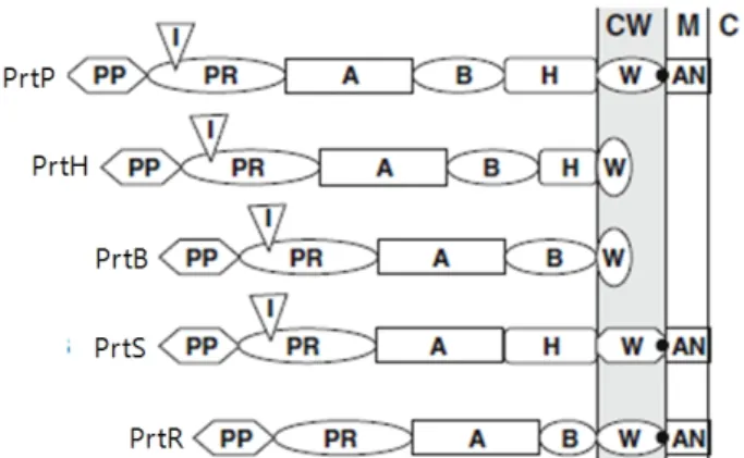
유제품에 이용되는 유산균의CEPs는 일반적으로 대략 2000 아미노산 잔기를 가지는 프리프로단백질(preproprotein)로 합 성된다. 이것은 몇 개의 구별되는 기능적 도메인(functional domain)으로 구성되어 있다(Fernandez-Espla 등, 2000; Siezen, 1999) (Fig. 2). 즉, N-터미널로부터 CEPs는 다음 도메인들을 포함한다.
1) 시그널 시퀀스(signal sequence, ~40 잔기);
CEP를 밖으로 배출시킴. 밖으로 CEP 배출 후 signal peptidase에 의해 제거됨
2) 프로도메인(prodomaine, PP, ~150 residues);
CEP의 maturation에 관여
3) 카탈릭 도메인(catalytic domain, PR, ~500 residues);
serine계 protease로서 catalytic triad (Asp, Ser, His) 부분 이 있어 기질에 작용
4) 삽입 도메인(Insert domain, ~150 residues);
CEPs의 기질 특이성을 조절함 5) A 도메인(A domain, ~400 residues);
아직까지 그 기능이 잘 알려져 있지 않으나, 기질이 결 합하는 부위로 알려짐
6) B 도메인(B domain, ~500 residues);
CEP 활성 및 특이성을 안정화와 관여 7) H 도메인(Helix domain, ~200 residues);
세포 바깥쪽A와 B 도메인의 위치를 조절하는데 관여
8) W domain(cell wall spacer domain, ~100 residues);
친수성이며 세포벽 스페이서로서 기능
B 도메인은 대부분의 CEPs에서 존재하는데 반해, S. ther- mophilus의 PrtS에서는 존재하지 않는다. PrtS의 W 도메인은 그람양성균에서 나타나는 세포벽 도메인의 전형적인 아미 노산 조성을 함유한다. 즉, Pro-Gly 그리고 Ser-Thr이 많이 존재한다. 반면에 H 도메인은 단지 PrtP(210 아미노산), PrtS (367 아미노산), PrtH(72 아미노산)에서만 존재한다. 단백질 가 수분해효소 PrtP와 PrtS에서, W 도메인은 AN 도메인(anchor domain)전에 존재한다. PrtP와 PrtS의 AN 도메인은 그람양 성균의 세포벽 표면에 존재하며, 전형적인 sorting signal을 수행하는데 관련하여 작용한다(Navarre과 Schneewind, 1994).
PrtR의 B 도메인은 다소 그 크기가 작으며, H와 I 도메인이 결여되어 있다. 그것의 W 도메인은 streptococci의 의해 표 현된 어떤 한 세포 표면의 항원과 좀 더 유사하며, AN 도메
Fig. 2. Schematic representation of CEPs of different LAB strains (model according to Siezen, 1999; Fernandez-Espla et al., 2000;
Savijoki et al., 2006).
CW: cell wall, M: membrane, C: cytoplasm, PP: preprodomain, PR: catalytic domain, I: insert domain, A: A domain, B: B domain, H: helix domain, W: cell wall spacer domain, black dot: sorting signal, and AN: anchor domain.
PrtP: Lactococcus lactis Lactobacillus paracasei, PrtS: Streptococcus thermophilus, PrtR: Lactobacillus rhamnosus, PrtH: Lactobacillus helveticus, PrtB: Lactobacillus bulgaricus.
인은 비 전형적sorting signal을 가지고 있다(Pastar 등, 2003).
그람양성균에는 세포벽 표면에 결합하는CEP의 세 가지
기전이 있다. 하나는 가장 일반적으로 peptidoglycan에서 AN 도메인의 sorting 아미노산 인자(sorting motif)인 LPXTG의 Thr의 공유결합에 의해 이루어지는 것이다. 이러한 기전으 로 CEP가 세포벽에 결합하는 것은 CEPs 중 PrtP와 PrtS이 다. 반면에 PrtR의 경우 sorting 아미노산 인자가 비전형적으 로 LPXTG 대신에 MPQAG로 Leu과 Met 잔기가 각각 Met 과 Ala으로 대체되었다(Pastar 등, 2003).
두 번째로는 W 도메인에서 직접 CEP가 결합되는 것이
다. W 도메인은 수소결합 및 전기적 상호작용에 의해 비공 유결합을 야기하는 매우 친수성이며, 양전하를 띠는 아미노
산들이 존재하는W 도메인에 의해 이루어진다. 이러한 경우
에 해당되는 유산균은 L. bulgaricus이다(Germond 등, 2003).
마지막으로 세 번째 기전은W 도메인에 위치하는 S-layer 도메인에 의해 이루어진다. L. helveticus 경우, PrtH은 sorting 아미노산 인자가 존재하지 않아 세포벽에 결합하기 위해 그
들은S-layer라 불리는 부분의 기전을 통하여 세포벽에 결합
한다(Sadat-Mekmene 등, 2011).
이러한 CEP가 세포 밖으로 유리되어 세포벽에 결합하는
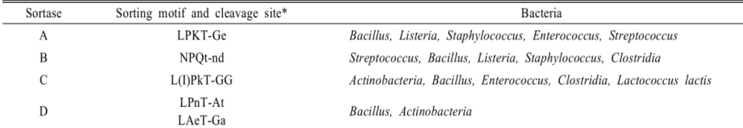
데 있어 또 다른 효소인 transpeptidasase인 sortase의 역할이 중요하다. Sortase 효소는 두 가지 기능을 하는데, 하나는 endopeptidase로서의 역할과 그리고 다른 하나는 transpeptidase 역할을 한다. 그래서 CEP의 AN 도메인에 있는 sorting 아미 노산 인자인 LPXTG를 이 sortase라는 효소가 endopeptidase 로서 작용하여 Thr과 Gly의 peptide 결합을 가수분해하고, Thr은 peptidoglycan에 연결시켜 결합하게끔 하는 작용을 한 다(Chang 등, 2012). 이러한 sortase는 그람양성균인 병원성 미생물의 세포벽에 있는 단백질의 결합 기전과 유사하다.
병원성 미생물을 포함한 그람양성균의sortase에 의해 인식
되는 세포벽에 위치한 단백질의sorting 아미노산 인자와 그
절단부위에 관한 것은Table 1에 나타나 있다. 여기에서 보듯이 각sortase는 특수한 sorting 인자를 가지는 단백질을 세포벽에 결합시킬 수 있다. 예를 들면 2개의 sortase A와 B에 대한 그 특
Table 1. Different classes of sortase in gram positive bacteria
Sortase Sorting motif and cleavage site* Bacteria
A LPKT-Ge Bacillus, Listeria, Staphylococcus, Enterococcus, Streptococcus
B NPQt-nd Streptococcus, Bacillus, Listeria, Staphylococcus, Clostridia
C L(I)PkT-GG Actinobacteria, Bacillus, Enterococcus, Clostridia, Lactococcus lactis
D LPnT-At
LAeT-Ga Bacillus, Actinobacteria
*Uppercase letters represent amino acids that are absolutely conserved.
(Comfort과 Clubb, 2004; Dramsi 등, 2005; Oxaran 등, 2010)
수한sorting 인자로 각각 LPXTG와 NPQTN는 Staphylococcus aureus에서 이미 동정 및 특정화되었다(Marraffini 등, 2006).
최근에Oxaran 등(2010)은 유산균 L. lactis IL1403종에서도 sortase C가 존재한다고 보고하였으며, 이 sortase C는 VYPK 을 포함한LPXTG를 가지고 있는 piline 단백질을 인식한다 고 하였다.
한편, CEP의 maturation에 관해 L. lactis 경우, prtP 유전자 는 세포막에 결합된 lipoprotein(PrtM)을 encoding하면서 전 사에 의해 진행된다. 이 PrtM은 PrtP의 autocatalytic maturation 을 위해 필수적이다(Haandrikman 등, 1989, 1991). 반면에 L.
helveticus, L. bulgaricus, 혹은 S. thermophilus에서는 어떠한 prtM 유전자도 CEP를 encoding하는 부근에서 동정되지 못 하였다(Fernandez-Espla 등, 2000; Siezen, 1999). 이는 다른
방식으로CEP가 이 유산균에서는 진행되어진다고 할 수 있
다. 한편 L. bulgaricus의 CEP의 maturation 과정에서 prtM과 같은chaperone은 요구되어지지 않는다고 보고되었다(Gilbert 등, 1996; Germond 등, 2003).
CEPs는 기질로 유청단백질보다는 casein을 더 선호한다.
Casein은 αs1-, αs2-, β-과 κ-casein로 나뉘는데, 각각은 많 은 수의proline 잔기를 함유하는데, 이것은 α-helices와 β- sheets의 형성을 방해하고, random coils의 형성을 증가시킨 다. 이러한 2차 구조 특성은 CEPs의 작용을 증가시키는 구 조로 변하게 되어CEPs의 접근을 용이하게 할 수 있다(Fox, 1989; Miclo 등 2012).
Lactococcus PrtP는 PI와 PIII type으로 나뉠 수 있는데, 이 것들은 αs1-, αs2-, β-과 κ-casein에 대한 그들의 기질 특이 성에 의해 구분된다(Kunji 등, 1996). PI type의 CEP는 β- casein에 대해 더 많이 분해를 시켜 100개 이상의 다른 oligo- peptide를 생산하는데, peptide의 크기는 4~30개 아미노산 잔기의 범위에 있다(Juillard 등, 1995). κ-casein은 이 효소 에 의해 잘 분해되지 않는다. 반면에 PIII type은 αs1-, αs2-, β-과 κ-casein을 동일하게 잘 분해할 수 있다(Pritchard과 Coolbear, 1993). L. helveticus의 경우, CEPs PI-, PIII-, inter- mediate PI/PIII-type과 어떤 새로운 종류의 기질 특이성이 보
고되었다(Sadat-Mekmene 등, 2011). 반면에 S. thermophilus 에서 분리된CEP인 PrtS는 PI/PIII-type 효소 중간의 기질 특 이성을 보여주었다(Fernandez-Espla 등, 2000).
2. Oligopeptide 운송 시스템
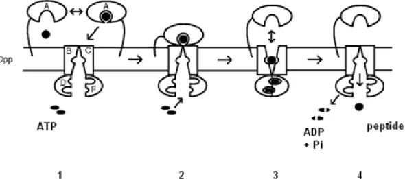
우유 단백질인casein 이용 두 번째 단계 즉 단백가수분해 효소 시스템의2번째 단계는 CEP에 의해 분해된 peptide의 운반에 있는데, 이 작용은 능동적으로 이루어진다. Opp 시스 템이라 불리는 기전에 의해 이루어진다(Doeven 등, 2005).
Opp 단백질은 원래 가지고 있는 ABC type transporter의 주 요transport system이다. L. lactis의 Opp 시스템은 다음과 같 이 구성되어 있다(Fig. 3).
1) OppA: oligopeptide를 고정
2) OppB et OppC: peptide 수송을 형성하는 세포막 단백질 3) OppD et OppF: ATP가 이용되는 ATPase 단백질
L. lactis의 Opp 시스템은 18개의 아미노산 잔기를 가지는 peptide까지 세포 내부로 운반할 수 있으며, 이러한 peptide의 특 성은 transport kinietics에 영향을 준다(Detmers 등, 1998; Juillard 등, 1998). 일반적으로 다른 유산균의 Opp 시스템은 그렇게 많 이 연구되어 있지는 않으나, S. thermophilus(Garault 등, 2002) 와L. bulgaricus(Peltoniemi 등, 2002)에서 Opp 시스템이 Lacto- coccus의 것과 유사한 조성과 양상을 보인다고 보고하였다. S.
thermophilus에서 OppA와 유사한 유전자를 encoding하여 oligo- peptide를 세포 내부로 운반하는데 이용되는데, 그것은 Opp 시스템 대신에 Ami 시스템이라 부른다(Garault 등, 2002).
Fig. 3. Oligopeptide (Opp) transport system model in gram positive bacteria (model according to Doeven et al., 2008). 1: Oligopeptide fixing (OppA), 2: OppA change conformation. ATP is going to fix to ATPase (OppD et F). 3: under ATP fixation, 2 permeases change conformation, oligopeptides are fixed to OppB and OppC. 4: ATP hydrolysis allows to internalize oligopeptide. All systems back to their initial conformation.
L. lactis MG1363와 IL1403 종에서 peptide 운반을 위한 또 다른 시스템이 동정되었는데, 이는 proton motive force(PMF)- driven dipeptide/tripeptide(DtpT)과 ATP-driven Dpp(DtpP) system 을 포함한다(Hagting 등, 1994; Foucaud 등, 1995). Dpp는 di-, tri-, 그리고 tetrapeptides를 수송할 수 있으며, 상대적으로 소 수성branched-chain amino acids(BCAAs)를 포함하며, tripeptides 에 가장 친화력을 보인다(Sanz 등, 2003). 반면에 DtpT는 좀 더 친수성을 띤 그리고 전하를 띤di 그리고 tripeptide 운반 에 적합하다(Hagting 등, 1994). L. helveticus 역시 PMF-driven DtpT를 encoding하는데, 이는 Lactococcus와 유사한 특수성을 지닌다(Nakajima 등, 1997).
3. 세포내 peptidases
우유 단백질을 이용하기 위한 유산균의proteolytic system 의 마지막 단계는 세포내로 들어온oligopeptide가 세포내 다 양한peptidase의 협동작용에 의해 더 분해되는 것이다(Kunji 등, 1996). 유산균에 의해 peptide의 이용은 그들의 peptide 운반 시스템의 특성과 그들의 세포내peptidase 조성의 이해 에 달려 있다.
Peptidase는 exopeptidase 혹은 endopeptidase로 구분되어 질 수 있다. Exopeptidase는 peptide 결합을 가수분해하는데 N-terminal 혹은 C-terminal의 polypeptide의 결합을 분해하고 아미노산이 유리되게 한다. 반면에 endopeptidase는 oligopeptide 중간의 peptide 결합을 분해한다.
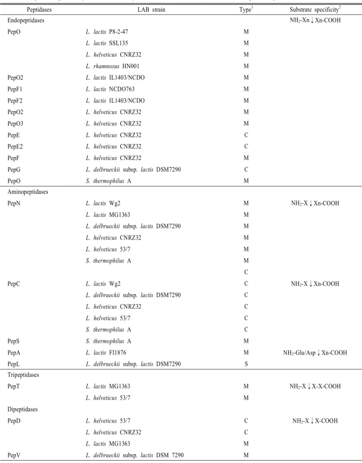
많은 수의 세포내peptidase들이 S. thermophilus 및 다양한 Lactococcus 그리고 Lactobacillus 종에서 생화학적으로 특 정화되었다(Table 2). 세포내 endopeptidases, 일반적인 amino-
Table 2. Peptidases genetically and biochemically characterized from LAB (Model according to Savijoki et al., 2006)
Peptidases LAB strain Type1 Substrate specificity2
Endopeptidases PepO
PepO2 PepF1 PepF2 PepO2 PepO3 PepE PepE2 PepF PepG PepO
L. lactis P8-2-47 L. lactis SSL135 L. helveticus CNRZ32 L. rhamnosus HN001 L. lactis IL1403/NCDO L. lactis NCDO763 L. lactis IL1403/NCDO L. helveticus CNRZ32 L. helveticus CNRZ32 L. helveticus CNRZ32 L. helveticus CNRZ32 L. helveticus CNRZ32
L. delbrueckii subsp. lactis DSM7290 S. thermophilus A
M M M M M M M M M C C M C M
NH2-Xn↓Xn-COOH
Aminopeptidases PepN
PepC
PepS PepA PepL
L. lactis Wg2 L. lactis MG1363
L. delbrueckii subsp. lactis DSM7290 L. helveticus CNRZ32
L. helveticus 53/7 S. thermophilus A
L. lactis Wg2 L. delbrueckii subsp. lactis DSM7290 L. helveticus CNRZ32
L. helveticus 53/7 S. thermophilus A S. thermophilus A L. lactis FI1876
L. delbrueckii subsp. lactis DSM7290
M M M M M M C C C C C C M M S
NH2-X↓Xn-COOH
NH2-X↓Xn-COOH
NH2-Glu/Asp↓Xn-COOH
Tripeptidases PepT
Dipeptidases PepD
PepV
L. lactis MG1363 L. helveticus 53/7
L. helveticus 53/7 L. helveticus CNRZ32 L. lactis MG1363
L. delbrueckii subsp. lactis DSM 7290
M M
C C M M
NH2-X↓X-X-COOH
NH2-X↓X-COOH
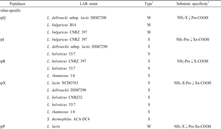
Table 2. Continued
Peptidases LAB strain Type1 Substrate specificity2
Proline-spesific PepQ
PepI
PepR
PepX
PepP
L. delbruecki subsp. lactis DSM7290 L. bulgaricus B14
L. bulgaricus CNRZ 397 L. bulgaricus CNRZ 397
L. delbrueckii subsp. lactis DSM7290 L. helveticus 53/7
L. helveticus CNRZ 397 L. helveticus 53/7 L. rhamnosus 1/6 L. lactis NCDO763 L. delbrueckii DSM7290 L. helveticus CNRZ32 L. helveticus 53/7 L. rhamnosus 1/6 S. thermophilus ACA-DC4 L. lactis
M M M S S S S S S S S S S S S M
NH2-X↓Pro-COOH
NH2-Pro↓Xn-COOH
NH2-Pro↓X-COOH
NH2-X-Pro↓Xn-COOH
NH2-X↓Pro-Xn-COOH
1 Catalytic class of peptidase according to sequence analysis or biochemical characterization.
2 The arrow indicates the cleavage site.
M; Metallopeptidase, C; cysteine-peptidase, and S; serine peptidase
peptidases(PepN과 PepC) 그리고 X-prolyl dipeptidyl amino- peptidase(PepX)는 oligoeptide에 작용하는 첫 번째 peptidase 이다.
Endopeptidase의 공통적 특성은 casein 자체를 분해할 수 있는 능력이 없다는 것이지만, casein 유래 peptide는 그들 내 부의peptide 결합을 분해할 수는 있다는 것이다. 예를 들면 αs1-casein f1-23 및 β-casein f193-209 단편은 유산균 starter 의endopeptidase가 가장 잘 분해를 할 수 있는 단편이다. αs1- casein f1-23 단편과 β-casein f203-209 단편의 post-proline에 대한 유일한 특이성이 non starter인 L. rhamnosus HN001의 PepO에 의해 증명되었으며, starter strain L. helveticus CNRZ32 에서 PepO2에 대해 증명되었다(Christensson 등, 2002; Chen 등, 2003).
L. lactis PepF는 아미노산 7~17 잔기를 가지는 peptide를 분해하는 것뿐만 아니라, 질소 공급원의 부족 상태에서 단 백질 전위에 중요하게 작용한다고 보고되었다(Monnet 등, 1994; Nardi 등, 1997). 광범위한 기질 특이성을 가지는 다양 한 유산균으로부터 특정화된 peptidase로는 metallopeptidase 인 PepN 그리고 cysteine peptidase PepC가 존재한다.
S. thermophilus의 경우, 15개의 peptidase가 기질을 이용하
여 동정되었는데, 3개의 oligoendopeptidase(PepO, PepEP 그 리고PepF), 4개의 aminopeptidase(AP1, PepC, PepN 그리고 PepA), 4개의 proline 관여 peptidase(PepX, Prd : prolidase, Prn : prolinase, PepP : aminopeptidase P) 그리고 4개의 di/tri pep- tidase(DP1, DP2, TP1 그리고 TP2)이다(Rul과 Monnet, 1997).
S. thermophilus KLDS3.0503은 12개의 기능을 하는 세포내 peptidase가 존재한다고 하였는데, 그것들은 PepO, PepA, PepN, PepC, PepS, PepB, PepQ, PepV, PepT, PepM, PepXP, PepP이 다. 이중 생화학적 및 유전학적으로 정제되고 특정화된 6개 의peptiddase는 다음과 같다. PepO(Chavagnat 등, 2000), PepA (Rul 등, 1995), PepN(Rul 등, 1994), PepC(Chapot-Chartier 등, 1994), PepS(Fernandez-Espla과 Rul, 1999), 그리고 PepX(Tsakalidou 등, 1998). 지금까지 carboxypeptidase 활성을 나타내는 유산 균은 보고되어 있지 않다.
결 론
유산균의 단백가수분해 시스템은 그들의 성장을 위해 질 소 공급원으로 우유 단백질을 이용하는데 아주 중요한 역할 을 한다. 이 시스템은 단백질인 casein의 분해, oligopeptide
의 운반 및 작은peptide와 아미노산으로 분해함으로써 유산 균이 이들을 흡수하여 이용하는 것이다. 또한 CodY이란 조 절 단백질을 통하여 단백가수분해 시스템이 조절된다.
단백가수분해 시스템의 많은 연구는L. lactis뿐만 아니라 다른 유산균에서도 진행되고 있다. 이들의 연구는 새로운 유산균의 개발 및 최종제품의 특성과 관련이 있다. 산업적
으로 생산비의 손실을 줄이기 위한 수단으로CEPs를 가지
고 있는 유산균을 선발 및 이용하여 발효시간을 단축시킬 수 있으며, 또한 발효과정 중 CEPs에 의해 다양한 생리기능 성 물질 및peptide의 생산 잠재력이 아주 크다고 볼 수 있 다. 이는 최종 제품에 물성학적 즉 조직감, 향미 등을 느끼 게 하여 소비자들이 선호하는 제품을 생산할 수 있어 그 커 다란 경제적 파급효과를 기대할 수 있을 것이라 본다.
따라서 본 연구는 유산균의 단백질가수분해시스템을 소 개하여 유산균의 우유단백질 이용에 대한 정보를 제공함으 로써 다양한 발효 유제품 및 기능성 신소재 식품 제조에 활 용될 것으로 기대된다.
참고문헌
1. Axelsson, L. 1998. Lactic acid bacteria: classification and physiology. In: Salminen S, von Wright A (eds) Lactic acid bacteria. Microbiology and functional aspects. Marcel Dekker, New York, pp 1-72.
2. Bolotin, A., Wincker, P., Mauger, S., Jaillon, O., Malarme, K., Weissenbach, J., Ehrlich, S. D. and Soroki, A. 2001.
The complete genome sequence of the lactic acid bacterium Lactococcus lactis ssp. lactis IL1403. Genome Res. 11:
731-753.
3. Bolotin, A., Quinquis, B., Renault, P., Sorokin, A., Ehrlich, S. D., Kulakauskas, S., Lapidus, A., Goltsman, E., Mazur, M., Pusch, G. D., Fonstein, M., Overbeek, R., Kyprides, N., Purnelle, B., Prozzi, D., Ngui, K., Masuy, D., Hancy, F., Burteau, S., Boutry, M., Delcour, J., Goffeau, A. and Hols, P. 2004. Complete sequence and comparative genome analysis of the dairy bacterium Streptococcus thermophilus.
Nat. Biotechnol. 22:1554-1558.
4. Chaillou, S., Champomier-Verges, M. C., Cornet, M., Crutz-Le, C., Dudez, A. M., Martin, V., Beaufils, S., Darbon-Rongere, E., Bossy, R., Loux, V. and Zagorec, M. 2005. The complete genome sequence of the meat-borne lactic acid bacterium Lactobacillus sakei 23K. Nat. Biotechnol. 23(12):1527-1533.
5. Chang, O. K. 2011. Caractérisation d'une forme extracellulaire soluble de la protéase PrtS chez Streptococcus thermophilus
4F44 Mise en évidence et détermination de ses sites de coupure sur les caséines. Ph D Thesis, Institut National Polytechnique de Lorraine.
6. Chen, Y. S., Christensen, J. E., Strickland, M. and Steele, J. L. 2003. Identification and characterization of Lactobacillus helveticus PepO2, an endopeptidase with post-proline specificity.
Appl. Environ. Microbiol. 69:1276-1282.
7. Christensen, J. E., Dudley, E. G., Pederson, J. A. and Steele, J. L. 1999. Peptidases and amino acid catabolism in lactic acid bacteria. Antonie Van Leeuwenhoek. 76:217-246.
8. Chang, O. K., Perrin, C., Galia, W., Saulnier, F., Miclo, L., Roux, E., Driou, A., Humbert, G. and Dary, A. 2012.
Release of the cell-envelope protease PrtS in the growth medium of Streptococcus thermophilus 4F44. International Dairy Journal 23(2):91-98.
9. Chapot-Chartier, M. P., Rul, F., Nardi, M. and Gripon, J. C.
1994. Gene cloning and characterization of PepC, a cysteine aminopeptidase from Streptococcus thermophilus, with sequence similarity to the eucaryotic bleomycin hydrolase. Eur. J.
Biochem. 224:497-506.
10. Chavagnat, F., Meyer, J. and Casey, M. 2000. Purification, characterisation, cloning and sequencing of the gene encoding oligopeptidase PepO from Streptococcus thermophilus A.
FEMS Microbiology Letters. 191:79-85.
11. Christensson, C., Bratt, H., Collins, L. J., Coolbear, T., Holland, R., Lubbers, M. W., O’Toole, P. W. and Reid, J.
R. 2002. Cloning and expression of an oligopeptidase, PepO, with novel specificity from Lactobacillus rhamnosus HN001 (DR20). Appl. Environ. Microbiol. 68:254-262.
12. Comfort, D. and Clubb, R. T. 2004. A comparative genome analysis identifies distinct sorting pathways in Gram-positive bacteria. Infect. Immun. 72:2710-2722.
13. Delorme, C., Bartholini, C., Luraschi, M., Pons, N., Loux, V., Almeida, M., Guédon, E., Gibrat, J. F. and Renault, P.
2011. Complete genome sequence of the pigmented Strepto- coccus thermophilus strain JIM8232. J. Bacteriol. 193(19):
5581-5582.
14. den Hengst, C. D., van Hijum, S. A., Geurts, J. M., Nauta, A., Kok, J. and Kuipers, O. P. 2005a. The Lactococcus lactis CodY regulon: identification of a conserved cis- regulatory element. J. Biol. Chem. 280:34332-34342.
15. den Hengst, C. D., Curley, P., Larsen, R., Buist, G., Nauta, A., van Sinderen, D., Kuipers, O. P. and Kok, J. 2005b.
Probing direct interactions between CodY and the oppD
promoter of Lactococcus lactis. J. Bacteriol. 187:512-521.
16. Detmers, F. J., Kunji, E. R., Lanfermeijer, F. C., Poolman, B. and Konings, W. N. 1998. Kinetics and specificity of peptide uptake by the oligopeptide transport system of Lactococcus lactis. Biochemistry. 37:16671-16679.
17. Doeven, M. K., Kok, J. and Poolman, B. 2005. Specificity and selectivity determinants of peptide transport in Lactococcus lactis and other microorganisms. Mol. Microbiol. 57:640-649.
18. Dramsi, S., Trieu-Cuot, P. and Bierne, H. 2005. Sorting sortases: a nomenclature proposal for the various sortases of Gram-positive bacteria. Res. Microbiol. 156:289-297.
19. Fernandez-Espla, M. D., Garault, P., Monnet, V. and Rul, F. 2000. Streptococcus thermophilus cell wall-anchored pro- teinase: release, purification, and biochemical and genetic characterization. Appl. Environ. Microbiol. 66:4772-4778.
20. Fernandez-Espla, M. D. and Rul, F. 1999. PepS from Stre- ptococcus thermophilus - A new member of the amino- peptidase T family of thermophilic bacteria. Eur. J. Biochem.
263:502-510.
21. Fox, P. F. 1989. The milk protein system. In: Fox P (ed) Developments in dairy chemistry, vol. 4. Elsevier Applied Science, London, pp 1-53.
22. Garault, P., Le Bars, D., Besset, C. and Monnet, V. 2002.
Three oligopeptide-binding proteins are involved in the oligopeptide transport of Streptococcus thermophilus. J. Biol.
Chem. 277:32-39.
23. Germond, J. E., Delley, M., Gilbert, C. and Atlan, D. 2003.
Determination of the domain of the Lactobacillus delbrueckii subsp. bulgaricus cell surface proteinase PrtB involved in attachment to the cell wall after heterologous expression of the prtB gene in Lactococcus lactis. Appl. Environ. Microbiol.
69:3377-3384.
24. Gilbert, C., Atlan, D., Blanc, B., Portalier, R., Germond, G. J., Lapierre, L. and Mollet, B. 1996. A new cell surface pro- teinase: sequencing and analysis of the prtB gene from Lactobacillus debrueckii subsp. bulgaricus. J. Bacteriol. 178:
3059-3065.
25. Guedon, E., Renault, P., Ehrlich, D. and Delorme, C. 2001a.
Transcriptional pattern of genes coding for the proteolytic system of Lactococcus lactis and evidence for coordinated regulation of key enzymes by peptide supply. J. Bacteriol.
183:3614-3622.
26. Guedon, E., Serror, P., Ehrlich, S. D., Renault, P. and Delorme, C. 2001b. Pleiotropic transcriptional repressor CodY senses
the intracellular pool of branched-chain amino acids in Lactococcus lactis. Mol. Microbiol. 40:1227-1239.
27. Haandrikman, A., Kok, J., Laan, H., Soemitro, S., Ledeboer, A., Konings, W. and Venema, G. 1989. Identification of a gene required for maturation of an extracellular lactococcal serine proteinase. J. Bacteriol. 171:2789-2794.
29. Haandrikman, A., Kok, J. and Venema, G. 1991. Lactococcal proteinase maturation protein PrtM is a lipoprotein. J.
Bacteriol. 173:4517-4525.
30. Hagting, A., Kunji, E., Leenhouts, K., Poolman, B. and Konings, W. 1994. The di- and tripeptide transport protein of Lactococcus lactis. A new type of bacterial peptide transporter. J. Biol.
Chem. 269:11391-11399.
31. Ham, J. S., Lee, T. H., Byun, M. J., Lee, K. T., Kim, M. K., Han, G. S., Jeong, S. G., Oh, M. H., Kim, D. H. and Kim, H. B. 2011. Complete genome sequencing of Bifidobacterium longum subsp. longum KACC 91563. J. Bacteriol. 193(18):
5044.
32. Holck, A. and Naes, H. 1992. Cloning, sequencing and expression of the gene encoding the cell-envelope-associated proteinase from Lactobacillus paracasei subsp. paracasei NCDO 151. J. Gen. Microbiol. 138:c1353-1364.
33. Juillard, V., Laan, H., Kunji, E., Jeronimus-Stratingh, C. M., Bruins, A. and Konings, W. 1995. The extracellular PI-type proteinase of Lactococcus lactis hydrolyzes β-casein into more than one hundred different oligopeptides. J. Bacteriol.
177:3472-3478.
34. Juillard, V., Guillot, A., Le Bars, D. and Gripon, J. C. 1998.
Specificity of milk peptide utilization by Lactococcus lactis.
Appl. Environ. Microbiol. 64:1230-1236.
35. Kok, J. and de Vos, W. M. 1994. The proteolytic system of lactic acid bacteria. In: Gasson, M., De Vos, W. (eds) Genetics and biotechnology of lactic acid bacteria. Blackie Academic & Professional, Glasgow, pp 169-210.
36. Kok, J., Leenhouts, K. J., Haandrikman, A. J., Ledeboer, A. M. and Venema, G. 1988. Nucleotide sequence of the cell wall proteinase gene of Streptococcus cremoris Wg2.
Appl. Environ. Microbiol. 54:231-238.
37. Korhonen, H. and Pihlanto, A. 2003. Food-derived bioactive peptides-opportunities for designing future foods. Curr.
Pharm. Des. 9:1297-1308.
38. Kunji, E. R. S., Mierau, I., Hagting, A., Poolman, B. and Konings, W. N. 1996. The proteolytic systems of lactic acid bacteria. Antonie Van Leeuwenhoek. 70:187-221.
39. Leroy, F. and Devuyst, L. 2004. Lactic acid bacteria as functional starter cultures for the food fermentation industry.
Trends Food Sci. Technol. 15:67-78.
40. Marraffini, L. A., DeDent, A. C. and Schneewind, O. 2006.
Sortases and the art of anchoring proteins to the envelopes of gram-positive bacteria. Microbiol. Mol. Biol. Rev. 70:
192-221.
41. Miclo, L., Roux, E., Genay, M., Brusseaux-Lorson, E., Poirson, C., Jameh, N., Perrin, C. and Dary, A. 2012. Variability of hydrolysis of β, αs1- and αs2-caseins by 10 strains of Streptococcus thermophilus and resulting bioactive peptides.
J. Agric. Food Chem. 60(2):554-565.
42. Mierau, I., Kunji, E. R., Venema, G. and Kok, J. 1997. Casein and peptide degradation in lactic acid bacteria. Biotechnol.
Genet. Eng. Rev. 14:279-301.
43. Monnet, V., Nardi, M., Chopin, A., Chopin, M. C. and Gripon, J. C. 1994. Biochemical and genetic characterization of PepF, an oligopeptidase from Lactococcus lactis. J. Biol.
Chem. 269:32070-32076.
44. Nakajima, H., Hagting, A., Kunji, E. R., Poolman, B. and Konings, W. N. 1997. Cloning and functional expression in Escherichia coli of the gene encoding the di- and tripeptide transport protein of Lactobacillus helveticus. Appl. Environ.
Microbiol. 63:2213-2217.
45. Nardi, M., Renault, P. and Monnet, F. 1997. Duplication of the pepF gene and shuffling of DNA fragments on the lactose plasmid of Lactococcus lactis. J. Bacteriol. 179:
4164-4171.
46. Navarre, W. W. and Schneewind, O. 1994. Proteolytic cleavage and cell wall anchoring at the LPXTG motif of surface proteins in gram positive bacteria. Mol. Microbiol. 14:
115-121.
47. Nouaille, S., Ribeiro, L. A., Miyoshi, A., Pontes, D., Le Loir, Y., Oliveira, S. C., Langella, P. and Azevedo, V. 2003.
Heterologous protein production and delivery systems for Lactococcus lactis. Genet. Mol. Res. 2:102-111.
48. Oxaran, V., Ledue-Clier, F., Longin, C., Herry, J. M., Briandet, R., Juillard, V. and Piard, J. C. (2010). Rôles des sortases A et C de Lactococcus lactis dans l’oligomérisation de pilines. 17ème Colloque du CBL. Nancy. France.
49. Pastar, I., Tonic, I., Golic, N., Kojic, M., van Kranenburg, R., Kleerebezem, M., Topisirovic, L. and Jovanovic, G. 2003.
Identification and genetic characterization of a novel pro- teinase, PrtR, from the human isolate Lactobacillus rhamnosus
BGT10. Appl. Environ. Microbiol. 69:5802-5811.
50. Pederson, J. A., Mileski, G. J., Weimer, B. C. and Steele, J. L. 1999. Genetic characterization of a cell envelope-associated proteinase from Lactobacillus helveticus CNRZ32. J. Bacteriol.
181:4592-4597.
51. Peltoniemi, K., Vesanto, E. and Palva, A. 2002. Genetic characterization of an oligopeptide transport system from Lactobacillus delbrueckii subsp. bulgaricus. Arch. Microbiol.
177:457-467.
52. Poolman, B., Kunji, E., Hagting, A., Juillard, V. and Konings, W. 1995. The proteolytic pathway of Lactococcus lactis. J.
Appl. Bacteriol. 79:65-75.
53. Pritchard, G. G. and Coolbear, T. 1993. The physiology and biochemistry of the proteolytic system in lactic acid bacteria.
FEMS Microbiol. Rev. 12:179-206.
54. Rul, F. and Monnet, V. 1997. Presence of additional peptidases in Streptococcus thermophilus CNRZ 302 compared to Lactococcus lactis. J Appl. Microbiol. 82:695-704.
55. Rul, F., Gripon, J. C. and Monnet, V. 1995. St-PepA, a Streptococcus thermophilus aminopeptidase with high speci- ficity for acidic residues. Microbiology. 141:2281-2287.
56. Rul, F., Monnet, V. and Gripon, J. C., 1994. Purification and characterization of a general aminopeptidase (St-PepN) from Streptococcus salivarius ssp. thermophilus CNRZ 302. J.
Dairy Sci. 77:2880-2889.
57. Sanz, Y., Toldra, F., Renault, P. and Poolman, B. 2003. Spe- cificity of the second binding protein of the peptide ABC- transporter (Dpp) of Lactococcus lactis IL1403. FEMS Microbiol. Lett. 227:33-38.
58. Savijoki, K., Ingmer, H. and Varmanen, P. 2006. Pro- teolytic systems of lactic acid bacteria. Appl. Microbiol.
Biotechnol. 71:394-406.
59. Saxelin, M., Tynkkynen, S., Mattila-Sandholm, T. and de Vos, W. 2005. Probiotic and other functional microbes:
from markets to mechanisms. Curr. Opin. Biotechnol. 16:
204-211.
60. Sadat-Mekmene, L., Genay, M., Atlan, D., Lortal, S. and Gagnaire, V. 2011. Original features of cell-envelope pro- teinases of Lactobacillus helveticus. A review. Int. J. Food Microbiol. 146(1):1-13.
61. Siezen, R. J. 1999. Multi-domain, cell-envelope proteinases of lactic acid bacteria. Antonie Van Leeuwenhoek. 76: 139- 155.
62. Stefanitsi, D., Sakellaris, G. and Garel, J. R. 1995. The pre-
sence of two proteinases associated with the cell wall of Lactobacillus bulgaricus. FEMS Microbiol. Lett. 128:53-58.
63. Tsakalidou, E., Anastasiou, R., Papadimitriou, K., Manolopoulou, E. and Kalantzopoulos, G. 1998. Purification and charac- terisation of an intracellular X-prolyl-dipeptidyl aminopeptidase
from Streptococcus thermophilus ACA-DC 4. J. Biotechnol.
59:203-211.
(Received 2012. 11. 2 / Accepted 2012. 11. 15)