Basal cell adenoma is an uncommon benign neoplasm of the salivary gland, accounting for 2% of all primary salivary gland tumors. Histologically, it consists of monomorphic basaloid epithelial cells without a myxo- chondroid stromal component, which distinguishes it from the pleomorphic adenoma (1, 2). It occurs most fre- quently in the parotid gland, uncommonly in the oral cavity, upper and lower lip, hard palate, and sub- mandibular gland (3).
There have been few reports of basal cell adenoma oc- curring in the parapharyngeal space (3-5). Here we re- port a case of basal cell adenoma presenting as a para- pharyngeal mass that was incidentally detected.
Case Report
A 35-year-old woman visited our institute with a com- plaint of headache and dizziness and a brain MRI was
performed. On MR imaging, a portion of a well-circum- scribed solid mass was incidentally found in the left parapharyngeal space (Fig. 1A-C). The patient was transferred to an otorhinolaryngologic specialist for evaluation and treatment of the parapharyngeal mass.
The patient had no subjective symptoms for the para- pharyngeal mass. Furthermore, the mass could not be palpated on physical examination.
For further evaluation of the mass, a neck CT scan was performed and showed a 3×2×3 cm sized, well-de- fined, inhomogeneously enhanced solid mass in the left parapharyngeal space at the level of the naso-orophar- ynx. The mass was confined to the parapharyngeal space, obliterating the parapharyngeal fat, and was sepa- rated from the deep lobe of the parotid gland by a fat plane and displaced the internal carotid artery posterolat- erally (Fig. 2A, B). There were no abnormally enlarged lymph nodes in both sides of the neck. We suggested the possibility of a pleomorphic adenoma arising from minor salivary gland or a schwannoma to a clinician.
The mass was extirpated by the transcervical ap- proach below the mandibular angle and was easily sepa- rated from the deep lobe of the parotid gland. On gross pathological examination, the mass was yellowish white and palpated hard. Hemorrhage or necrosis in the mass
J Korean Radiol Soc 2007;56:439-442
─ 439 ─
Basal Cell Adenoma Presenting as a Parapharyngeal Space Mass: A Case Report1
Seung-Bae Hwang, M.D., Gyung-Ho Chung, M.D., Chong-Soo Kim, M.D.
1Department of Diagnostic Radiology, Chonbuk National University Medical School, 634-18 Keumam-Dong, Chonju-shi, Chonbuk, 561-712, South Korea.
Received December 27, 2006 ; Accepted March 19, 2007
Address reprint requests to : Gyung-Ho Chung, M.D., Department of Diagnostic Radiology, Chonbuk National University Medical School, 634- 18 Keumam-dong, Chonju-shi, Chonbuk 561-712, South Korea.
Tel. 82-63-250-1177 Fax. 82-63-272-0481 E-mail: [email protected]
Basal cell adenoma is a rare benign epithelial neoplasm of the salivary gland that oc- curs most frequently in the parotid glands. However, there have been few reports on basal cell adenoma arising from the minor salivary glands in the parapharyngeal space. Here we report a case of basal cell adenoma presenting as a parapharyngeal mass in a 35-year-old woman.
Index words :Salivary gland, neoplasms Pharynx
Head and neck
was not revealed. On histological examination, the tu- mor cells revealed eosinophilic cytoplasm, indistinct cell borders and round to oval nuclei, distributed in tubular and trabecular patterns with prominent peripheral pal- isading (Fig. 3A). Immunohistiochemical staining for cy- tokeratin was positive in the central portion of the tu- mor cell nests, which is indicative of ductal epithelial differentiation (Fig. 3B) and staining for smooth muscle actin was positive in the palisading cells at the periphery of the tumor cell nests, which is indicative of myoep- ithelial differentiation (Fig. 3C). Staining for GFAP (glial fibrillary acidic protein) was negative in the tumor cells.
The final diagnosis was a basal cell adenoma, and the tu- mor was considered to have originated in the minor sali- vary gland in the parapharyngeal space.
Discussion
Salivary gland tumors represent less than 3% of all neoplasms of the head and neck. Basal cell adenoma is one of the rare, benign salivary gland tumors, account- ing for only 2% of all tumors arising from the salivary glands and is mainly located in the parotid gland (3).
Basal cell adenoma originally was classified as part of the category of monomorphic adenomas, which is an ob- solete term. However, basal cell adenoma was recog- nized as a histological distinct entity in 1991 by the World Health Organization (WHO) (1-3). Histologically, it is a benign tumor composed of basaloid cells organized with a prominent basal cell layer, and is sharply delineat-
Seung-Bae Hwang, et al: Basal Cell Adenoma Presenting as a Parapharyngeal Space Mass
─ 440 ─
A B
Fig. 2. A, B. A contrast-enhanced axial (A) and coronal (B) CT scan show 3×2
×3 cm sized, inhomogeneously en- hanced solid mass with a central low density area. The mass obliterates the parapharyngeal fat and is separated from the deep lobe of the parotid gland by a fat plane.
A B C
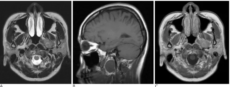
Fig. 1. A 35-year-old woman presented with headache and dizziness; routine brain MR imaging was performed.
A--C. Axial T2-weighted (A) and sagittal T1-weighted (B) images show a well circumscribed, round mass in the left parapharyngeal space, displacing the internal carotid artery posteriorly. On a contrast-enhanced T1-weighted image (C), the mass is inhomoge- neously enhanced.
ed from the stroma by a basement membrane. There is characteristic palisading in the peripheral portion of the tumor cell nests. A myxochondroid stromal component, as seen in pleomorphic adenoma or a mesenchymal component should be absent. It is also positive for cytok- eratin, smooth muscle actin, and S-100 protein by im- munohistiochemical staining (2, 4, 6).
Basal cell adenoma can be divided into four subtypes based on their morphologic pattern: trabecular, solid, tubular, and membranous, and the most frequent type is the solid variant (6, 7). Malignant transformation of basal cell adenoma is rare but has been suggested by some researchers, giving rise to basaloid cell carcinomas (basal cell adenocarcinoma, adenoid cystic carcinoma) and, less commonly, non-basaloid carcinoma (salivary duct carcinoma, adenocarcinoma) (1, 8).
Basal cell adenoma typically appears as a solitary,
slowly growing, asymptomatic mass usually in the parotid gland or the minor salivary gland of the other parts. Surgical extirpation of the tumor has been cura- tive. Although recurrence is rare, the membranous sub- type, which is a hereditary variety of basal cell adeno- ma, has been reported to have a 25-37% recurrence rate in some reports (1-3, 8).
Imaging features of basal cell adenomas are rarely re- ported because of their low prevalence. General imag- ing findings on previously reported cases have demon- strated that these tumors occur mainly in the parotid gland and have well-demarcated margins, solid or cystic components, and homogeneous or heterogeneous en- hancement. These imaging findings are not characteris- tic of a basal cell adenoma.
Grossly, basal cell adenoma is solid or cystic, or gray or white and does not usually show intratumoral hemor-
J Korean Radiol Soc 2007;56:439-442
─ 441 ─ A
C
B
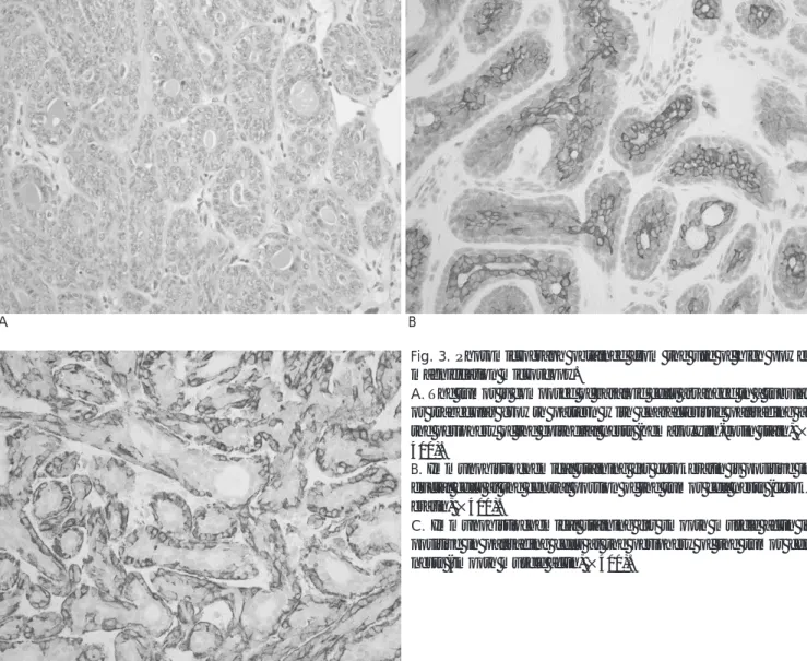
Fig. 3. Photomicrograph obtained from the use of high power magnification microscopy.
A. The tumor is composed of basaloid cells arranged in a tubular or trabecular growth pattern with characteristic palisading at the periphery of the epithelial nests (hematoxylin-eosin stain, × 400).
B. Immunohistiochemical staining for cytokeratin is positive in ductal cells at the central portion of the tumor cell nests (cytok- eratin, ×400).
C. Immunohistiochemical staining for smooth muscle actin is positive in palisading cells at the periphery of the tumor cell nests (smooth muscle actin, ×400).
대한영상의학회지 2007;56:439-442
부인두 공간에서 생긴 기저세포선종: 증례 보고1
1전북대학교 의과대학 영상의학과
황 승 배・정 경 호・김 종 수
기저세포선종은 이하선에서 주로 발생하는 드문 양성 상피 기원 타액선 종양이다. 그러나 부인두공간에 있는 소 타액선에서 발생한 기저세포선종의 보고는 거의 없었다. 이에 저자들은 35세 여자에서 부인두공간 종괴로 나타난 기저세포선종의 증례를 보고한다.
rhage or calcification. However, Jeong et al. (3) reported that basal cell adenoma arising in the parapharyngeal space shows intratumoral hemorrhage on MR imaging.
Basal cell adenoma has a characteristic vascular pattern in which small capillaries and venules are prominent in the adenoma, and these vascular structures can cause intratumoral hemorrhage (3, 9). Chawla et al. (1) have described CT features of basal cell adenomas arising in the parotid glands that correlated with histopathological findings. The tumor showed heterogeneous enhance- ment on contrast-enhanced CT in 11 of 14 cases (79%), with stellate-shaped low-density areas in three tumors, linear non-enhancing bands in three tumors and cystic areas in five tumors. In our case, the central low-density area in the tumor was seen on contrast-enhanced CT but we could not verify the hypodense area in the tumor pathologically. Some reports have demonstrated the hy- pointense rim of the tumor capsule on T2-weighted MR images due to tumor encapsulated by fibrous connective tissue (2, 3).
The parapharyngeal space contains mainly fatty tis- sue, lymphatics, and minor salivary gland tissue, and is actually a potential space located lateral to the upper pharynx and extends from the skull base to the hyoid bone. This space may be the source of salivary gland tu- mors, as it was in this case (3). Pleomorphic adenoma originated from the minor salivary gland is the most common primary lesion arising in the parapharyngeal space. Basal cell adenoma occurs in the parapharyngeal space very rarely. According to our own investigation, only three cases have been reported (3-5).
The major differential diagnosis for parapharyngeal tumors includes other tumors of minor salivary gland origin, including the pleomorphic adenoma, Warthin tu- mor and other low-grade malignant tumors, nerve
sheath tumors, and paragangliomas. It is difficult to make a differential diagnosis from other minor salivary gland tumors in the parapharyngeal space.
Schwannoma or paraganglioma is usually located in the carotid space; therefore, it displaces the internal carotid artery anteromedially. Paraganglioma is a hypervascu- lar tumor and is characterized by flow signal void within the mass on MR imaging.
In conclusion, basal cell adenoma is a rare benign ep- ithelial neoplasm originating from salivary gland tissue and we report here on a very rare case of a basal cell adenoma arising in the parapharyngeal space. Although it has low prevalency and no specific imaging features, we think that it should be considered in the differential diagnosis of parapharyngeal space tumors or salivary gland origin tumors.
References
1. Chawla AJ, Tan TY, Tan GJ. Basal cell adenomas of the parotid gland: CT scan features. Eur J Radiol 2006;58:260-265
2. Jang M, Park D, Lee SR, Hahm CK, Kim Y, Kim Y, et al. Basal cell adenoma in the parotid gland: CT and MR findings. AJNR Am J Neuroradiol 2004;25:631-635
3. Jeong AK, Lee HK, Kim SY, Cho KJ. Basal cell adenoma in the parapharyngeal space: MR findings. Clin Imaging 2001;25:392-395 4. Hemachandran M, Lal A, Vaiphei K. Basal cell adenoma-an un-
usual presentation. Ann Diagn Pathol 2003;7:292-295
5. Orabi AA, Riad MA, O’Regan MB. Stylomandibular tenotomy in the transcervical removal of large benign parapharyngeal tumours.
Br J Oral Maxillofac Surg 2002;40:313-316
6. Hiranuma T, Kagamiuchi H, Kitamura R. A basal cell adenoma of the sublingual gland. Int J Oral Maxillofac Surg 2003;32:566-567 7. Batsakis JG, Luna MA. Basaloid salivary carcinoma. Ann Otol
Rhinol Laryngol 1991;100:785-787
8. Leegaard T, Lindeman H. Salivary gland tumours: clinical picture and treatment. Acta Otolaryngol Suppl 1969;263:155-159
9. Triest WE, Fried MP, Stanievich JF. Membranous basal cell adeno- ma of the hypopharynx. Arch Otolaryngol 1983;109:774-777 Seung-Bae Hwang, et al: Basal Cell Adenoma Presenting as a Parapharyngeal Space Mass
─ 442 ─