단일 다발 전방 십자인대 재건술 후 대퇴골 터널의 위치와 대퇴골 터널 기울기의 상관관계
이주홍 • 박 혁 • 정성엽
전북대학교 의학전문대학원 전북대학교병원 정형외과학교실, 임상의학연구소
Correlation between Femoral Tunnel Location in Three-Dimensional Computed Tomography and Femoral Tunnel Angle in Plain
Radiographs after Single-Bundle Anterior Cruciate Reconstruction
Ju-Hong Lee, M.D., Hyuk Park, M.D. , and Seong-Yup Jeong, M.D.
Department of Orthopedic Surgery, Chonbuk National University Hospital,
Research Institute of Clinical Medicine, Chonbuk National University Medical School, Jeonju, Korea
Purpose: The purpose of this study is to determine correlation between femoral tunnel angle in the coronal plane on a simple radiograph
and femoral tunnel location in the sagittal plane on three-dimensional computed tomography (3D-CT).Materials and Methods: The subjects included 42 patients who underwent 3D-CT after the operation out of 70 cases of anterior cruciate
ligament reconstruction using quadriceps tendon-patelllar bone autograft from April, 2009 to June, 2011. Measurement of the femoral tunnel angle was based on the anatomical axis of the femur in antero-posterior (AP) and Rosenberg views; femoral tunnel location was described as a proportional percentage on the medial surface of the lateral femoral condyle in the 3D-CT image; then the correlation between femoral tunnel angle and femoral tunnel location was analyzed retrospectively.Results: Femoral tunnel angle was 41.5
o±6.8o (range: 29.7o−53.9o) on AP radiographs, and 34.9o±6.9o (range: 23.8o−46.5o) on Rosenberg views. The femoral tunnel was located 36.9%±11.3% from posterior, and 38.1%±6.5% from proximal on the 3D-CT image. On plain AP radiographs, femoral tunnel angle and femoral tunnel location showed negative correlation (p<0.001, rho=-0.498), and, in comparison with Rosenberg view, they showed negative correlation (p=0.006, rho=-0.416). Twenty three patients (53.5%) had femoral tunnel in the anatomical location. Their femoral tunnel angle on AP radiographs was 43.3o±6.1o, while the femoral tunnel angle of patients who had femoral tunnel in non-anatomical locations was 38.4o±6.4o (p=0.004). In the Rosenberg picture, similar difference was observed between the two groups (p=0.012).Conclusion: On AP radiographs and Rosenberg views, femoral tunnel angle showed significant correlation with the femoral tunnel
location on the 3D-CT image, and the group who had femoral tunnel location in the anatomical range showed a relatively higher femoral tunnel angle.Key words: anterior cruciate ligament reconstruction, femoral tunnel angle, femoral tunnel location, three-dimensional computed
tomography서 론
비해부학적인 위치에 형성된 대퇴 터널이 전방십자인대 재건술 실패의 가장 흔한 원인 중 하나로 알려져 있다.1,2) 최근 십자인대 재건술의 임상적 결과를 높일 것으로 생각되는 정상 해부학적 위
Copyright © 2014 by The Korean Orthopaedic Association
“This is an Open Access article distributed under the terms of the Creative Commons Attribution Non-Commercial License (http://creativecommons.org/licenses/by-nc/3.0/) which permits unrestricted non-commercial use, distribution, and reproduction in any medium, provided the original work is properly cited.”
The Journal of the Korean Orthopaedic Association Volume 49 Number 1 2014 Received June 18, 2013 Revised October 15, 2013
Accepted December 23, 2013 Correspondence to: Hyuk Park, M.D.
Department of Orthopedic Surgery, Chonbuk National University Medical School, 20 Geonji-ro, Deokjin-gu, Jeonju 561-712, Korea
TEL: +82-63-250-1760 FAX: +82-63-271-6538 E-mail: [email protected]
치로의 재건에 대한 관심이 높아졌다.
대퇴골 터널의 위치는 슬관절 운동역학 회복에 중요한 영향을 미치며,3-5) 최근 보고되는 해부학적 및 생역학적 연구 결과들을 토대로 대퇴골의 해부학적 부착부에 이식건을 위한 골 터널을 정 확히 위치시키는 것이 권장되고 있다.
전방십자인대 재건술 후 대퇴골 터널의 위치를 확인하는 가장 정확한 방법은 3차원 컴퓨터 단층 촬영(three-dimensional com- puted tomography, 3D-CT)으로 알려져 있다.6-8) 그러나 3D-CT는 단순 방사선 사진에 비해 경제적인 부담이 있고, 검사 시행 과정 이 불편하며, 보다 많은 방사선에 노출된다는 단점을 가지고 있 어 최근 45o 굴곡 후전방 단순 방사선 사진을 이용하여 대퇴골 터 널의 위치를 확인하는 방법이 제시되고 있다.9)
본 연구는 슬부 단순 방사선 사진에서 대퇴골의 해부학적 축에 대해 관상면상 대퇴골 터널의 기울기가 클수록 3D-CT의 시상면 상 대퇴골 터널의 위치가 보다 해부학적인 위치에 존재할 것이라 는 가설하에서 대퇴골 터널의 기울기와 3D-CT상에서 대퇴골 터 널의 위치와의 연관성을 분석하였고 사용된 단순 방사선 사진 간
의 유용성을 평가하였다.
대상 및 방법
2009년 4월부터 2011년 6월까지 시행한 자가 대퇴사두건을 이용 한 단일 다발 전방십자인대 재건술 70예 중 술 후 3D-CT 촬영을 시행한 42예(남자 36명, 여자 6명)를 대상으로 하였다. 평균 나이 는 33.7±12.2세(15-46세)였다(Table 1). 120o 이상 슬관절을 굴곡 한 상태에서 두 개의 전내측(anteromedial, AM) 삽입구와 Trans- portal Guide® (Arthrex, Naples, FL, USA)를 이용하여 우측 슬관절 기준 10시 또는 그 이하, AM 다발과 후외측(posterolateral, PL) 다 발 사이, 그리고 대퇴외과의 후방 피질골로부터 약 4 mm 정도가 남게 대퇴골 터널을 만들었다. 이식물의 골편은 대퇴골 터널에 위치 후 7×20 mm 금속 간섭 나사를 이용하여 약 120o 굴곡 위에 서 고정하였다. 본 연구는 전북대학교병원 연구윤리 심의위원회 (institutional review board)의 승인(2012-10-003-001)을 받고 시행 되었다.
1. 대퇴 터널 기울기
수술 후 전후방 및 체중부하 45o 굴곡 후전방 방사선 사진에서 대 퇴골의 해부학적 축을 기준으로 대퇴골 터널 기울기를 측정하였 다. 대퇴골의 해부학적 축은 대퇴 간부의 근위부와 원위부에 대 퇴 과상간 축과 평행한 두 선을 긋고, 두 선의 중심을 이은 선으로 정의했고, 대퇴골 터널의 기울기는 대퇴골의 해부학적 축과 대퇴 골 터널의 중심선이 이루는 각을 측정하였다(Fig. 1).
Figure 1. The femoral tunnel angle (*) is measured from the longitudinal axis of the femoral tunnel (x) and anatomical axis (y) of the femur on antero-posterior (A) and Rosenberg views (B).
Table 1. Patient Information
Variable Value
Case (male/female) 42 (36/6)
Age (yr) 33.7 (15−46)
Acute (<3 ms)/chronic 29/13 Elapsed time to surgery (mo) 11.4 (0.3−175.0) Follow-up (mo) 5.0 (2−19) Values are presented as number or median (range).
Figure 2. Femoral tunnel location was described as a proportional percentage (from proximal to distal and posterior to anterior) on the medial surface of the lateral femoral condyle in the three-dimensional computed tomography image. The circle showed position of the graft in femoral tunnel.
2. 3차원 컴퓨터 단층 촬영상 대퇴골 터널의 위치
Forsythe 등6)이 제시한 대퇴골 터널의 위치를 3D-CT상 기술하 는 방법에 따라서 시상면에서 근위부에서 원위부를 백분율로 표 시(proximal-distal [PD] position)하였고, 후방부에서 전방부를 백 분율로 표시(posterior-anterior [PA] position)하여 2차원적인 위치 를 나타내었다(Fig. 2). PD position은 대퇴과간 절흔의 가장 근위 부에서 가장 원위부까지의 거리 중 대퇴골 터널의 위치를 백분율 로 나타냈고 PD position의 방향은 대퇴골의 해부학적 축과 평행 하였다. PA position은 대퇴골 외과 내측벽의 후방으로부터 대퇴 과간 절흔의 가장 전방부까지의 거리 중 대퇴골 터널의 위치를 백분율로 계산한 것이다. 3D-CT에서 대퇴 터널 내에서의 이식물 위치는 간섭 나사의 후방에 이식물이 위치하는 공간의 중심을 기 준으로 하였다(Fig. 2).
대퇴골 터널의 해부학적 범위는 Forsythe 등6)이 Anatomic Co- ordinate Axes Method를 이용하여 측정한 결과 평균 PA position 은 AM 터널과 PL 터널이 각각 23.1%±6.1% (범위: 16.3%-36.4%), 15.3%±4.8% (범위: 8.9%-24.3%)였고, 평균 PD position은 AM 터 널과 PL 터널이 각각 28.2%±5.4% (범위: 20.1%-36.2%), 58.1%±
7.1% (범위: 50.2%-73.1%)였고, PA position과 PD position의 95%
신뢰구간인 11.9%-27.3%, 24.4%-63.0%를 해부학적 범위로 하여 본 연구의 환자를 분류하였다.
3. 통계 분석
연구 자료의 통계 분석은 SPSS Windows version 12.0 (SPSS Inc., Chicago, IL, USA) 통계프로그램을 이용하였다. 통계적인 유의성 을 결정하기 위해 대퇴골 터널의 기울기와 PD position, 대퇴골 터 널의 기울기와 PA position을 Pearson 상관계수를 이용하여 분석 하고, 방사선 사진상 대퇴골 터널 기울기에 따른 3D-CT에서 대 퇴골 터널위치 분석을 위해 receiver operative characteristic (ROC) curve를 사용하였고, 유의수준은 0.05 이하로 하였다.
결 과
전후방 방사선 사진 상에서 대퇴골의 해부학적 축을 기준으로 대 퇴골 터널 기울기는 평균 41.5o±6.8o (범위: 29.7o-53.9o), Rosenberg
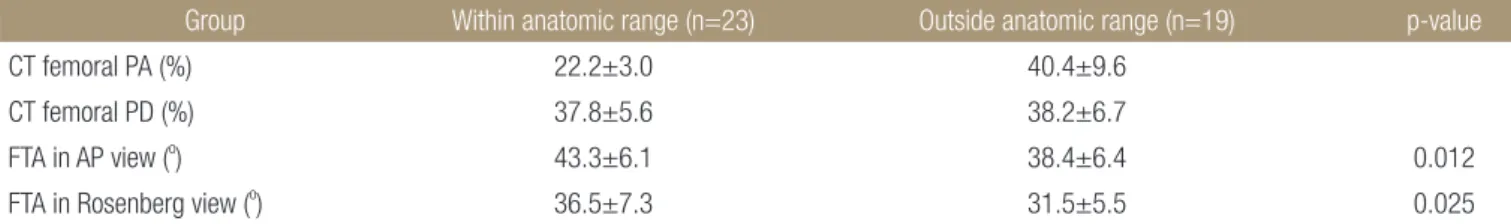
사진에서는 34.9o±6.9o (범위: 23.8o-46.5o)였다. 3D-CT 영상에서 대퇴골 터널은 후방에서 36.9%±11.3%, 근위에서 38.1%±6.5%에 위치하였다. 전후방 방사선 사진에서 대퇴골 터널의 기울기와 대 퇴골 터널의 PA position은 음의 상관관계(p<0.001, rho=-0.498) 를 보였지만, PD position은 통계적인 의미가 없었다(p=0.351, rho=-0.148). Rosenberg 사진과의 비교에서도 PA position은 음의 상관관계(p=0.006, rho=-0.416)를 보였고, PD position은 통계적 의 미가 없었다(p=0.174, rho=0.272)(Table 2). 해부학적 위치에 대 퇴골 터널이 존재한 환자는 23예(53.5%)로 전후방 방사선 사진 상 대퇴골 터널의 기울기는 43.3o±6.1o였고, 비해부학적인 위치 에 존재한 환자에서의 기울기는 38.4o±6.4o로 두 군 간에 차이 를 보였다(p=0.004). Rosenberg 사진에서는 해부학적 위치에 존 재하는 환자에서 기울기는 36.5o±7.3o, 비해부학적 위치인 환자 에서 기울기는 31.5o±5.5o였고 두 군 간에 통계적 차이를 보였다 (p=0.012)(Table 3).
ROC curve를 통한 분석에서 Rosenberg 사진에서 대퇴골 터널 의 기울기는 34.3o를 기준으로 기준보다 큰 경우 민감도 73.7%, 특 이도 65%로 해부학적인 위치의 가능성이 높았고(p=0.045), 전후 방 방사선 사진에서 기준점은 찾을 수 없었다(p=0.38).
고 찰
해부학적 전방십자인대 재건술은 전방십자인대를 원래의 용적, 콜라겐 방향과 해부학적 부착 위치를 복원함으로써 얻는 기능적 인 회복을 의미한다.10) Woo 등11)은 전방십자인대 재건술 후 생 역학 연구에서 단일 다발 재건술이 전방 안정성의 회복에는 효
Table 3. Femoral Tunnel Position on Three-Dimensional CT
Group Within anatomic range (n=23) Outside anatomic range (n=19) p-value
CT femoral PA (%) 22.2±3.0 40.4±9.6
CT femoral PD (%) 37.8±5.6 38.2±6.7
FTA in AP view (o) 43.3±6.1 38.4±6.4 0.012
FTA in Rosenberg view (o) 36.5±7.3 31.5±5.5 0.025
Values are presented as mean±standard deviation. CT, computed tomography; PA, posterior to anterior; PD, posterior to distal; FTA, femoral tunnel angle; AP, antero-posterior.
Table 2. Relation between CT Position and Femoral Tunnel Angle on X-ray
CT position (%) FTA in AP view FTA in Rosenberg view
r p-value r p-value
Femoral PA -0.498 <0.001 -0.416 0.006 Femoral PD -0.148 0.351 0.174 0.272 CT, computed tomography; FTA, femoral tunnel angle; AP, antero- posterior; PA, posterior to anterior; PD, posterior to distal.
과적이지만 회전 안정성을 복원하기 힘들다고 보고하였다. 그러 나 최근 여러 논문에서 단일 다발 재건술을 해부학적 위치로 재 건을 했을 경우 Lachman 및 축 이동 검사에서 보다 좋은 결과를 얻었고, 회전 안정성도 향상됨을 보고하고 있다.12-14) 비해부학 적 위치에 대퇴골 터널을 재건했을 경우 발생 가능한 합병증으 로 Johnson 등15)은 해부학적 위치보다 전방에 대퇴골 터널이 형 성되면 슬관절 굴곡 시 과부하가 발생하여 관절운동범위에 제한 이 생긴다고 하였으며, Ristanis 등16)은 대퇴골 터널이 12시에 가 깝게 수직방향으로 형성되면 회전 불안정성이 발생할 수 있다고 하였다. 또한 너무 후방에 대퇴골 터널이 형성되어도 대퇴골 후 벽에 골절 위험성이 있어 이식건의 고정에 어려움이 발생할 수 있다.
본 연구에서는 전후방 슬관절 사진과 Rosenberg 사진상에서 대 퇴골 터널의 기울기와 3D-CT상에서의 대퇴골 터널의 위치의 상 관관계를 찾아 단순 방사선 사진으로 대퇴골 터널의 위치를 예측 이 가능할 것이라는 가설하에 연구를 진행하였고, 연구 결과 해 부학적인 위치에 대퇴골 터널이 형성된 환자에서 전후방 슬관절 사진과 Rosenber 사진에서 대퇴골 터널의 기울기가 유의하게 증 가하였다(Table 3). 이는 해부학적 터널을 기준으로 했을 때 대퇴 골 터널의 기울기가 증가할수록 시상면상의 대퇴골 터널의 위치 가 해부학적인 위치에 있으려는 경향을 확인할 수 있었다. 대퇴 골 터널의 PA position과 대퇴골 터널의 기울기(전후방 슬관절 사 진과 Rosenberg 사진)의 연관성을 분석한 결과 PA position은 통 계적으로 유의한 음의 상관관계를 보였는데(Table 2), 이는 대퇴 골 터널이 전방에 위치할수록 대퇴골 터널의 기울기가 작아진다 는 의미이며, 대퇴골 터널의 기울기가 작아진다는 의미는 대퇴골 터널이 수직에 가깝게 형성되었다는 것을 말하며, Table 3을 참고 할 때 시상면상 비해부학적인 위치에 대퇴골 터널이 형성되는 경 향이 있다고 생각할 수 있다.
Bernard 등17)은 슬관절 측면 단순 방사선 사진으로 대퇴골 터 널의 위치를 측정하였지만 여러 연구에서 측면 단순 방사선 사진 만으로 대퇴골 터널의 위치를 확인하는 것은 신뢰도가 낮다고 보 고하고 있다. Hoser 등18)도 CT와 단순 방사선 사진으로 골 터널의 위치를 비교한 논문에서 단순 방사선 사진으로 골 터널의 위치를 확인하는 것은 신뢰도가 낮고, 특히 비해부학적 위치에 재건되었 을 경우는 대퇴골 천정 쪽에 터널이 위치하여 단순 방사선 사진 에서 더 확인이 힘들다고 보고하여, 대퇴골 터널 위치의 신뢰도 를 높이기 위해 CT를 사용한 방법들을 소개하고 있다. 본 연구에 서는 3D-CT상에서의 대퇴골 터널의 위치의 기술은 Forsythe 등6) 이 제시한 Anatomic Coordinate Axes Method를 따랐다. 이 방법은 슬관절을 90o 굴곡시킨 상태에서 3D-CT상의 대퇴골 터널 위치를 기술하여 관절경 시야와 유사하고 대퇴골의 해부학적 축과 평행 또는 수직인 축을 기준으로 하여 이해가 쉽다는 장점을 가지지 만 과거에 사용되지 않은 방법으로 기존 연구들과의 비교가 힘들
다는 단점을 가진다. Bernard 등17)이 슬관절 측면 단순 방사선 사 진에서 Blumensaat line을 기준으로 측정했던 것과 같은 방법으로 3D-CT상 대퇴골 터널 위치를 기술한 quadrant method가 있고, 이 는 대퇴골의 해부학적 축과 다른 방향으로 기술만으로 위치의 이 해가 어렵고, CT의 시상면상 Blumensaat line을 확인하는 것이 쉽 지 않다는 단점을 가지고 있다.
Illingworth 등9)은 전방십자인대 재건술 후 골 터널의 위치를 평 가하는 단순하고 효과적인 방법으로 Rosenberg 사진에서 대퇴골 의 해부학적 축을 기준으로 한 대퇴골 터널의 기울기가 감소할수 록, 즉 대퇴골 터널이 32.7o 이하일 때 시상면상에서 대퇴골 터널 이 보다 전방에 위치하는 경향이 있고, 결국 비해부학적 위치에 대퇴골 터널을 재건했을 가능성이 높다고 제시하였다. 본 연구에 서는 슬관절 단순 전후방 촬영과 Rosenberg 사진에서 측정한 대 퇴골 터널 기울기 모두 감소할수록 비해부학적 위치에 대퇴골 터 널을 재건했을 가능성이 높았으며, 대퇴골 터널이 해부학적으로 재건되었을 때 대퇴골 터널 기울기가 단순 전후방 촬영에서 평균 43.3o±6.1o, Rosenberg 사진에서 평균 36.5o±7.3o였다. Rosenberg 사진에서 34.3o보다 큰 경우 해부학적 위치에 대퇴골 터널을 재건 했을 가능성이 높았으나 환자의 수가 적어 민감도와 특이도가 높 지 않았으며 앞으로 보다 많은 환자를 이용한 분석이 필요할 것 으로 생각된다.
본 연구는 현재까지 측정된 환자의 수가 적었다는 점, 측정방 법 및 전방십자인대의 해부학적 위치의 불확실성으로 인한 한계 를 가지고 있다. 전방십자인대 재건술에서 중요한 대퇴골 터널 의 위치를 단순 방사선 사진으로 보다 쉽게 검사하고자 한 Illing- worth 등9)의 논문에서 사용한 Rosenberg 사진은 대퇴골의 내회전 이 적다는 장점을 가지지만 45o 굴곡 위를 일관되게 유지하는 것 이 용이하지 않아 촬영자에 따라서 굴곡각도에 영향을 줄 수 있 고, 결국 굴곡 각도가 촬영할 때마다 다르다면 대퇴골의 회전 및 대퇴골 터널의 각도에 영향을 줄 수 있어 최종 측정값의 신뢰도 를 떨어뜨릴 수 있다. 따라서 단순 방사선 사진 촬영 시 재현성 이 떨어지는 Rosenberg 사진보다 촬영이 쉽고 일관된 영상을 얻 기 용이한 슬관절 단순 전후방 촬영에서 대퇴골의 해부학적 축을 기준으로 대퇴골 터널의 경사각을 측정하였다. 정확한 대퇴 터널 위치를 확인하기 위해서는 3D-CT 촬영이 필요하겠지만 외래에 서 촬영이 용이하고 경제적이다는 장점을 가진 전후방 슬부 방사 선 사진을 시행하였고, 이는 대퇴골이 내회전된다는 단점을 가지 지만 본 연구결과에서는 Rosenberg 사진에 비해서 대퇴골 터널 위치와 연관성이 유사하지만 근소하게 높았다.
다른 연구의 한계점으로 전방십자인대 재건술에서 해부학적 위치는 많은 연구가 진행 중이지만6-8,19) 각 논문 간에 측정방법이 달라서 일관된 해부학적 위치를 찾는 데는 한계를 보였다. 본 연 구에서는 Illingworth 등9)의 연구와 비교를 위해 Anatomic Coor- dinate Axes Method를 이용하여 전방십자인대의 해부학적 위치
를 측정하였고, 또한 같은 전방십자인대의 해부학적 범위를 기 준으로 연구를 진행하였으나 일관된 전방십자인대의 해부학적 위치를 알기 위해서는 좀 더 많은 연구가 필요할 것으로 생각된 다.
결 론
전후방 슬부 방사선 및 Rosenberg 사진에서 대퇴골의 해부학적 축을 기준으로 한 대퇴골 터널의 기울기는 3D-CT 영상에서의 대퇴골 터널 위치와 높은 연관성을 보였고, 해부학적 부착부 범 위 내에 대퇴골 터널이 위치한 군에서 관상면상 대퇴골 터널 기 울기가 상대적으로 증가하였다. 따라서 전후방 슬부 방사선 및 Rosenberg 사진에서 대퇴골 터널의 해부학적 위치 여부를 평가할 때는 관상면상의 대퇴골 터널 기울기 정도가 간접적인 기준이 될 수 있을 것으로 생각된다.
REFERENCES
1. Scopp JM, Jasper LE, Belkoff SM, Moorman CT 3rd. The effect of oblique femoral tunnel placement on rotational con- straint of the knee reconstructed using patellar tendon auto- grafts. Arthroscopy. 2004;20:294-9.
2. Wetzlera MJ, Getelman MH, Friedman MJ, Bartolozzi AR.
Revision anterior cruciate ligamentsurgery: etiology of fail- ures. Oper Tech Sports Med. 1998;6:64-70.
3. Heming JF, Rand J, Steiner ME. Anatomical limitations of transtibial drilling in anterior cruciate ligament reconstruc- tion. Am J Sports Med. 2007;35:1708-15.
4. Loh JC, Fukuda Y, Tsuda E, Steadman RJ, Fu FH, Woo SL.
Knee stability and graft function following anterior cruciate ligament reconstruction: comparison between 11 o'clock and 10 o'clock femoral tunnel placement. 2002 Richard O'Connor Award paper. Arthroscopy. 2003;19:297-304.
5. Yagi M, Wong EK, Kanamori A, Debski RE, Fu FH, Woo SL.
Biomechanical analysis of an anatomic anterior cruciate liga- ment reconstruction. Am J Sports Med. 2002;30:660-6.
6. Forsythe B, Kopf S, Wong AK, et al. The location of femoral and tibial tunnels in anatomic double-bundle anterior cruci- ate ligament reconstruction analyzed by three-dimensional computed tomography models. J Bone Joint Surg Am.
2010;92:1418-26.
7. Kopf S, Forsythe B, Wong AK, et al. Nonanatomic tunnel po- sition in traditional transtibial single-bundle anterior cruciate ligament reconstruction evaluated by three-dimensional com-
puted tomography. J Bone Joint Surg Am. 2010;92:1427-31.
8. Lertwanich P, Martins CA, Asai S, Ingham SJ, Smolinski P, Fu FH. Anterior cruciate ligament tunnel position measurement reliability on 3-dimensional reconstructed computed tomog- raphy. Arthroscopy. 2011;27:391-8.
9. Illingworth KD, Hensler D, Working ZM, Macalena JA, Tashman S, Fu FH. A simple evaluation of anterior cruciate ligament femoral tunnel position: the inclination angle and femoral tunnel angle. Am J Sports Med. 2011;39:2611-8.
10. van Eck CF, Lesniak BP, Schreiber VM, Fu FH. Anatomic single- and double-bundle anterior cruciate ligament recon- struction flowchart. Arthroscopy. 2010;26:258-68.
11. Woo SL, Kanamori A, Zeminski J, Yagi M, Papageorgiou C, Fu FH. The effectiveness of reconstruction of the anterior cruciate ligament with hamstrings and patellar tendon. A ca- daveric study comparing anterior tibial and rotational loads. J Bone Joint Surg Am. 2002;84:907-14.
12. Bedi A, Musahl V, Steuber V, et al. Transtibial versus antero- medial portal reaming in anterior cruciate ligament recon- struction: an anatomic and biomechanical evaluation of surgi- cal technique. Arthroscopy. 2011;27:380-90.
13. Jepsen CF, Lundberg-Jensen AK, Faunoe P. Does the position of the femoral tunnel affect the laxity or clinical outcome of the anterior cruciate ligament-reconstructed knee? A clinical, prospective, randomized, double-blind study. Arthroscopy.
2007;23:1326-33.
14. Sadoghi P, Kröpfl A, Jansson V, Müller PE, Pietschmann MF, Fischmeister MF. Impact of tibial and femoral tunnel position on clinical results after anterior cruciate ligament reconstruc- tion. Arthroscopy. 2011;27:355-64.
15. Johnson DL, Swenson TM, Irrgang JJ, Fu FH, Harner CD.
Revision anterior cruciate ligament surgery: experience from Pittsburgh. Clin Orthop Relat Res. 1996;325:100-9.
16. Ristanis S, Giakas G, Papageorgiou CD, Moraiti T, Stergiou N, Georgoulis AD. The effects of anterior cruciate ligament re- construction on tibial rotation during pivoting after descend- ing stairs. Knee Surg Sports Traumatol Arthrosc. 2003;11:360- 5.
17. Bernard M, Hertel P, Hornung H, Cierpinski T. Femoral in- sertion of the ACL. Radiographic quadrant method. Am J Knee Surg. 1997;10:14-21.
18. Hoser C, Tecklenburg K, Kuenzel KH, Fink C. Postoperative evaluation of femoral tunnel position in ACL reconstruction:
plain radiography versus computed tomography. Knee Surg
Sports Traumatol Arthrosc. 2005;13:256-62.
19. Pinczewski LA, Salmon LJ, Jackson WF, von Bormann RB, Haslam PG, Tashiro S. Radiological landmarks for placement
of the tunnels in single-bundle reconstruction of the anterior cruciate ligament. J Bone Joint Surg Br. 2008;90:172-9.
단일 다발 전방 십자인대 재건술 후 대퇴골 터널의 위치와 대퇴골 터널 기울기의 상관관계
이주홍 • 박 혁 • 정성엽
전북대학교 의학전문대학원 전북대학교병원 정형외과학교실, 임상의학연구소
목적: 단순 방사선 사진에서 대퇴골 터널 기울기와 3차원 컴퓨터 단층 촬영(three-dimensional computed tomography, 3D-CT)의 대퇴골 터널 위치의 상관관계를 알아보고자 한다.
대상 및 방법: 2009년부터 2011년까지 시행한 전방십자인대 재건술 후 3D-CT 촬영을 시행한 42예를 대상으로 하였다. 전후방 및 Rosenberg 사진에서 대퇴골의 해부학적 축을 기준으로 대퇴골 터널 기울기를 측정하였고 3D-CT에서 대퇴골 터널 위치를 표시하여 측정값 간의 연관성을 분석하였다.
결과: 전후방 방사선 사진에서 대퇴골 터널 기울기는 41.5o±6.8o, Rosenberg 사진에서는 34.9o±6.9o였다. 3D-CT 영상에서 대퇴골 터널 위치는 후방에서 36.9%±11.3%, 근위에서 38.1%±6.5%였다. 대퇴골 터널 기울기와 위치는 전후방 방사선 사진에서 음의 상관 관계(p<0.001, rho=-0.498), Rosenberg 사진에서도 음의 상관관계(p=0.006, rho=-0.416)를 보였다.
결론: 전후방 방사선 및 Rosenberg 사진에서 대퇴골 터널 기울기는 3D-CT 영상에서의 대퇴골 터널 위치와 높은 연관성을 보였고, 해부학적 범위 내에 대퇴골 터널이 위치한 군에서 관상면상 대퇴골 터널 기울기가 상대적으로 증가하였다.
색인단어: 전방십자인대 재건술, 대퇴골 터널 기울기, 대퇴골 터널 위치, 3차원 컴퓨터 단층촬영
접수일 2013년 6월 18일 수정일 2013년 10월 15일 게재확정일 2013년 12월 23일 책임저자 박 혁
전주시 덕진구 건지로 20, 전북대학교 의학전문대학원 정형외과학교실 TEL 063-250-1760, FAX 063-271-6538, E-mail [email protected]
Copyright © 2014 by The Korean Orthopaedic Association
“This is an Open Access article distributed under the terms of the Creative Commons Attribution Non-Commercial License (http://creativecommons.org/licenses/by-nc/3.0/) which permits unrestricted non-commercial use, distribution, and reproduction in any medium, provided the original work is properly cited.”