The elbow is the most commonly dislocated joint in chil- dren. However, a simultaneous dislocation of the elbow and a translocation of the proximal radio-ulnar joint is a rare injury. To the best of the our knowledge, there has only been one such case treated with a closed method5).
We experienced a case of proximal radio-ulnar transloca- tion combined with a posterior elbow dislocation, which was treated with a closed reduction. This case stresses the importance of considering this type of injury using a care- ful investigation of radiographs and the earliest reduction using a closed method.
CASE REPORT
A ten-year-old girl fell from a desk onto her outstretched left hand. She complained of severe pain in her left elbow, which was swollen, bruised and deformed. The circulation
in her left hand was normal, but the sensation in the two ulnar digits and the strength of abduction and adduction of the finger was decreased. The anteroposterior and lateral radiographs indicated a simple posterior dislocation of the elbow (Fig. 1). Therefore, traction was applied to the elbow under intravenous sedation and the elbow appeared to be reduced with a clunk. However, the flexion and extension were slightly restricted, and the forearm was fixed in pro- nation. The radiographs taken after the closed reduction revealed a translocation of both forearm bones, such that the ulna articulated with the capitellum and the radial head articulated with the trochlea (Fig. 2). A second closed reduc- tion was performed under fluoroscopic control. It was pos- sible to relocate both bones by supination of the wrist and direct pressure over the medial side of the radial head and the lateral side of the olecranon (Fig. 3). Full flexion and extension were then possible and the normal range of rota- tion was restored. Initially, the elbow was immobilized for one week in a long arm splint at 90 degrees flexion and full supination, due to swelling, and was then immobilized in a long arm cast in the same position for an additional three
582 J. of Korean Orthop. Assoc.
2004; 39: 582-5
582
Proximal Radio-Ulnar Translocation Associated with Elbow Dislocationon
-Case Report-
Bong-Jin Lee, M.D., Sung-Rak Lee, M.D., and Dong-Hwan Shin, M.D.
Department of Orthopaedic Surgery, Halla General Hospital, Jeju, Korea
582 582 Address reprint requests to
Dong-Hwan Shin, M.D.
Department of Orthopaedic Surgery, Halla General Hospital, 1963-2 Yeon-dong, Jeju 690-170, Korea
Tel: +82.64-740-5111, Fax: +82.64-743-3110 E-mail: [email protected]
A translocation of the proximal radius and ulna combined with a posterior dislocation of the elbow is quite rare. To the best of our knowledge, the only case with this condition, who had been treated using a closed method was reported by MacSween in 1978. This paper reports a ten-year-old girl who fell from a desk onto her outstretched left hand. The initial radiographs showed a simple posterior dislocation of the elbow. However, the radiographs taken after the closed reduction revealed a translocation of both forearm bones. It was possible to relocate both bones using a closed method, and the patient recovered from the associated ulnar nerve palsy at five weeks post-trauma. At the follow-up examination three months post-trauma, the nerve was found to be fully regenerated using electromyography and a nerve conduction study, and the patient regained the full range of elbow motion without pain. When a posteri- or dislocation of the elbow occurs, close attention is needed in order to detect the combined transloca- tion of the proximal radio-ulnar joint. If treated early, a closed reduction leads to a good result. However, a careful physical examination and a thorough investigation of the radiographs are necessary.
Key Words: Proximal radio-ulnar joint, Translocation, Closed reduction
Proximal Radio-Ulnar Translocation Associated with Elbow Dislocationon 583
weeks. At the time of cast removal, the patient was instruct- ed to perform an active range of motion exercise of the elbow.
At five weeks post-trauma, the patient appeared to have recovered from the ulnar nerve palsy. At seven weeks post- trauma, full flexion was attained, but 30 degrees of exten- sion, 20 degrees of pronation and 30 degrees of supination were lost. At the follow-up examination three months post- trauma, the nerve was found to be fully regenerated using electromyography and a nerve conduction study, and the patient had regained pain-free full range of motion of the elbow (Fig. 4). At the two years follow-up, the patient had normal elbow function, and was able to play various sports.
DISCUSSION
In 1978, MacSween5)reported the first case of a transpo- sition of the radius and ulna associated with dislocation of
the elbow in a child. His patient was treated using closed method, but he reported a 10 degree loss of pronation at one year after the injury.
A translocation of the both forearm bones might occur as a result of two mechanisms: The primary case occurs simul- taneously with elbow dislocation, while the secondary case develops after a closed reduction of the elbow dislocation.
The latter case is referred to an iatrogenic translocation.
The possibility of an iatrogenic proximal radio-ulnar trans- location was first suggested by Harvey and Tchelebi4). They reported that in a pure lateral or posterolateral dislocation of the elbow, which is diagnosed as a posterior dislocation, a reduction by traction and pronation might force the radi- al head to cross anteriorly over the ulna, resulting in a trans- located position.
Eklof3)suggested that a translocation of the proximal radioulnar joint combined with a dislocation of the elbow
′′
. .
Fig. 1.Initial radiographs were read as a posterior dislocation of the elbow.
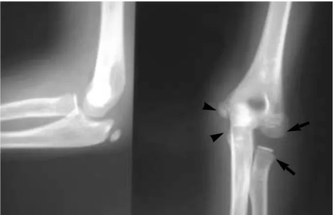
Fig. 2.Radiographs taken after the first closed reduction reveal a translocation of both forearm bones. The arrows show the radi- al head and capitellum and the arrowheads show the proximal ulna and trochlea.
L
L
Fig. 3.Radiographs taken after the second closed reduction show an anatomical reduction of the elbow. The arrows indicate the reduced radiocapitellar joint and the arrowheads indicate the ulnohumeral joint.
Fig. 4.At 3 months post-trauma, the patient regained her full range of supination and pronation.
584 Bong-Jin Lee∙Sung-Rak Lee∙Dong-Hwan Shin
might occur when the elbow is extended and pronated, as with the injury described in this report. During dislocation, the capsule and ligaments are disrupted to the point that the bones of the forearm cross each other and become locked in hyperpronation.
In our case, during the first closed reduction, it was believed there was a reduction and the characteristic ‘clunk’ was heard, but in fact only the posterior dislocation of the elbow was re- duced and the translocation of both forearm bones remained.
Carey1)described this unsuccessful closed reduction as a
‘pseudoreduction’, in that the flexion and extension were improved, but the forearm remained fixed in almost com- plete pronation1).
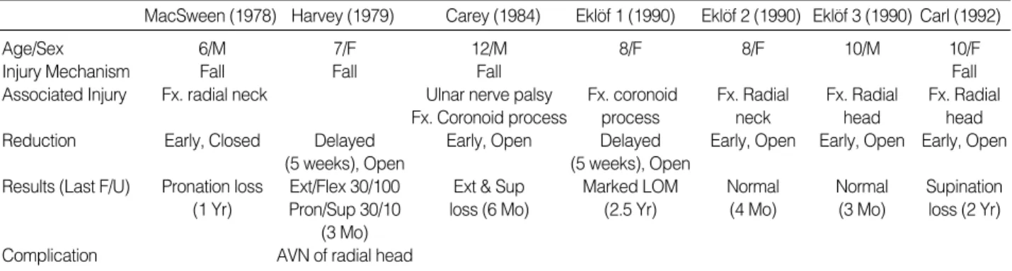
In all reports except for the one reported by MacSween5), the closed reductions failed because the interposed soft tis- sues prevented reductions3)and the radial head divot mechan- ically locked onto the medial aspect of the proximal ulna2). The details of the previous reports are summarized in Table 1. The associated injuries are a radial head or neck fracture, a coronoid process fracture, and a ulnar nerve palsy. The com- plications are a limited range of motion and avascular necro- sis of the radial head.
It may be difficult to recognize this type of injury at the initial presentation, and the method of treatment remains uncertain. The best indication for a diagnosis is the reversed
position of the bones of the proximal forearm. However, this unexpected anatomical relationship is easily overlooked.
Therefore, a careful physical examination and thorough in- vestigation of the radiographs are necessary to avoid serious complications. When a lateral or posterolateral dislocation is recognized, traction and forced pronation should be avoid- ed in order to prevent an iatrogenic translocation of both forearm bones.
It is believed that if detected early, a closed reduction, with careful pathoanatomical considerations would be successful in most cases of a proximal radio-ulnar translocation.
REFERENCES
1. Carey RP: Simultaneous dislocation of the elbow and the proximal radio-ulnar joint. J Bone Joint Surg, 66-B: 254-256, 1984.
2. Carl A, Prada S and Teixeira K: Proximal radioulnar transposi- tion in an elbow dislocation. J Orthop Trauma, 6: 106-109, 1992.
3. Eklof O, Nybonde T and Karlsson G: Luxation of the elbow complicated by proximal radio-ulnar translocation. Acta Radiol, 31:
145-146, 1990.
4. Harvey S and Tchelebi H: Proximal radio-ulnar translocation.
A case report. J Bone Joint Surg, 61-A: 447-449, 1979.
5. MacSween WA: Transposition of radius and ulna associated with dislocation of the elbow in a child. Injury, 10: 314-316, 1978.
. .
′′
M, Male; F, Female; Fx, Fracture; F/U, Follow-up; Yr, Year; Mo, Month; Ext, Extension; Flex, Flexion; Pron, Pronation; Sup, Supination; LOM, Limited range of motion; AVN, Avascular necrosis.
MacSween (1978) Harvey (1979) Carey (1984) Eklof 1 (1990) Eklof 2 (1990) Eklof 3 (1990) Carl (1992)
Age/Sex 6/M 7/F 12/M 8/F 8/F 10/M 10/F
Injury Mechanism Fall Fall Fall Fall
Associated Injury Fx. radial neck Ulnar nerve palsy Fx. coronoid Fx. Radial Fx. Radial Fx. Radial
Fx. Coronoid process process neck head head
Reduction Early, Closed Delayed Early, Open Delayed Early, Open Early, Open Early, Open
(5 weeks), Open (5 weeks), Open
Results (Last F/U) Pronation loss Ext/Flex 30/100 Ext & Sup Marked LOM Normal Normal Supination
(1 Yr) Pron/Sup 30/10 loss (6 Mo) (2.5 Yr) (4 Mo) (3 Mo) loss (2 Yr)
(3 Mo)
Complication AVN of radial head
Table 1.Summary of the clinical data of previous case reports of a proximal radio-ulnar translocation combined with an elbow dislocation
. . . .
. .
Proximal Radio-Ulnar Translocation Associated with Elbow Dislocationon 585
주관절 후방 탈구에 동반된 근위 요척 관절의 교차전위(translocation)는 매우 희귀한 손상이다. 이를 도수 정복술로 치 료한 경우는 1978년 MacSween의 1예만이 보고되어 있다. 저자들은 팔꿈치를 편 상태로 탁자에서 떨어진 10세 여아에서 처음에는 단순한 주관절 후방 탈구로 생각하고 도수정복을 하였으나, 도수정복 후 촬영한 방사선 사진 상 요골과 척골의 교차전위가 있음을 확인하고, 재차 도수정복을 시도하여 성공하였다. 수상 5주에 동반되었던 척골 신경 마비는 임상적으 로 회복되었으며, 수상 3개월 후 시행한 근전도 및 신경전도 검사 상 신경은 완전한 회복을 보였고, 주관절의 관절운동 범 위도 완전히 정상화되었다. 주관절 후방 탈구를 진단 시에는 동반된 근위 요척 관절의 동반 손상에 유념하여, 이학적 검사 와 방사선 판독을 신중히 하여야 하며, 조기치료 시 도수정복 만으로도 좋은 결과를 보인다고 생각한다.
색인 단어: 근위 요척 관절, 교차전위(Translocation), 도수정복
주관절 탈구에 동반된 근위 요척 관절의 교차전위 -증례 보고-
이봉진ㆍ이성락ㆍ신동환
제주 한라병원 정형외과