A malignant fibrous histiocytoma (MFH) most fre- quently develops in the deep fascia or skeletal muscle of the limbs, retroperitoneum and abdominal cavity (1-3).
Primary breast MFH is extremely rare, and a few such cases have been reported that have described the imag- ing findings of breast MFH (3-5).
We report the imaging findings of primary breast MFH in a 53-year-old woman, including the mammog- raphy, US and MRI. To the best of our knowledge, this is the first case report with the MR imaging features of MFH of the breast.
Case Report
A 53-year-old woman was admitted to our hospital due to fever and a rapidly growing huge tender mass in her right breast. Eleven months prior to visiting our hos- pital, the patient had undergone a vacuum-assisted breast biopsy (VABB) at a local clinic for an incidentally detected mass during screening breast US (Fig. 1A), and no abnormality was seen on mammography. The histol- ogy findings of the VABB were no definite malignancy, and the findings suggested a fibroadenomatous change.
Three months after the VABB, follow-up US revealed a 1cm sized irregular anechoic lesion (Fig. 1B). Aspiration was done for the palpable lump at the biopsy-site one month afterward and brownish fluid was noted, so this was considered as a slow resolving post-biopsy hematoma or recurrent fluid collection. But on a retro- spectively review, the mass was considered to be resid- ual tumor, and after 5 months, the mass started growing again (Fig. 1C). Four months after this, the patient was
Imaging Findings of Malignant Fibrous Histiocytoma of the Breast: A Case Report1
Ji Na Kim, M.D., Shin Ho Kook, M.D., Hyoun Joo Kwag, M.D., Yoon Jung Choi, M.D., Jin Hee Sohn, M.D.2, Yong Lai Park, M.D.3, Jin Hyo Kim, M.D.4
1Department of Radiology, Kangbuk Samsung Hospital, Sungkyunkwan University School of Medicine
2Department of Pathology, Kangbuk Samsung Hospital, Sungkyunkwan University School of Medicine
3Department of General Surgery, Kangbuk Samsung Hospital, Sungkyunkwan University School of Medicine
4You and Me Surgery, Jeonju
Received September 11, 2009 ; Accepted September 15, 2009
Address reprint requests to : Yoon Jung Choi, M.D., Department of Radiology, Kangbuk Samsung Hospital, Sungkyunkwan University School of Medicine, 108, Pyung-dong, Jongno-gu, Seoul 110-746, Korea.
Tel. 82-2-2001-1031 Fax. 82-2-2001-1030 E-mail: [email protected]
A malignant fibrous histiocytoma (MFH) is the most common soft tissue sarcoma en- countered during adulthood, but the breast is not a common site of involvement for MFH. Several investigators have reported the histopathological and biological features of a MFH involving the breast, but only a few reports have focused on the imaging findings of breast MFHs. To emphasize the importance of arriving at a preoperative di- agnosis for the treatment implications, we report here the imaging findings, including the mammography, US and MRI findings, for a MFH of the breast of a 53-year-old woman who presented with a rapid growing huge mass in the right breast.
Index words :Histiocytoma, Malignant Fibrous Breast
Ultrasonography
Magnetic Resonance Imaging
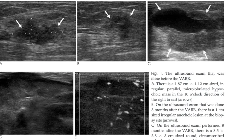
transferred to our hospital via the emergency room due to progressive growth of the lesion and the patient had had a high fever that had lasted for three days. The mass was firm and extremely large, and mammography could not be performed. An US exam done in our hospital re- vealed a well-defined huge heterogeneous mixed echoic mass with variable sized cystic areas (Fig. 1D) and high vascularity was seen in the solid portion on power Doppler US (Fig. 1E). We assessed the lesion to be BI- RADS category 5 and an US-guided core biopsy was per- formed. The histopathology revealed an unclassified malignant tumor and the differential diagnosis included malignant myoepithelial tumor, sarcomatoid carcinoma or sarcoma. Pre-operative breast MRI was performed with a 1.5-T MR imaging system (Intera, Ver. 11.1.4.3;
Philips, Best, the Netherlands). On the MR images, a 12
×6×16 cm sized, smoothly well-defined multi-lobulat- ed mass had replaced almost the entire right breast. The mass showed intermediate signal intensity on the T1- weighted images and central, variable, mixed heteroge- neous (from very high to very low) signal intensity and peripheral intermediate signal intensity on the T2-
weighted images (Figs. 2A, B). A gadolinium-enhanced dynamic scan showed the early rapid peripheral irregu- lar thick rim enhancement of the mass (Fig. 2C).
The patient underwent a simple mastectomy and ipsi- lateral axillary lymph nodes dissection. On the macro- scopic examination, the breast was mostly replaced by the tumor mass; the mass had a relatively well-delineat- ed and lobulated margin and the tumor measured 16 × 12 × 6 cm. The cut surface was grayish yellow with a
“fish flesh” appearance, and necrosis and hemorrhage were noted (Fig. 3A). The histopathological findings showed diffuse proliferations of atypical spindle cells with a whorling pattern (Fig. 3B). The tmor cells exhibit- ed round to oval vesicular nuclei with plump eosinophilic or foamy granular cytoplasm (Fig. 3C). A diffuse positive reaction was seen on CD10 immunos- taining. The expression of other epithelial markers (MOC31, CK7 and CK20), HMB-45, c-Kit, estrogen, progesterone receptors and Her-2 were negative. The staining for p53 was positive. According to the histologi- cal and immunohistochemical staining results, this case was finally diagnosis as a malignant tumor, and espe-
A B
D E
Fig. 1. The ultrasound exam that was done before the VABB.
A. There is a 1.87 cm × 1.12 cm sized, ir- regular, parallel, microlobulated hypoe- choic mass in the 10 o’clock direction of the right breast (arrows).
B. On the ultrasound exam that was done 3 months after the VABB, there is a 1 cm sized irregular anechoic lesion at the biop- sy site (arrows).
C. On the ultrasound exam performed 9 months after the VABB, there is a 3.5 × 2.8 × 3 cm sized round, circumscribed mixed echoic mass with posterior en- C
cially a high-grade malignant fibrous histiocytoma after ruling out other malignancies. We retrospectively re- viewed the vacuum-assisted breast biopsy specimen, and there was only loose connective tissue, suggesting neither definite malignancy nor fibroadenoma.
Discussion
Primary malignancies of the breast stromal elements and sarcomas of a mesenchymal origin account for less than 1% of all breast neoplasm. To the best of our knowledge, 28 cases of a breast primary MFH have
A B
C
Fig. 3. On the gross specimen, the tumor mass has a relatively well-delineated and lobulating margin, and the mass measures 16×12×6 cm.
A. The cut surface is grayish yellow with a “fish flesh” appear- ance and necrosis and hemorrhage (arrows).
B. The histopathology findings of the breast MFH (H & E stain,
×200, ×400) show a diffuse proliferation of atypical spindle cells with a whorling pattern (arrows).
C. The tumor cells exhibit round to oval vesicular nuclei with plump eosinophilic or foamy granular cytoplasm (arrows).
A B C
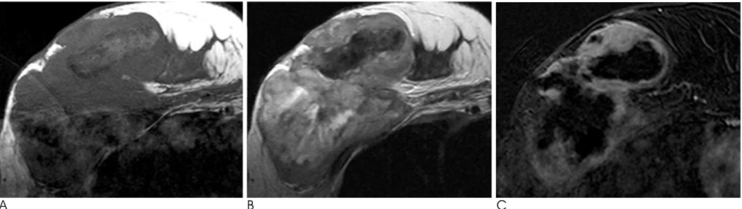
Fig. 2. MR images of the right breast.
A, B. There is a 12 cm sized, smoothly well-defined multilobulated intermediate signal intensity mass on the T1 weighted image (A) and the T2 weighted MR image (B). The mass shows central variable, mixed heterogeneous (from very high to very low) signal in- tensity and peripheral intermediate signal intensity.
C. The Gd-enhanced scan shows the early rapid irregular thick peripheral rim enhancement of the mass.
been reported in the English medical literature.
Secondary MFH of the breast can be derived from ma- lignant transformation according to several predisposing factor, including radiation therapy, surgery, a burn scar and other malignancy such as a malignant phyllodes tu- mor. Almost all the cases of secondary MFH of the breast have developed after receiving radiation therapy for breast cancer treatment, and usually within 10 to 30 years after a long latent period (1, 2, 6).
In our case, the patient acknowledged the presence of a palpable mass four months after receiving a VABB, which is too short a time to have developed a secondary MFH. Considering that the patient did not have a histo- ry of undergoing radiation therapy, which is the most frequent reported predisposing factor, we think that the disease in this case is a primary breast MFH and the VABB was the VABB was performed during an inter- vening episode of disease progression or the biopsy missed the tumor tissue. Generally, soft-tissue MFHs manifest as a painless, progressive growing palpable mass over several months and they are sometimes ac- companied with a large hematoma or intratumoral hem- orrhage (1, 2), like what occurred in our case. Fever is not a common symptom of patients with MFH, but our patient had fever that was probably due to the extensive tumoral necrosis.
Histologically, MFHs are divided into five subtypes that include storiform-pleomorphic, myxoid, giant cell, inflammatory and angiomatoid (1-3, 5). Primary and secondary soft-tissue MFHs, including breast MFHs, have a poor prognosis without any relation to the sub- types. In our case, the subtype could not be classified as the diagnosis was made after all of the other beast sarco- mas were ruled out according to the negative expression of specific markers and the lack of a prominent cellular type.
According to the previous reports of the imaging find- ings of breast MFHs and MFHs at other sites (1, 2, 4, 5, 7), US depicts a well-defined heterogeneous solid mass
and the hypoechoic areas representing necrosis. The CT images reveal a well-defined large lobulated mass with iso- or hypodense areas that correspond to myxoid le- sions or necrosis. Tumors may have areas of high atten- uation hemorrhage. The solid component is enhanced after intravenous contrast injection. MR imaging of gen- eral MFHs shows low to intermediate signal intensity on the T1-weighted images, heterogenous high signal inten- sity on the T2-weighted images and enhancement of the solid component on the Gd-enhanced MR images.
Extensive areas of hemorrhage can obscure an underly- ing neoplasm. We performed MRI instead of CT, and MRI showed findings that were similar to those of the above mentioned general MFHs.
A primary breast MFH is rare malignant neoplasm, but one should consider the possibility of this tumor when a patient presents with a progressive growing breast mass with variable solid and cystic foci of hemor- rhage and necrosis.
References
1. Murphey MD, Gross TM, Rosenthal HG. From the archives of the AFIP. Musculoskeletal malignant fibrous histiocytoma: radiologic- pathologic correlation. Radiographics 1994;14:807-826
2. Son E, Park J, Jeon H, Cho S. Malignant fibrous histiocytoma (MFH) in axilla. Yonsei Med J 2004;45:736-738
3. Oh SJ, Kim KM, Hong TH, Park WC, Kim JS, Jung SS. Giant cell malignant fibrous histiocytoma of the breast: a case report. J Korean Med Sci 2004;19:477-480
4. Hocevar M, Marinsek ZP, Zidar A. Myxofibrosarcoma of the breast as an unusual variant of malignant fibrous histiocytoma: re- port of a case. Surg Today 2004;34:752-754
5. Kijima Y, Umekita Y, Yoshinaka H, Taguchi S, Owaki T, Funasako Y, et al. Stromal sarcoma with features of giant cell ma- lignant fibrous histiocytoma. Breast Cancer 2007;14:239-244 6. Ugurlu K, Turgut G, Kabukcuoglu F, Ozcan H, Sanus Z, Bas L.
Malignant fibrous histiocytoma developing in a burn scar. Burns 1999;25:764-767
7. Ajisaka H, Maeda K, Uchiyama A, Miwa A. Myxoid malignant fi- brous histiocytoma of the breast: report of a case. Surg Today 2002;
32:887-890
대한영상의학회지 2010;62:295-299
유방에 발생한 악성 원발성 섬유성 조직구종의 영상학적 소견: 증례 보고1
1성균관대학교 의과대학 강북삼성병원 영상의학과
2성균관대학교 의과대학 강북삼성병원 병리과
3성균관대학교 의과대학 강북삼성병원 외과
4유앤미외과
김지나∙국신호∙곽현주∙최윤정∙손진희2∙박용래3∙김진효4
악성 섬유성조직구종은 성인에서 발생하는 가장 흔한 연조직육종이나 유방에서 발생하는 경우는 드물다. 지금까 지 외국문헌에 유방에서 발생한 악성 섬유성조직구종의 병리조직학적 소견에 대한 몇몇 보고가 있었으나 영상소견 을 포함한 경우는 극히 드물며, 특히 MR 영상을 함께 보고한 예는 없었다. 저자들은 점진적으로 크기가 증가해온 커다란 우측 유방 종괴를 주소로 내원한 53세 여자 환자에서 우측 유방 대부분에 종양의 침윤을 보이고 종양 내부에 출혈성 괴사와 액와림프절 전이를 동반한 1예를 경험하였기에 유방촬영술(Mammography), 초음파, MR 영상 소 견과 조직 병리 소견을 함께 보고하는 바이다.