submit.radiology.or.kr J Korean Soc Radiol 2012;66(1):7-10
7 INTRODUCTION
Uremia is a syndrome of clinical and metabolic abnormali- ties that develops in parallel with deteriorating renal function and brain involvement. Uremic encephalopathy (UE) is a well-known complication of uremia (1). The movement dis- order seen in uremic patients typically consists of asterixis and myoclonus, which may be related to cortical dysfunction.
Several recent studies have been reported with acute move- ment disorder accompanying akinetic, parkinsonian syn- dromes associated with bilateral basal ganglia (BG) lesions in uremic patients (2). However, to the best of our knowledge, cerebellar vermis (CV) involvement in UE is very rare, and only one case report in a non-diabetic hemodialysis patient with this condition was reported in the Korean literature (3).
We describe a rare case of diabetic UE showing symmetrical BG and CV lesions on magnetic resonance imaging (MRI).
CASE REPORT
A 50-year-old man presented with dysarthria and hand trem-
bling, that had persisted for 3 days, and uncontrollable invol- untary swinging or dancing-like arm movements, which had been noted for 1 day. Examination of his medical history showed that he had a 20-year history of hypertension and chronic re- nal failure and had been diagnosed with diabetes mellitus 2 months previously. He had received regular hemodialysis treatment 3 times a week for 20 years.
The patient’s vital signs were stable; his blood pressure was 130/90 mm Hg; body temperature, 36.6°C; heart rate, 72 bpm;
and respiration rate, 20 breaths/min. An examination of his mental status showed drowsiness, and neurologic examina- tion showed slurred speech, negative cerebellar function test, and negative myoclonus without motor weakness. Laboratory findings revealed metabolic acidosis (pH 7.17), elevated levels of blood urea nitrogen (BUN; 30 mg/dL) and serum creati- nine (Cr; 5.1 mg/dL), as well as normal blood glucose levels (124 mg/dL).
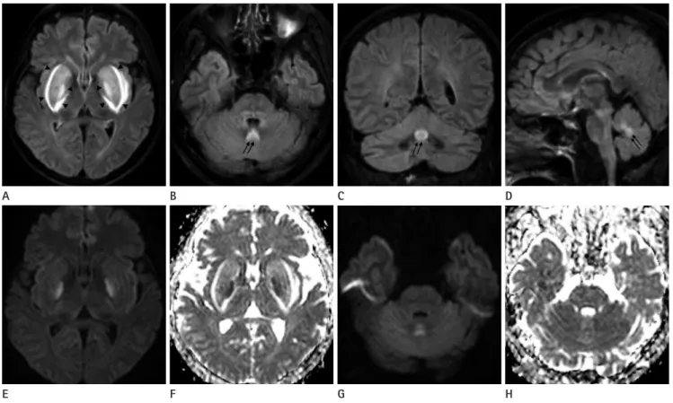
Initial brain MRI showed symmetrically increased signal intensities involving the entire BG region, and T2-weighted, and the fluid attenuated inversion recovery (FLAIR) images showed a brightly hyperintense rim extending to the internal
Case Report
pISSN 1738-2637
J Korean Soc Radiol 2012;66(1):7-10
Received August 31, 2011; Accepted September 29, 2011 Corresponding author: Seung Young Lee, MD Department of Radiology, Chungbuk National University Hospital, 410 Seongbong-ro, Heungdeok-gu, Cheongju 361-711, Korea.
Tel. 82-43-269-6472 Fax. 82-43-269-6479 E-mail: [email protected]
Copyrights © 2012 The Korean Society of Radiology
Uremic encephalopathy (UE) is a well-known complication of uremia, but its patho- physiology remains unknown. It is widely reported that in UE, the bilateral basal ganglia (BG) shows hyperintensities on T2/fluid attenuated inversion recovery magnetic resonance imaging (MRI), but cerebellar lesions are extremely rare, with to the best of our knowledge, only one case reported to date. We describe the findings from computed tomography and MRI for typical BG and cerebellar vermis lesions.
Index terms
Uremic Encephalopathy Basal Ganglia
Cerebellar Vermis Intracerebral Hemorrhage
Cerebellar Lesions of Uremic Encephalopathy on MRI in Hemodialyzed Diabetic Patient: A Case Report
당뇨성 신부전 환자에서 발생한 소뇌 병변을 동반한 요독성 뇌병증: 1예
Min Chul Kil, MD, Seung Young Lee, MD, Sang-Hoon Cha, MD, Bum Sang Cho, MD, Min Ho Kang, MD
Department of Radiology, Chungbuk National University Hospital, Cheongju, Korea
Cerebellar Lesions of Uremic Encephalopathy on MRI in Hemodialyzed Diabetic Patient
submit.radiology.or.kr
J Korean Soc Radiol 2012;66(1):7-10
8
Brain CT performed immediately showed massive intracere- bral hemorrhage (ICH) in the left BG and mild ICH in the right BG (Fig. 2). The patient was transferred to a long-stay in- stitution but showed no improvements on hospital day 28.
DISCUSSION
In uremia, brain involvement is a common complication (1).
In cases of this complication, the imaging findings are very characteristic, and there are two types of radiological manifes- tations of UE: cortical (2) and BG involvement (1, 3-6).
A case of UE with reversible bilateral BG involvement was first reported by Okada et al. in a patient with chronic glomer- ulonephritis (4). They presented the MRI findings, which dem- onstrated decreased signal intensity on T1-weighted images and increased signal intensity on T2-weighted images; the le- sions resolved after dialysis.
Wang and Cheng (5) presented a group of 12 patients with and external capsules. Further, three-dimensional FLAIR im-
ages showed increased signal intensity involving the CV. Dif- fusion-weighted imaging (DWI) showed localized hyperin- tensities with reduced apparent diffusion coefficient (ADC) values in the globus pallidus (GP) on both sides, suggestive of cytotoxic edema. However, in the CV, it showed hyperintensi- ty without signal change on ADC (Fig. 1).
Intensified daily hemodialysis combined with supportive therapy was administered, but the symptoms did not improve.
The patient was in a confused state 3 days after admission, immediately after hemodialysis. At this point, laboratory find- ings revealed further elevated BUN (74.7 mg/dL) and Cr (10.1 mg/dL) levels and reduced blood glucose level (54 mg/dL).
Follow-up brain computed tomography (CT) was performed, and the extent of the hypodense areas corresponding to the lesions shown on initial MRI seemed to have increased.
The patient suddenly presented with weakness on the right side and a semi-comatose mental status 9 days after admission.
A
E
B
F
C
G H
D
Fig. 1. Uremic encephalopathy in a 50-year-old man.
Three-dimensional FLAIR images (A-D) show hyperintensities in the entire BG extending to the internal and external capsules, representing the
“Lentiform fork” (arrowheads) and CV (arrows). DWI (E) shows hyperintensities with reduced ADC values (F) in bilateral golobus pallidus. In the CV, DWI (G) showes hyperintensities without signal change on ADC (H).
Note.-ADC = apparent diffusion coefficient, BG = basal ganglia, CV = cerebellar vermis, DWI = diffusion-weighted imaging, FLAIR = fluid atten- uated inversion recovery, GP = globus pallidus
Min Chul Kil, et al
submit.radiology.or.kr J Korean Soc Radiol 2012;66(1):7-10
9
used for movement. And, the other function of the CV is the control of muscle tone and level of force. Also, the CV affects oc- ular movement, although, in our case, T2-weighted and FLAIR images showed hyperintensity in the CV, and the patient pre- sented without disturbance of ocular movement, including nystagmus, ocular motor apraxia, rapid eye movement disor- der, or hypotonia.
A and ADC map showed diffusion restriction (hyperintensity on DWI and hypointensity on ADC map) only in the GP, but not in the putamen, external capsule, internal capsule and CV.
This finding indicates that cytotoxic and vasogenic edema may simultaneously affect the BG and CV. Previously reported cases also showed that diffusion restriction was observed only in the GP on DWI (6, 8). Thus, DWI findings may play a key role in distinguishing UE from BG lesions caused by other conditions.
Ertl-Wagner et al. (9) suggested that BG hemorrhage may be caused by the “breakthrough” mechanism. Loss of autoregula- tion due to acidosis leads to severe BG edema. Correction of the metabolic derangement results in a decrease in the intra- cranial pressure and an increase in vascular reactivity. The initial loss and subsequent increase in reactivity probably dis- rupts the blood-brain barrier, which is the so-called “break- through” mechanism, and this leads to bilateral edema of the BG and finally to hemorrhagic transformation (9). In our case, bilateral but asymmetrical ICH in the BG (massive on the left and mild on the right, as shown by CT) developed, as was re- ported in 2 previous cases (9, 10). Considering that the patient’s blood pressure was relatively stable in the range of 130/80- end-stage renal disease who developed acute movement dis-
orders. They also found bilateral BG lesions ranging from fo- cal putaminal and pallidal lesions to widespread involvement of the entire BG with perifocal edema.
Although no definite pathophysiology has yet been estab- lished, metabolic acidosis and long-term hyperglycemia in uremic patients are believed to play an important role as risk factors in the development of BG lesions. The cellular func- tions of the BG in such uremic patients are already compro- mised by long-term diabetes mellitus through microangio- pathic changes or energy utilization failure. Since these cells are particularly susceptible to toxic and metabolic insults, when they were further exposed to markedly elevated levels of ure- mic toxins, their regional cellular metabolism and/or vascular autoregulation collapse, and tissue damage and focal edema ensue (1, 5).
Bilateral symmetrical BG lesions are typically caused by dif- fuse systemic or metabolic disturbances. Bilateral symmetrical BG T2/FLAIR hyperintensities on MRI are non-specific and re- main non-diagnostic in the absence of a clinical setting. There- fore, the differential diagnosis includes hypoxic injury, mito- chondrial disease, vasculitis, carbon monoxide intoxication, hypoglycemia, extrapontine myelinolysis, Wilson’s disease, Creutzfeldt-Jakob disease, deep venous thrombosis and so on (7). Kumar and Goyal (8) assert that the “Lentiform Fork” sign is a unique radiologic picture presented by metabolic acidosis in a patient with UE. The lateral arm of the fork, constituted by the edematous external capsule, extended from the anterior end of the putamen at the rostral end of the frontal horn of the lateral ventricle to the “stem” of the fork, which is formed by the fusion of edematous external and internal capsules at the infero-posterior end of the putamen. The medial arm extended from the stem up to one-third of the medial edge where it split into two branches engulfing the GP. In our case, a bright hy- perintense rim extending into the internal and external cap- sules surrounding the swollen BG was seen on T2-weighted and FLAIR images, representing the “Lentiform Fork.” The
“Lentiform Fork” sign is not pathognomonic but a specific imaging finding in UE, so it is important with regard to dif- ferential diagnosis (8).
The main responsibility of the CV is proprioception, which is the ability to recognize the relative positioning of body parts
A B
Fig. 2. Follow-up brain CT performed on hospital day 9.
Follow-up brain CT (A, B) shows ICH (arrows) in the BG (massive on the left and mild on the right) and increased extent of hypodense area corresponding to the lesions shown on initial MRI in the BG and CV.
Note.-BG = basal ganglia, CV = cerebellar vermis, ICH = intracerebral Hemorrhage
Cerebellar Lesions of Uremic Encephalopathy on MRI in Hemodialyzed Diabetic Patient
submit.radiology.or.kr
J Korean Soc Radiol 2012;66(1):7-10
10
sis patient. Korean J Nephrol 2006;25:1061-1066
4. Prüss H, Siebert E, Masuhr F. Reversible cytotoxic brain edema and facial weakness in uremic encephalopathy. J Neurol 2009;256:1372-1373
5. Wang HC, Cheng SJ. The syndrome of acute bilateral basal ganglia lesions in diabetic uremic patients. J Neurol 2003;
250:948-955
6. Yoon CH, Seok JI, Lee DK, An GS. Bilateral basal ganglia and unilateral cortical involvement in a diabetic uremic patient. Clin Neurol Neurosurg 2009;111:477-479
7. Lim CC. Magnetic resonance imaging findings in bilateral basal ganglia lesions. Ann Acad Med Singapore 2009;38:
795-798
8. Kumar G, Goyal MK. Lentiform Fork sign: a unique MRI picture. Is metabolic acidosis responsible? Clin Neurol Neurosurg 2010;112:805-812
9. Ertl-Wagner B, Jansen O, Schwab S, Sartor K. Bilateral basal ganglion haemorrhage in diabetic ketoacidotic coma: case report. Neuroradiology 1999;41:670-673
10. Terzi M, Akkaya O, Onar M. Pure sensory stroke due to bilat- eral basal ganglion hemorrhage: a case report. Turk Neuro- surg 2010;20:406-408
150/90 mm Hg when he was in the hospital, we believe the hemorrhage was caused not by hypertension but by the hemor- rhagic transformation due to UE.
In summary, bilateral BG lesions are the most common ra- diologic finding in UE, but they are not specific or pathogno- monic. Because of their differentiating ability, DWI findings and the “Lentiform fork” sign are important for differential diagnosis. Lastly, clinicians and radiologists should be well- aware of the rare features of UE including CV involvement and hemorrhagic transformation.
REFERENCES
1. Lee EJ, Park JH, Ihn Y, Kim YJ, Lee SK, Park CS. Acute bilat- eral basal ganglia lesions in diabetic uraemia: diffusion- weighted MRI. Neuroradiology 2007;49:1009-1013 2. Lee PH, Shin DH, Kim JW, Song YS, Kim HS. Parkinsonism
with basal ganglia lesions in a patient with uremia: Evi- dence of vasogenic edema. Parkinsonism Relat Disord 2006;12:93-96
3. Sim SR, Lee SH, Jahng JH, Lim JY, Choi YK, Bae KS, et al.
Uremic encephalopathy associated with bilateral basal ganglia and cerebellar lesion in a non-diabetic hemodialy-
당뇨성 신부전 환자에서 발생한 소뇌 병변을 동반한 요독성 뇌병증: 1예
길민철 · 이승영 · 차상훈 · 조범상 · 강민호
요독성 뇌병증은 요독증의 잘 알려진 합병증의 하나이지만, 그 병태생리는 잘 알려져 있지 않다. 요독성 뇌병증에서 MRI 소견은 T2 강조 영상과 fluid attenuated inversion recovery 영상에서 양측 기저핵에 높은 신호 강도를 보이는 것으로 많 이 보고가 되어 있으나, 소뇌 병변을 동반한 경우는 매우 드물게 보고가 되어있다. 이에 본 저자들은 소뇌 병변을 동반한 요독성 뇌병증의 CT와 MRI 영상 소견을 보고하고자 한다.
충북대학교병원 영상의학과