Anomalous systemic arterial supply to the lungs has been reported in some cases of congenital heart disease, congenital lung diseases such as bronchopulmonary se- questration and hypogenetic lung syndrome, and in ap- parently normal heart and lung (1, 2). The latter has been described as the rarest form of the condition (3).
The basal segments of the left lower lobe are most fre- quently involved, and this anomaly is therefore referred to as the systemic arterial supply to normal basal seg- ments of the left lower lobe (3, 4). We report a case of this anomaly, describing its characteristic radiographic and operative findings.
Case Report
A five-year-old girl whose growth and development
were normal was admitted to hospital due to intermit- tent periumbilical pain. Physical examination was unre- markable except for the presence of a grade 3/6 continu- ous systolic murmur along the left lower sternal border.
The murmur was first noted at 7 months of age, but no further investigation was performed. Laboratory find- ings were normal, though an electrocardiogram demon- strated bilateral ventricular hypertrophy.
Posteroanterior chest radiography revealed retrocar- diac nodular and tubular densities, partially obliterating the descending thoracic aorta (Fig. 1A). The radiograph- ic findings were initially misinterpreted as pneumonia, but further careful review of these and comparison with the patient’s clinical information suggested possible sys- temic arterial supply to the normal lung. To confirm or refute this, chest CT scanning was performed: a con- trast-enhanced spiral scan showed an anomalous sys- temic artery arising from the descending thoracic aorta and branching into the normal pulmonary parenchyma of the left lower lobe, and the dilated, tortuous left infe- rior pulmonary vein draining into the left atrium, par- tially obliterating the descending thoracic aorta (Figs.
1B, C). At lung window setting, the left interlobar pul- monary artery distal to the origin of the superior seg-
J Korean Radiol Soc 2003;48:193-196
─ 193 ─
Systemic Arterial Supply to the Normal Basal Segments of the Left Lower Lobe: Case Report1
Min-Jeong Kim, M.D., Hyun Woo Goo, M.D., Seung Il Park, M.D.2, Chong Hyun Yoon, M.D.
Systemic arterial supply to the normal basal segments of the left lower lobe is a rare congenital anomaly which has characteristic radiologic features. The lung supplied by the anomalous systemic artery has a normal bronchial tree. We recently encountered a case in which the diagnosis of systemic arterial supply to the normal basal segments of the left lower lobe was strongly suggested by the chest radiographic, CT, and MRI find- ings. The diagnosis was subsequently confirmed by left lower lobectomy.
Index words : Lung, congenital malformation Lung, CT
Lung, MR
Children, cardiovascular system
1Department of Radiology, Asan Medical Center, University of Ulsan College of Medicine
2Department of Thoracic Surgery, Asan Medical Center, University of Ulsan College of Medicine
Received June 17, 2002 ; Accepted September 25, 2002
Address reprint requests to : Chong Hyun Yoon, M.D., Department of Radiology, Asan Medical Center, University of Ulsan College of Medicine, 388-1 Poongnap-dong, Songpa-gu, Seoul 138-040, Korea.
Tel. 82-2-3010-4362 Fax. 82-2-476-4719 E-mail: [email protected]
mental artery was absent, but the bronchial tree was normal (Fig. 1D). A CT scan also depicted crowding of dilated vessels, with ground-glass attenuation in the left basal lung (Fig. 1E). Chest MR imaging was performed as preoperative work-up: a contrast-enhanced MR an- giogram, like the CT image described above, showed an anomalous systemic artery arising from the descending thoracic aorta (Fig. 1F), and a dilated left inferior pul- monary vein draining into the left atrium.
At thoracostomy, an anomalous systemic artery was found to originate from the descending thoracic aorta, supplying the common basal segments of the left lower lobe via the pulmonary ligament. The veins of the left basal segments drained into the left atrium via the large inferior pulmonary vein. Two small branches of the left pulmonary artery were seen to supply the superior seg- ment of the left lower lobe, but no branch of the left pul- monary artery supplying the basal segments was found.
Min-Jeong Kim, et al: Systemic Arterial Supply to the Normal Basal Segments of the Left Lower Lobe
─ 194 ─
A B
C D
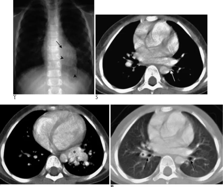
Fig. 1. A 5-year-old girl with systemic arterial supply to normal basal segments of the left lung.
A. Chest radiograph shows retrocardiac nodular and tubular densities (arrowheads), partially obliterating the descending thoracic aorta (arrow).
B. Contrast-enhanced CT scan (mediastinal window) obtained at the level of the left atrium shows an anomalous systemic artery (arrow) arising from lateral aspect of the descending thoracic aorta.
C. CT scan (mediastinal window) obtained at ventricular level shows the dilated inferior pulmonary vein (asterisk), obliterating the descending thoracic aorta.
D. CT scan (lung window) obtained at the level of the origin of the basal segmental bronchi (arrows) reveals that the left interlobar pulmonary artery is absent. The right interlobar pulmonary artery (asterisk) is present immediately lateral to the basilar bronchial trunk.
A left lower lobectomy was performed, and subsequent pathologic examination showed that the bronchial trees and pulmonary parenchyma were normal.
Discussion
Anomalous systemic arterial supply to the normal basal segments of the lower lobe of the lung is a rare congenital abnormality, and is regarded by some au- thors as part of the broad spectrum of bronchopul- monary sequestration (1-5). Some, on the other hand, believe this entity is a congenital vascular anomaly that is distinct from bronchopulmonary sequestration (2).
This, defined as abnormal lung tissue that has no nor- mal continuity with the tracheobronchial tree and is supplied by an anomalous systemic artery, is of two types, intra- or extralobar, a classification which de- pends on its pleural covering and venous drainage sys- tem (6). In contrast, anomalous systemic arterial supply to the normal basal segments of the lower lobe of the lung has a normal connection to the bronchial tree and the normal pulmonary parenchyma (5-7). The etiology of systemic arterial supply to the normal lung is un- known, but it is thought that the persistence of an em- bryonic connection between the aorta and the pul- monary parenchyma leads to this anomaly (1-4, 7).
Most patients are asymptomatic, and the anomaly is dis- covered at chest radiography for the evaluation of car- diac murmur, as in our case. Occasionally, blood flow through the aberrant vessel is enough to result in signifi- cant volume overload of the left-sided cardiac cham- bers, and congestive heart failure (8).
The anomalous systemic artery supplying the basal segments of the left lower lobe and the large inferior pulmonary vein appear at chest radiography as an ill-de- fined area of nodular and tubular opacity, and confusion with pneumonia or pulmonary vascular malformation such as arteriovenous malformation may arise. Some authors have claimed that the origin or tortuosity of the anomalous artery is responsible for the focal obscurity of the descending aorta (2-4, 9). In our case, it was at- tributed to the dilated inferior pulmonary vein adjacent to the descending thoracic aorta.
Contrast-enhanced spiral CT of the chest depicted the dilated and tortuous vascular structures, with ground- glass attenuation, in the left lower lobe, and the anom- alous systemic artery originated from the descending thoracic aorta. The ground-glass attenuation in the in- volved lung is thought to represent increased pul- monary perfusion. CT also provided information about the normal bronchial tree and pulmonary parenchyma, which was histologically confirmed. Another important
J Korean Radiol Soc 2003;48:193-196
─ 195 ─
E F
Fig. 1. E. CT scan (lung window) obtained at the level of the basal lungs shows crowding of dilated vessels with ground-glass atten- uation in left basal lung, resulting from hyperemia, in comparison with the contralateral lung and the ligular division of the left up- per lobe.
F. Contrast-enhanced MR angiographic images also show the anomalous systemic artery arising from lateral aspect of the descend- ing thoracic aorta (arrow).
CT finding was the absence of the interlobar pulmonary artery distal to the origin of the superior segmental artery. These CT findings facilitate distinction from clas- sic bronchopulmonary sequestration (2, 4, 6, 7).
Although MR imaging can also demonstrate an anom- alous systemic artery, it is not easy to determine confi- dently whether the lung tissue supplied by a systemic artery is normal. We therefore believe, as reported by others (2, 4, 6, 7) that contrast-enhanced spiral CT of the chest is the mainstay for evaluation of systemic arterial supply to the normal basal segments.
If clinical symptoms such as hemoptysis and conges- tive heart failure are not present, treatment is conserva- tive, though for the correction of left-to-right shunt, surgery is always imperative. An anomalous systemic artery may simply be ligated if there is pulmonary sup- ply to the involved segments of the lung. If, on the other hand, the aberrant artery represents the sole source of blood flow, lobectomy is required (2, 5, 8). In our case, contrast-enhanced CT demonstrated that an anomalous systemic artery arising from the descending aorta pro- vided the only blood flow to the normal basal segments of the left lower lobe, and left lower lobectomy was therefore planned and performed on the basis of the CT findings.
In conclusion, systemic arterial supply to the normal basal segments of the left lower lobe should be included in the differential diagnosis when a chest radiograph de- picts retrocardiac nodular density with focal obscurity
of the descending thoracic aorta. We believe that con- trast-enhanced CT is indispensable for correct diagnosis and proper treatment.
References
1. Ellis K. Fleischner lecture. Developmental abnormalities in the systemic blood supply to the lungs. AJR Am J Roentgenol 1991;156:669-679
2. Miyake H, Hori Y, Takeoka H, Takuma M, Kawagoe T, Mori H.
Systemic arterial supply to normal basal segments of the left lung:
characteristic features on chest radiography and CT. AJR Am J Roentgenol 1998;171:387-392
3. Kirks DR, Kane PE, Free EA, Taybi H. Systemic arterial supply to normal basilar segments of the left lower lobe. AJR Am J Roentgenol 1976;126:817-821
4. Kurosaki Y, Kurosaki A, Irimoto M, Kuramoto K, Itai Y. Systemic arterial supply to normal basal segments of left lower lobe: CT findings. J Comput Assist Tomogr 1993;17:857-861
5. Tao CW, Chen CH, Yuen KH, Huang MH, Li WY, Perng RP.
Anomalous systemic arterial supply to normal basilar segments of the lower lobe of the left lung. Chest 1992;102:1583-1585
6. Do KH, Goo JM, Im JG, Kim KW, Chung JW, Park JH. Systemic arterial supply to the lungs in adults: spiral CT findings.
RadioGraphics 2001;21:387-402
7. Ashizawa K, Ishida Y, Matsunaga N, Otsuji H, Sakamoto I, Hayashi K. Anomalous systemic arterial supply to normal basal segments of left lower lobe: characteristic imaging findings. J Comput Assist Tomogr 2001;25:764-769
8. Yabek SM, Burstein J, Berman WJr, Dillon T. Aberrant systemic arterial supply to the left lung with congestive heart failure. Chest 1981;80:636-637
9. Ernst SM, Bruschke AV. An aberrant systemic artery to the right lung with normal pulmonary tissue. Chest 1971;60:606-608 Min-Jeong Kim, et al: Systemic Arterial Supply to the Normal Basal Segments of the Left Lower Lobe
─ 196 ─
대한방사선의학회지 2003;48:193-196
정상 좌하엽 폐저분절의 체동맥 혈액 공급: 증례 보고1
1울산대학교 의과대학 서울아산병원 방사선과학교실
2울산대학교 의과대학 서울아산병원 흉부외과학교실
김민정・구현우・박승일2・윤종현
정상 좌하엽 폐저분절의 체동맥 혈액 공급(systemic arterial supply to the normal basal segments of the left lower lobe)은 정상 기관지와 연결을 보이며 체동맥으로부터 혈류공급을 받는 폐실질의 한 부분으로 특징적인 방사선학적 소 견을 보이는 드문 선천성 기형이다. 저자들은 단순흉부촬영, 전산화단층촬영, 그리고 자기공명영상으로 하행 흉부 대동 맥에서 혈액공급을 받는 정상 좌하엽 폐저분절의 체동맥 혈액 공급으로 진단하였으며 수술소견 상 확진된 1예를 경험 하였기에 이를 보고하고자 한다.