Human Urine-derived Stem Cells Seeded Surface Modified Composite Scaffold Grafts for Bladder Reconstruction in a Rat Model
We conducted this study to investigate the synergistic effect of human urine-derived stem cells (USCs) and surface modified composite scaffold for bladder reconstruction in a rat model. The composite scaffold (Polycaprolactone/Pluronic F127/3 wt% bladder submucosa matrix) was fabricated using an immersion precipitation method, and heparin was immobilized on the surface via covalent conjugation. Basic fibroblast growth factor (bFGF) was loaded onto the heparin-immobilized scaffold by a simple dipping method. In maximal bladder capacity and compliance analysis at 8 weeks post operation, the USCs-
scaffoldheparin-bFGF group showed significant functional improvement (2.34 ± 0.25 mL and 55.09 ± 11.81 μL/cm H2O) compared to the other groups (2.60 ± 0.23 mL and
56.14 ± 9.00 μL/cm H2O for the control group, 1.46 ± 0.18 mL and 34.27 ± 4.42 μL/cm H2O for the partial cystectomy group, 1.76 ± 0.22 mL and 35.62 ± 6.69 μL/cm H2O for the scaffold group, and 1.92 ± 0.29 mL and 40.74 ± 7.88 μL/cm H2O for the
scaffoldheparin-bFGF group, respectively). In histological and immunohistochemical analysis, the USC-scaffoldheparin-bFGF group showed pronounced, well-differentiated, and organized smooth muscle bundle formation, a multi-layered and pan-cytokeratin-positive
urothelium, and high condensation of submucosal area. The USCs seeded scaffoldheparin-bFGF
exhibits significantly increased bladder capacity, compliance, regeneration of smooth muscle tissue, multi-layered urothelium, and condensed submucosa layers at the in vivo study.
Keywords: Bladder Regeneration; Surface Modified Scaffold; Urine-derived Stem Cells;
Basic Fibroblast Growth Factor 2 Jun Nyung Lee,1* So Young Chun,2*
Hyo-Jung Lee,2 Yu-Jin Jang,3 Seock Hwan Choi,1 Dae Hwan Kim,4 Se Heang Oh,5 Phil Hyun Song,6 Jin Ho Lee,7 Jong Kun Kim,8 and Tae Gyun Kwon1
1Department of Urology, Kyungpook National University School of Medicine, Daegu; 2Bio-Medical Research Institute, Kyungpook National University Hospital, Daegu; 3Department of Neural Development and Disease, Korea Brain Research Institute, Daegu;
4Department of Laboratory Animal Research Support Team, Yeungnam University, Daegu; 5Department of Nanobiomedical Science & WCU Research Center, Dankook University, Cheonan; 6Department of Urology, Yeungnam University College of Medicine, Daegu; 7Department of Advanced Materials, Hannam University, Daejeon; 8Department of Emergency Medicine, Kyungpook National University School of Medicine, Daegu, Korea
* Jun Nyung Lee and So Young Chun contributed equally to this work.
Received: 1 June 2015 Accepted: 2 September 2015 Address for Correspondence:
Tae Gyun Kwon, MD
Department of Urology, Kyungpook National University School of Medicine, 130 Dongdeok-ro, Jung-gu, Daegu 41944, Korea Tel: +82.53-200-2671, Fax: +82.53-200-3029
E-mail: [email protected]
Funding: This research was supported by Basic Science Research Program through the National Research Foundation of Korea (NRF) funded by the Ministry of Science, ICT & Future Planning (grant number) (2014R1A1A3049460); (NRF-2014M3A9D3033887);
funded by the Ministry of Education (2015R1D1A3A03020378);
and supported by a grant of the Korea Health Technology R&D Project through the Korea Health Industry Development Institute (KHIDI), funded by the Ministry of Health & Welfare (HI14C1642)
http://dx.doi.org/10.3346/jkms.2015.30.12.1754 • J Korean Med Sci 2015; 30: 1754-1763
INTRODUCTION
Bladder augmentation remains one of the greatest surgical challenges in the field of urology. Conventional bladder reconstruction using gastrointestinal tissue is associat- ed with a series of complications (mucus production, bacterial colonization, electro- lyte imbalances, or malignancy), significant morbidity, and functional alterations (1).
Tissue engineering technique can circumvent many of these limitations and therefore has become considered as a potential alternative for bladder reconstruction.
The chief obstacle over the years to reconstructing the bladder through regeneration has been the absence of an ideal biomaterial that can provide a structurally intact low- pressure reservoir, serve as a scaffold for the healing and regeneration of the bladder wall, and retain normal function until it is replaced by host tissues (2,3). Until recently, three classes of biomaterials have been extensively investigated for bladder tissue engi- neering. Naturally derived materials, e.g. collagen (4) and alginate (5), acellular tissue matrices, e.g., bladder submucosa (BSM) (6) and small intestinal submucosa (7), and synthetic polymers, e.g., polyglycolic acid (PGA) (8), polylactic acid (PLA) (9), and poly- lactic-co-glycolic acid (PLGA) (10). However, none of them could provide required re- sults for ideal biomaterials. In an effort to fabricate a scaffold that can be applied to morphological and functional bladder reconstruction, we have been worked to devel- op a composite scaffold composed with synthetic and natural derived biomaterial. As a synthetic biomaterial, polycaprolactone (PCL) was investigated as a potential substi- Cell Therapy & Organ Transplantation
tute for bladder tissue reconstruction because of its flexibility, biocompatibility, stability, and resistance to resorption (11). We could fabricate a blend of PCL with Pluronic F127 (F127) and bladder submucosa matrix (BSM), which has been shown to be a non-immunogenic, non-cytotoxic collagen-rich membrane that can be rapidly replaced by native tissues (12).
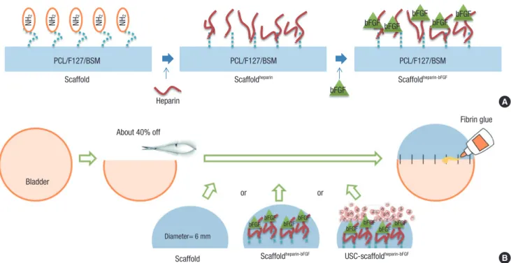
In our previous work, the PCL/F127/3 wt% BSM composite scaffold exhibited significantly enhanced hydrophilicity, the surface was easily immobilized, and there was no evidence of teratoma formation in vivo (12). Based on our previous results, we fabricated an advanced scaffold containing a specific growth factor to enhance proliferation of smooth muscle cells and uro- thelial cells, which are the two essential cell types for functional bladder regeneration. Basic fibroblast growth factor (bFGF) has been shown to stimulate the proliferation and survival of both smooth muscle and urothelial cells (13), thus suggesting the benefits of bFGF-loaded scaffolds for urological tissue engi- neering applications. Therefore, to fabricate a scaffold for bFGF delivery, we covalently conjugated heparin to the surface of a scaffold to form a heparin-immobilized scaffold, which was then loaded with bFGF (scaffoldheparin-bFGF) (14).
Tissue engineering of bladder also requires ideal cell source.
Autologous cells are one of the most popularly used cells with the advantages including, decreased infection, require no im- munosuppression reagents, and do not require histocompati- bility matching (15). However, autologous bladder cell harvest procedures are surgically invasive, and the prolonged cell ex- pansion methods are expensive, subject to contamination, and unrealistic for routine clinical use. Alternative sources of healthy and abundant bladder tissue are desired for optimal bladder engineering and bladder regeneration. Skeletal muscle cells, bone marrow stromal cells, embryonic, and parthenogenetic stem cells are all currently under investigation as potential fu- ture alternatives, however these modalities are in their infancy.
For the purpose of this study, we turned to a stem cell source from urine, i.e., urine-derived stem cells (USCs). USCs have been proposed as an alternative stem cell source for urological tissue reconstruction since they have mesenchymal stem cell characteristics and have demonstrated the capacity to differen- tiate into a variety of urological cell lineages (16).
In this study, we hypothesized that a USC-seeded scaffold- heparin-bFGF graft would promote regeneration of the bladder wall. To investigate the synergic effect of the USC-scaffoldhepa- rin-bFGF in terms of smooth muscle and urothelial layer re- generation, the scaffoldheparin-bFGF was characterized by quantifying the amounts of immobilized heparin and loaded bFGF released, and assessing the biocompatibility and differ- entiation of USCs in vitro. In addition, the potential of the USC- scaffoldheparin-bFGF for bladder reconstruction was evaluat- ed in vivo using a rat model.
MATERIALS AND METHODS
Fabrication of the heparin-immobilized bFGF-loaded scaffold (scaffoldheparin-bFGF)
PCL (MW 80,000 Da; Sigma-Aldrich, St. Louis, MO, USA), tetra- glycol (glycofurol; Sigma-Aldrich), Pluronic F127 (MW 12,500 Da; BASF, Ludwigshafen, Germany), and BSM were used for scaffold fabrication. BSM was prepared as described in our pre- vious report, and the formulation consisting of 3 wt% BSM of the polymer base was chosen (12). The PCL/F127/BSM scaffold was prepared using an immersion precipitation method (17). A PCL pellet/Pluronic F127 powder mixture (95/5 [w/w]) was dis- solved in tetraglycol (12 wt%), and BSM powder was evenly mixed with the polymer solution. The mixed solutions were poured into a polytetrafluoroethylene mold (70 × 70 × 0.4 μL) and then directly immersed in water for 1 hr at room tempera- ture. After additional washing and vacuum drying, the PCL/
F127/BSM composite scaffold was sterilized using ethanol.
Heparin (MW 18,000 Da; Sigma-Aldrich) was immobilized to the scaffold to bind bFGF to the scaffold. The scaffold was pre-wet with 70% (v/v) ethanol, washed with deionized water, and hydrated with a 0.1 M 2-(N-morpholino) ethanesulfonic acid (MES) buffer (pH 5.5). The carboxylic acid groups of hepa- rin were activated using 1-ethyl-3-(3-dimethylaminopropyl) carbodiimide hydrochloride (EDC) and N-hydroxylsuccinimi- de (NHS) for 4 hrs. The scaffold was soaked in 10 mL of the he- parin solution with gentle agitation at 48°C for 6 hr. The hepa- rin-immobilized scaffold was rinsed with the 0.1 M MES buffer (pH 5.5) solution and phosphate buffered saline (PBS, pH 7.4) (18). The heparin-immobilized scaffold was soaked in PBS con- taining 100 ng/mL of bFGF (Peprotech, Rocky Hill, NJ, USA) and 1 mg/mL of bovine serum albumin (Sigma-Aldrich) for 4 hr with gentle agitation, and then the scaffold was washed with PBS (Fig. 1).
Determination of heparin and bFGF content
Heparin content on the scaffold surface was determined by the toluidine blue colorimetric method (19,20). The scaffold was placed in 1 mL of 0.2% NaCl solution, then 1 mL of toluidine blue solution was added, and inner air was removed. After 10 min of vibration, 2 mL of hexane was added and mixed by vor- texing. The absorbance of the aqueous layers at 631 nm was de- termined by a UV spectrophotometer. Immobilized heparin was visualized by labeling with fluorescein isothiocyanate (FITC, 1 mg/mL) at 4°C for 3 hr. The FITC-labeled scaffold was cryosectioned (20 μm) and observed under a confocal laser- scanning microscope (LSM510, Carl Zeiss, Oberkochen, Ger- many).
For the bFGF affinity and release test, the scaffoldheparin-bFGF
was immersed in 1 mL of PBS containing 1% bovine serum al- bumin at 37°C for 28 days. The solution was collected and re-
placed with fresh medium. The amount of bFGF was deter- mined using a Quantikine Immunoassay kit, according to the manufacturer’s instructions (Human bFGF Quantikine ELISA kit, R&D Systems, Minneapolis, MN, USA). An unmodified heparin scaffold was used as a control (n = 3).
Scaffoldheparin-bFGF surface morphology, biocompatibility, and effects on cell differentiation
Urine samples from the upper urinary tract were obtained from a 52-yr-old female patient. A volume of 100 mL of each urine sample was centrifuged, and the cell pellets were washed with PBS. The cells were cultured in mixed medium consisting of ke- ratinocyte serum-free medium and progenitor cell medium (Gibco-Invitrogen, Grand Island, NY, USA) in a 1:1 ratio (21).
The surface morphology of the scaffolds and adherent cells on the scaffolds were assessed using a field emission scanning electron microscope (FE-SEM, S-4300, Hitachi, Hitachi-shi, Ja- pan). The USC-loaded scaffolds were fixed in a 4% paraformal- dehyde (PFA, Sigma-Aldrich) solution at 30°C for 45 min, fol- lowed by washing with dH2O, drying, and coating with gold.
The specimens were examined by FE-SEM at an acceleration voltage of 10 kV.
To analyze the cell adherence efficiency for the scaffoldheparin-
bFGF, USCs (1 × 10⁴ cells) were seeded on the scaffold (6 mm di- ameter, 0.4 mm thickness) and incubated overnight at 37°C and 50 rpm. After one day, the scaffolds were treated with DNA lysis buffer consisting of 0.1% sodium dodecyl sulfate (SDS, v/v), 1 mM ethylenediaminetetraacetic acid (EDTA), and 100 mM Tris–
HCl (pH 7.4). The unmodified scaffold (not loaded with bFGF) was used as a control. Samples were frozen and thawed repeat- edly, and then incubated overnight. Total DNA in the samples was measured using a fluorescent DNA quantitation kit (Bio- Rad, Richmond, VA, USA), according to the manufacturer’s in- structions. DNA concentration measurements were confirmed using confocal microscopy (Axio Observer. Z1, Carl Zeiss).
To measure cell viability and proliferation, 5 × 10³ cells were seeded on the scaffolds (10 mm diameter, 0.4 mm thickness), and a CCK-8 assay (Dojindo, Tokyo, Japan) was performed, ac- cording to the manufacturer’s instructions. The unmodified scaf- fold was used as a control. For myogenic differentiation, USCs were seeded on the scaffoldheparin-bFGF in Dulbecco’s modified Eagle medium (DMEM) containing non-essential amino acids, glutamine, and 15% fetal bovine serum (FBS, Gibco-Invitrogen).
At approximately 95% confluence, 3 μM 5-aza-2´-deoxycytidine (Sigma-Aldrich) and 5 ng/mL of transforming growth factor β (TGF-β, Peprotech) were added to the culture medium for 24 hr and the cells were cultured up to 14 days. For urothelial cell dif- ferentiation, USCs were cultured with supernatant medium col- lected from human bladder urothelial cell cultures (Lonza, Walk- ersville, MD, USA) and cultured for 14 days.
Flow cytometric evaluation of cells (passage 3) was perform- ed for mesenchymal stem cell markers (CD44, CD90, and CD105), smooth muscle cell markers (α-SM actin, Capponin I), and uro- thelium markers (pan-CK, CK19) (BD Biosciences, San Jose, CA, USA), according to the manufacturer’s instructions. For re- al-time PCR, total RNA was extracted with an RNeasy kit (Qia-
bFGF
NH2 NH2 NH2 NH2 NH2
bFGF
bFGF bFGF bFGF
PCL/F127/BSM PCL/F127/BSM
PCL/F127/BSM
Scaffoldheparin-bFGF
Scaffoldheparin Scaffold
bFGF
Heparin A
Fig. 1. Schematic diagram of the scaffold fabrication and operation procedures. (A) Procedures for fabrication of the heparin-immobilized bFGF-loaded scaffolds (Scaffoldheparin-
bFGF) consisting of Polycaprolactone/Pluronic F127/bladder submucosa matrix (PCL/F127/BSM). (B) Bladder reconstruction operation procedure using the various scaffolds.
About 40% off
Diameter= 6 mm Scaffold
Fibrin glue
or or
Scaffoldheparin-bFGF USC-scaffoldheparin-bFGF
Bladder
B
bFGFbFGF bFGFbFGF
bFGF bFGF bFGF
bFGF
gen, Hilden, Germany), according to the manufacturer’s instruc- tions. A total of 2 μg of RNA was used for cDNA synthesis using cDNA Reverse Transcription kits (Applied Biosystems, Warring- ton, UK). The primers were designed with Primer Express Soft- ware (Applied Biosystems), and were listed in Table 1. To ana- lyze the data, the 2-△△Ct method of relative quantification was adapted to estimate the copy numbers.
Urodynamic study, histology, and immunohistochemistry of reconstructed bladders
Twenty-five rats were divided to 5 groups: 1) control group, sham operated; 2) partial cystectomy group, approximately 40% de- fect was created in the dome of the bladder wall; 3) scaffold group, the unmodified scaffold was attached after partial cys- tectomy; 4) scaffoldheparin-bFGF group, the heparin-immobilized bFGF-loaded scaffold was attached after partial cystectomy;
and 5) USC-scaffoldheparin-bFGF group, scaffoldheparin-bFGF combined with 1 × 10⁴ USCs was attached after partial cystectomy. The single-layer scaffold (disk form, diameter 6 mm) was sutured as a patch onto the defect of the bladder with 7-0 Vicryl sutures.
Omentum was loosely wrapped over the graft and fixed with fi- brin glue (Greenplast, Greencross, Seoul, Korea) (Fig. 1B).
An urodynamic study (filling cystometry) was performed on 5 animals from each group, at 8 weeks post-operation. Two ani- mals with bladder calculi (scaffold group; n = 1, scaffold heparin-bFGF; n = 1) were excluded from the urodynamic data collection. The bladder was filled with PBS, and maximal capacity was defined as the volume of infusion that triggered the first leakage of urine.
Compliance was defined as maximal capacity/(pressure which triggered the first leakage) - (baseline pressure). Then, the entire bladder was removed, fixed in formalin, and the cross-sectional area of bladders divided in half was measured using ImageJ (http:
//imagej.net/). The bladder samples were embedded in paraffin and cut into 5 μm sec-tions that were stained using hematoxylin and eosin (H&E) and for immunohistochemical (IHC) staining.
The regenerated smooth muscle and urothelial cell layers were identified by α-SM actin and pan-cytokeratin antibodies (Sig- ma-Aldrich), immune reaction was analyzed with cytotoxic T cell marker (CD8), and seeded human USCs on the scaffold were detected with human nuclei-specific antibody (HuNu, BD Biosciences).
Statistical analysis
All experiments were performed at least in triplicate on sepa- rate days. A t-test and a one-way analysis of variance (ANOVA) of Tukey’s test were used for statistical analysis. All values are expressed as the mean ± SD. Results are representative of at least three experiments.
Ethics statement
The study protocol using the cells from human urine was re- viewed and approved by the institutional review board of Kyung- pook National University Hospital (IRB No. KNUH 2012-10- 018). Informed consent was obtained from the patient regard- ing urine sampling and use of cells. All experimental proce- dures using rats were reviewed and approved by the institu- tional animal care and use committee of Yeungnam University College of Medicine (YUMC-AEC2013-003).
RESULTS
Characteristics of the heparin-immobilized scaffold Fig. 1A illustrates the procedures followed to functionalize the scaffold surface with primary amine groups and the subse- quent heparin immobilization, followed by bFGF loading. The carboxylic acids exposed on the surface were used to produce surface amine groups, and the heparin was immobilized to the Table 1. Primer sequences
Markers Symbol Full name Sequences
Stem cell
marker OCT4 Octamer-binding tran-
scription factor 4 5´-TCAGCCAAACGACCATCTGC 5´-GCTTGATCGCTTGCCCTTCT SSEA4 Stage specific
embryonic antigen 4
5´-TCCCAGGTTCAAGCGATTCTC 5´-CCAACATGGTGAAACGCAGTC NANOG Nanog 5´-GCATCCGACTGTAAAGAATCTTCA
5´-CATCTCAGCAGAAGACATTTGCA ALP Alkaline
phosphatase 5´-ACGAGCTGAACAGGAACAACGT 5´-CACCAGCAAGAAGAAGCCTTTG
C-KIT c-kit 5´-GGCATCATGATCAAAAGTGTGAA
5´-CCCTCCTGGTCCACAGAACA Smooth
muscle cell PAX7 Paired box 7 5´-GCAAATTGCTGTCCTGCTCA 5´-TGAAAACTGGTCACATCTGCCT differentia-
tion marker MYOD Myoblast determination protein
5´-ACAGCGCGGTTTTTTCCAC 5´-AACCTAGCCCCTCAAGGTTCAG DESMIN Desmin 5´-GGAGAGGAGAGCCGGATCA
5´-GGGCTGGTTTCTCGGAAGTT MYOSIN Myosin 5´-AGGCGGAGAGGTTTTCCAA
5´-CTTGTAGTCCAAGTTGCCAGTCA α-SM
ACTIN Alpha smooth
muscle actin 5´-CAAGTGATCACCATCGGAAATG 5´-GACTCCATCCCGATGAAGGA Epithelial cell
differentia- tion marker
UP1a Uroplakin 1A 5´-CGCTGGTGCCTGGATTG 5´-GGCACCCACACCAAAACT UP1b Uroplakin 1B 5´-CAATTGCTGTGGCGTAAATGG
5´-ATAACACAGCATTGACGAGGCC UP2 Uroplakin 2 5´-TCGTGCCAGGAACCAAATTC
5´-GGATTCCATGTTCCTTCGAGG CK7 Cytokeratin-7 5´-GGAACTCATGAGCGTGAAGCT
5´-CCAGTGGAATTCATCACAGAGA- TAT
CK13 Cytokeratin-13 5´-GGATGCTGAGGAATGGTTCCA 5´-GCTCTGTCTTGCTCCGTGATCT CK18 Cytokeratin-18 5´-ATTGAGGAGAGCACCACAGTGG
5´-TCTCATGGAGTCCAGGTCGATC CK19 Cytokeratin-19 5´-CAGGTCAGTGTGGAGGTGGAT
5´-TCGCATGTCACTCAGGATCTTG PAN-CK Pan-cytokeratin 5´-GCCTCCTTGGCAGAAACAGAA 5´-GCACTCGGTTTCAGCTCGAAT Housekeeping
gene β-ACTIN β-actin 5´-ATCGTCCACCGCAAATGCT 5´-AAGCCATGCCAATCTCATCTTG
surface via covalent conjugation (22). A toluidine blue assay was used to determine the amount of immobilized heparin, which was shown to be 0.72 ± 0.11 μg/6 mm2 (Fig. 2A). The fab- ricated scaffold had different sized pores on the surface at front and rear side, small and large; the pore sizes were -100 and -200 μm, respectively, and the heparin distribution was homoge-
neous when visualized with FITC-conjugated heparin (Fig. 2B).
Affinity and release of loaded bFGF
The amount of bFGF loaded into the scaffoldheparin-bFGF at days 1, 3, 7, 14, 21, and 28 was 33.68 ± 0.29, 32.80 ± 0.39, 30.39 ± 0.33, 27.31 ± 0.25, 25.67 ± 0.24, and 26.03 ± 0.30 pg/mL, and for the
Fig. 2. Measurement of heparin, bFGF, and scaffoldheparin-bFGF biocompatibility. (A) Amount of immobilized heparin on the scaffold (n = 3). (B) Visualization of immobilized heparin on the scaffold. (C) Affinity and amount of bFGF released from the heparin-immobilized scaffold for 28 days. An unmodified heparin scaffold was used as a control. (D) Field emission scanning electron microscope images of scaffold morphology and cells attached to the scaffold. (E) Cell adhesion to the scaffolds was determined by measuring the DNA concentration with Hoechst 33258 staining in confocal images. (F) Biocompatibility analysis of the scaffolds. Ctrl, culture plate dish; scaffold, unmodified scaffold; scaffold-
heparin-bFGF, heparin-immobilized bFGF-loaded scaffold; USCs, urine derived stem cells; O.D., optical density. All data are presented as mean ± SD (*P < 0.01; †P < 0.05).
μg/6 mm2
Scaffoldheparin Scaffold
1.0 0.8 0.6 0.4 0.2 0
Amount of immobilized heparin
*
A
DNA concentration (ng)
Scaffoldheparin-bFGF
Scaffold Ctrl
12 11 10 9 8 7
Cell adhesion
†
E
×200 Interval 1.5 μm B
D
F
* *
*
†
pg/mL
1 3 7 14 21 28
Day 35
30 25 20 15 10 5 0
Affinity and amount of released bFGF
C Scaffold
Scaffoldheparin
†
†
O.D. 450 nm
1 3 5 7
2.5
2.0
1.5
1.0
0.5
0
Cell proliferation †
Ctrl Scaffold Scaffoldheparin-bFGF
O.D. 450 nm
3 6 12 24
0.45
0.40
0.35
0.30
†
†
†
Cell viability Ctrl Scaffold Scaffoldheparin-bFGF
Scaffold
×400 ×1,000 ×400 ×1,000
Scaffoldheparin
USC
Scaffold Scaffoldheparin-bFGF
×100 ×200 ×100 ×200
USC
(Hour)
(Day)
Fig. 3. Cell differentiation supported by the scaffoldheparin-bFGF. (A) USC differentiation (%) into smooth muscle and urothelial cells on the scaffoldheparin-bFGF and representative FACS images. (B) Real-time PCR analysis of stem cell, myogenic, and urothelial lineage markers at days 0 and 14. *P < 0.01.
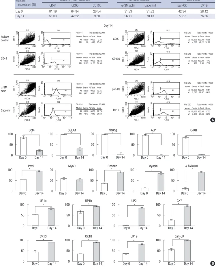
Markers expression (%)
Mesenchymal stem cell Smooth muscle Urothelium
CD44 CD90 CD105 α-SM actin Caponin I pan-CK CK19
Day 0 81.19 64.94 26.54 31.83 31.82 42.34 28.12
Day 14 51.03 42.22 9.50 96.71 70.13 77.87 76.66
Day 14
Isotype control
α-SM actin
Caponin I CD44
CD90
CD105
pan-CK
CK19
A
B Day 0 Day 14
100
50
0
Oct4*
Day 0 Day 14 100
50
0
Pax7*
Day 0 Day 14 100
50
0
UP1a
*
Day 0 Day 14 100
50
0
CK13
*
Day 0 Day 14 100
50
0
UP1b
*
Day 0 Day 14 100
50
0
CK18
*
Day 0 Day 14 100
50
0
UP2
*
Day 0 Day 14 100
50
0
CK19
*
Day 0 Day 14 100
50
0
CK7*
Day 0 Day 14 100
50
0
pan-CK
* Day 0 Day 14
100
50
0
MyoD
Day 0 Day 14 100
50
0
Desmin
*
Day 0 Day 14 100
50
0
Myosin
*
Day 0 Day 14 100
50
0
α-SM actin
* Day 0 Day 14
100
50
0
SSEA4
*
Day 0 Day 14 100
50
0
Nanog
*
Day 0 Day 14 100
50
0
ALP*
Day 0 Day 14 100
50
0
C-KIT
*
File: 015 Total events: 10,000 Marker Events % Total Mean
All 10,000 100.00 2.38 M1 20 0.20 15.62
File: 017 Total events: 10,000 Marker Events % Total Mean
All 10,000 100.00 108.98 M1 4,222 42.22 251.82
File: 018 Total events: 10,000 Marker Events % Total Mean
All 10,000 100.00 5.43 M1 950 9.50 17.38
File: 019 Total events: 10,000 Marker Events % Total Mean
All 10,000 100.00 36.51 M1 7,787 77.87 45.12
File: 020 Total events: 10,000 Marker Events % Total Mean
All 10,000 100.00 47.32 M1 7,666 76.66 60.17 File: 016 Total events: 10,000
Marker Events % Total Mean All 10,000 100.00 18.52 M1 5,103 51.03 31.95
File: 013 Total events: 10,000 Marker Events % Total Mean
All 10,000 100.00 73.45 M1 9,671 96.71 75.67
File: 014 Total events: 10,000 Marker Events % Total Mean
All 10,000 100.00 17.79 M1 7,013 70.13 21.69 SSC-H 0 1,000
015
0 1,000 PSC-H
Counts 0 150 015
100 101 102 103 104 FL2-H
M1
SSC-H 0 1,000 016
0 1,000 PSC-H
Counts 0 150 016
100 101 102 103 104 FL2-H
M1
SSC-H 0 1,000 017
0 1,000 PSC-H
Counts 0 150 017
100 101 102 103 104 FL2-H
M1
SSC-H 0 1,000 018
0 1,000 PSC-H
Counts 0 150 018
100 101 102 103 104 FL2-H
M1
SSC-H 0 1,000 019
0 1,000 PSC-H
Counts 0 150 019
100 101 102 103 104 FL2-H
M1
SSC-H 0 1,000 020
0 1,000 PSC-H
Counts 0 150 020
100 101 102 103 104 FL2-H
M1 SSC-H 0 1,000
013
0 1,000 PSC-H
Counts 0 140 013
100 101 102 103 104 FL2-H
M1
SSC-H 0 1,000 014
0 1,000 PSC-H
Counts 0 140
014
100 101 102 103 104 FL2-H
M1
heparin unmodified scaffold was 25.02 ± 0.29, 24.49 ± 0.07, 25.14 ± 0.40, 24.19 ± 0.23, 23.89 ± 0.12, and 24.19 ± 0.30 pg/mL, respectively (Fig. 2C). The initial concentration of bFGF on the heparin-immobilized scaffold revealed a high affinity and time-dependent release compared to the unmodified scaffold, and from day 21, the bFGF concentration became a basal level around 25.0 pg/mL. The unmodified scaffold showed rare af- finity for bFGF, which means that heparin acts an effective liker to load of bFGF on the scaffold.
Scaffoldheparin-bFGF morphology and biocompatibility The scaffolds exhibited a well-fabricated fibrous and porous structure, with large surface area (Fig. 2D). The pores had an av- erage diameter of 50.8 ± 8.4 μm. Analysis of cell adhesion based on DNA concentration showed that adhesion to the scaffoldhepa-
rin-bFGF was significantly higher than to the other surfaces (Fig.
2E). After culturing the cells for periods of 3, 6, 12, and 24 hr and 1, 3, 5, and 7 days, the viability and proliferation of USCs showed more viable cells present on the scaffoldheparin-bFGF (Fig. 2F); from day 3 of the culture, the absorbance value was significantly high- er for the scaffoldheparin-bFGF than that of the other surfaces.
Differentiation into smooth muscle and urothelial cells on the scaffoldheparin-bFGF
When USCs were cultured on the scaffoldheparin-bFGF with smooth muscle and urothelium induction media, the cells were shown to express smooth muscle and urothelium surface markers at day 14 (Fig. 3A). The number of α-SM actin, Caponin I, pan-CK, and CK19-positive cells increased by 3.04, 2.20, 1.84, and 2.73 times, respectively, while, mesenchymal stem cell markers for CD44, CD90, and CD105 decreased by 0.63, 0.65, and 0.36 times, respectively.
The FACS results were confirmed with gene analysis through real-time PCR (Fig. 3B). At day 14 of differentiation, the cells ex- hibited significantly reduced expression of stem cell markers Oct4, SSEA4, Nanog, ALP, and c-Kit, while, markers indicative of smooth muscle cell (Pax7, MyoD, Desmin, Myosin, and α-SM actin) and urothelium cell (UP1a, UP1b, UP2, Ck7, Ck13, Ck18, Ck19, and pan-CK) differentiation were significantly increased.
Urodynamic study, histology, and immunohistochemistry All rats for the in vivo study survived until the scheduled time of sacrifice, and there was no significant change in body weight or anastomosis problems observed during the experiment. The maximal bladder capacity and compliance at 8 weeks post op- eration was 2.60 ± 0.23 mL and 56.14 ± 9.00 μL/cm H2O for the control group, 1.46 ± 0.18 mL and 34.27 ± 4.42 μL/cm H2O for the partial cystectomy group, 1.76 ± 0.22 mL and 35.62 ± 6.69 μL/cm H2O for the scaffold group, 1.92 ± 0.29 mL and 40.74 ± 7.88 μL/cm H2O for the scaffoldheparin-bFGF group, and 2.34 ± 0.25 mL and 55.09 ± 11.81 μL/cm H2O for the USC-scaffoldheparin-bFGF
group (Table 2).
There was no diverticulum on the implanted portions for any group, which were covered on the outer surface by connective tissue, and 2 animals (scaffold group; n = 1, scaffoldheparin-bFGF
group; n = 1) were observed to have bladder calculi formation.
Gross image analysis showed that the USC-scaffold combina- tion resulted in a reconstructed bladder with better shape and volume. The mean cross-sectional area of the reconstructed bladders were 16.85 ± 1.21, 7.87 ± 1.37, 9.67 ± 0.87, 11.19 ± 0.87, and 15.71 ± 1.34 mm2 for the control, partial cystectomy, scaf- fold, scaffoldheparin-bFGF, and USC-scaffoldheparin-bFGF groups, respec- tively (Table 2) (Fig. 4A).
H&E and IHC analysis (Fig. 4B) showed that the USC-scaf- foldheparin-bFGF group exhibited pronounced, well-differentiated, and organized smooth muscle bundle formation, while other groups exhibited thin muscle layer regeneration consisting of fibroblasts and connective tissue. With regard to the urotheli- um, a multi-layered and pan-cytokeratin-positive urothelium was observed at the reconstructed area for most of the groups, except for the scaffold group. The condensation of submucosal area was notably high in the USC-scaffoldheparin-bFGF. The scaffold group showed enhanced CD8 lymphocyte accumulation, while the USC-scaffoldheparin-bFGF showed scant accumulation of CD8- positive cells. The seeded human USCs were not detected at the USC-scaffoldheparin-bFGF group at week 8.
DISCUSSION
Current research suggests that the use of biomaterial based scaffolds seeded with autologous urothelial and smooth mus- cle cells is the best option for bladder tissue engineering (23).
But, biomaterial based scaffolds lead to a number of complica- tions, and autologous cells are often difficult to obtain. There- fore, we investigated the synergistic effect of a combination of USCs and heparin-immobilized bFGF-loaded scaffolds. This approach would provide development of novel composite bio- material based scaffold and cell source.
Previously, we fabricated a scaffold containing BSM that we reported to have amine groups on the surface (12), and heparin was covalently immobilized to the exposed amine groups on the scaffold surface. Heparin is a highly sulfated glycosamino- glycan, which has binding affinity and activity maintaining of various growth factors (24). Because of these specific interac- tions with growth factors, heparin has been widely used in fab- rication of bioactive matrices for growth factor delivery (25,26), and thus, we attempted bFGF-loading of our scaffold via hepa- rin-immobilization. Comparison of a scaffold without surface modification to a heparin-immobilized scaffold showed higher bFGF loading for the modified scaffold, which was attributed to the ionic interactions that occurred between the positively charged bFGF protein (at physiological pH) and negatively
charged heparin (27,28). The observed sustained release pat- tern for the scaffoldheparin-bFGF was also likely caused by specifi- cally bound bFGF on the heparin-immobilized surface (14).
In addition to a biocompatible scaffold, an appropriate cell source is also an essential requirement for tissue regeneration.
USCs may play an important role in experimental research for urological organ regeneration and other urological-based ap- plications, as they represent an optimal source due to their ca- pacity for differentiation to myogenic and urothelial lineages
(16). In the present study, the heparin-immobilized bFGF-load- ed surface supported USC behavior, including attachment, via- bility, proliferation, and differentiation. To analyze the effects of the scaffoldheparin-bFGF on cell viability and proliferation, USCs were cultured with the two different types of scaffolds. The pro- liferation of USCs cultured on the scaffoldheparin-bFGF was signifi- cantly higher than on the unmodified scaffold. In addition, the USC-scaffoldheparin-bFGF was shown to enhance myogenic and urothelial differentiation rates, indicating that the scaffoldheparin- Table 2. Measurement of cross-sectional area and urodynamic results, including maximal bladder capacity and compliance
Parameters Control Partial cystectomy Scaffold Scaffoldheparin-bFGF USC-scaffoldheparin-bFGF
Cross-sectional area, mm2 (mean ± SD) 16.85 ± 1.21 7.87 ± 1.37 9.67 ± 0.87 11.19 ± 0.87 15.71 ± 1.34 Maximal bladder capacity, mL (mean ± SD) 2.60 ± 0.23 1.46 ± 0.18 1.76 ± 0.22 1.92 ± 0.29 2.34 ± 0.25 Compliance, µL/cm H2O (mean ± SD) 56.14 ± 9.00 34.27 ± 4.42 35.62 ± 6.69 40.74 ± 7.88 55.09 ± 11.81 Fig. 4. Morphological and immunohistochemical (IHC) analysis. (A) Morphological analysis of retrieved bladder. (B) Analysis of α-SM actin, pan-CK, CD8, and HuNu expression with IHC. α-SM actin, α-smooth muscle actin; pan-CK, pan-cytokeratin; CD8, cluster of differentiation 8; HuNu, Human nuclei-specific antibody. Control, sham operated; partial cystectomy, approximately 40% defect was created in the dome of the bladder wall; scaffold, unmodified scaffold was attached after partial cystectomy; scaffoldheparin-bFGF, the heparin-immobilized bFGF-loaded scaffold was attached after partial cystectomy; USC-scaffoldheparin-bFGF, scaffoldheparin-bFGF combined with USCs was attached after partial cystec- tomy. Scale bar = 50 μm. Magnification, 400 ×.
USC-scaffoldheparin-bFGF
Scaffoldheparin-bFGF
Scaffold Cystectomy
Control
A
B pan-CK
CD8
HuNu
USC-scaffoldheparin-bFGF
Scaffoldheparin-bFGF
Scaffold Cystectomy
Control α-SM actin
50 μm 50 μm 50 μm 50 μm 50 μm
50 μm 50 μm 50 μm 50 μm 50 μm
50 μm 50 μm 50 μm 50 μm 50 μm
50 μm
bFGF provides a suitable microenvironment for cell culture be- cause bFGF promotes mesenchymal cell proliferation (29) and induces extracellular matrix production (13), which may im- prove the local microenvironment for supporting implanted cell proliferation and differentiation.
For the in vivo bladder regeneration study, the USC-scaffold-
heparin-bFGF, scaffoldheparin-bFGF, and unmodified scaffold were im- planted into the partially cystectomized bladders of rats to eval- uate their potential for bladder tissue reconstruction. The im- ages of the bladder specimens after dissection showed that the implanted scaffolds were attached in the bladder and covered with fibrous tissue. For the unmodified scaffold, the thick fi- brous mass remained, and no expansion of the bladder volume was observed. The USC-scaffoldheparin-bFGF exhibited a thin fi- brous mass and expanded bladder volume, which are indica- tive of bladder reconstruction. The scaffoldheparin-bFGF (without cells) yielded only a moderate result. The observed fibrous mass can also be used as an indicator of the inflammation caused by the scaffold, as previously described in a study re- porting that fibrous tissue showed infiltration of neutrophils and macrophages (30). IHC analysis with the CD8 antibody showed that the unmodified scaffold exhibited an enhanced positive signal when compared to the other groups, indicating that the unmodified scaffold causes chronic inflammation, and surface modification and USCs could reduce this pathologic phenomenon.
The implanted scaffolds were not visible in the tissue sec- tions because they were dissolved during the specimen-pro- cessing step using xylene. The histological features of the im- planted grafts showed that the regenerated portions of the bladders in the USC-scaffoldheparin-bFGF group exhibited pro- nounced smooth muscle bundles, a multi-layered urothelium, condensed submucosa layer formation, and restored bladder volume. These anatomical reconstructions are essential for functional compliance. Other scaffold groups, however, showed only weak smooth muscle cell bundles, a thin urotheli- um, and loose submucosa regeneration at the graft. These re- sults suggest that the seeded USCs contributed to the tissue re- generation. While the seeded cells were expected to survive for 2 weeks in vivo (31), we observed that the exogenous cells in- troduced in vivo were effective for bladder regeneration in comparison to the cells recruited from surrounding host tissues or circulating blood flow. The USC regenerative mechanism was not specifically identified in this paper, however, based on previous reports, it is presumed to result from the paracrine ef- fects of tropic factors secreted from the USCs (31). Thus, the transplanted stem cells influenced the surrounding host cells, resulting in improved cell migration and differentiation into target cells.
In conclusion, the heparin-immobilized bFGF-loaded scaf- fold exhibits enhanced biocompatibility, and USCs seeded on
the scaffoldheparin-bFGF induces a synergistic effect, as indicated by increased bladder capacity, compliance, and histological tissue reconstruction signified by smooth muscle, urothelium, sub- mucosa layer regeneration, and reduced inflammation, in a partial cystectomy rat model. Therefore, we propose that USC- scaffoldheparin-bFGF would be an ideal strategy for bladder recon- struction.
DISCLOSURE
The authors have no potential conflicts of interest to disclose.
AUTHOR CONTRIBUTION
Conceived and designed the experiments: Lee JN, Chun SY.
Performed the experiments: Lee HJ, Jang YJ, Kim DH. Scaffold manufacturing: Oh SH, Lee JH. Analyzed the data: Song PH.
Drafting of the manuscript: Chun SY, Lee JN. Critical revision of the manuscript for important intellectual content: Kwon TG.
Statistical analysis: Lee JN. Obtaining funding: Kwon TG. Ad- ministrative, technical, or material support: Song PH, Kim DH.
Approval of the final manuscript: Kwon TG.
ORCID
Jun Nyung Lee http://orcid.org/0000-0002-6342-9846 So Young Chun http://orcid.org/0000-0003-4500-4956 Hyo-Jung Lee http://orcid.org/0000-0002-1179-0373 Yu-Jin Jang http://orcid.org/0000-0001-5660-3001 Seock Hwan Choi http://orcid.org/0000-0003-3796-2601 Dae Hwan Kim http://orcid.org/0000-0002-2083-0154 Se Heang Oh http://orcid.org/0000-0002-4635-6809 Phil Hyun Song http://orcid.org/0000-0002-3801-258X Jin Ho Lee http://orcid.org/0000-0002-1528-3416 Tae Gyun Kwon http://orcid.org/0000-0002-4390-0952
REFERENCES
1. Jednak R. The evolution of bladder augmentation: from creating a reser- voir to reconstituting an organ. Front Pediatr 2014; 2: 10.
2. Salem SA, Hwei NM, Bin Saim A, Ho CC, Sagap I, Singh R, Yusof MR, Md Zainuddin Z, Idrus RB. Polylactic-co-glycolic acid mesh coated with fibrin or collagen and biological adhesive substance as a prefabricated, degradable, biocompatible, and functional scaffold for regeneration of the urinary bladder wall. J Biomed Mater Res A 2013; 101: 2237-47.
3. Kim BS, Mooney DJ. Engineering smooth muscle tissue with a pre- defined structure. J Biomed Mater Res 1998; 41: 322-32.
4. Chen W, Shi C, Yi S, Chen B, Zhang W, Fang Z, Wei Z, Jiang S, Sun X, Hou X, et al. Bladder regeneration by collagen scaffolds with collagen binding human basic fibroblast growth factor. J Urol 2010; 183: 2432-9.
5. Lanza RP, Langer RS, Vacanti JP. Principles of tissue engineering. Amster- dam: Academic Press, 2013.
6. Geng HQ, Tang DX, Chen F, Wu XR, Zhou X. The bladder submucosa acellular matrix as a cell deliverer in tissue engineering. World J Pediatr 2006; 2: 57-60.
7. Zhang Y, Kropp BP, Lin HK, Cowan R, Cheng EY. Bladder regeneration with cell-seeded small intestinal submucosa. Tissue Eng 2004; 10: 181-7.
8. Zambon JP, de Sá Barretto LS, Nakamura AN, Duailibi S, Leite K, Mag- alhaes RS, Orlando G, Ross CL, Peloso A, Almeida FG. Histological changes induced by Polyglycolic-Acid (PGA) scaffolds seeded with autol- ogous adipose or muscle-derived stem cells when implanted on rabbit bladder. Organogenesis 2014; 10: 278-88.
9. Gomelsky A, Dmochowski RR. Tissue Engineering for Neurogenic Blad- der. In: Wein AJ, Andersson KE, Drake MJ, Dmochowski RR, editors.
Bladder dysfunction in the adult : the basis for clinical management.
New York, NY: Springer, 2014, p265-76.
10. Roth CC, Mondalek FG, Kibar Y, Ashley RA, Bell CH, Califano JA, Madi- hally SV, Frimberger D, Lin HK, Kropp BP. Bladder regeneration in a ca- nine model using hyaluronic acid-poly(lactic-co-glycolic-acid) nanopar- ticle modified porcine small intestinal submucosa. BJU Int 2011; 108:
148-55.
11. Yu DS, Lee CF, Chen HI, Chang SY. Bladder wall grafting in rats using salt-modified and collagen-coated polycaprolactone scaffolds: prelimi- nary report. Int J Urol 2007; 14: 939-44.
12. Jang YJ, Chun SY, Kim GN, Kim JR, Oh SH, Lee JH, Kim BS, Song PH, Yoo ES, Kwon TG. Characterization of a novel composite scaffold con- sisting of acellular bladder submucosa matrix, polycaprolactone and Pluronic F127 as a substance for bladder reconstruction. Acta Biomater 2014; 10: 3117-25.
13. Lee M, Wu BM, Stelzner M, Reichardt HM, Dunn JC. Intestinal smooth muscle cell maintenance by basic fibroblast growth factor. Tissue Eng Part A 2008; 14: 1395-402.
14. Yoon JJ, Chung HJ, Lee HJ, Park TG. Heparin-immobilized biodegrad- able scaffolds for local and sustained release of angiogenic growth factor.
J Biomed Mater Res A 2006; 79: 934-42.
15. Pigott JH, Ishihara A, Wellman ML, Russell DS, Bertone AL. Investiga- tion of the immune response to autologous, allogeneic, and xenogeneic mesenchymal stem cells after intra-articular injection in horses. Vet Im- munol Immunopathol 2013; 156: 99-106.
16. Chun SY, Kim HT, Lee JS, Kim MJ, Kim BS, Kim BW, Kwon TG. Charac- terization of urine-derived cells from upper urinary tract in patients with bladder cancer. Urology 2012; 79: 1186.e1-7.
17. Oh SH, Kim JR, Kwon GB, Namgung U, Song KS, Lee JH. Effect of sur- face pore structure of nerve guide conduit on peripheral nerve regenera- tion. Tissue Eng Part C Methods 2013; 19: 233-43.
18. Jonnalagadda SB, Gollapalli NR. Kinetics of reduction of toluidine blue
with sulfite-kinetic salt effect in elucidation of mechanism. J Chem Educ 2000; 77: 506.
19. Lee AC, Yu VM, Lowe JB 3rd, Brenner MJ, Hunter DA, Mackinnon SE, Sakiyama-Elbert SE. Controlled release of nerve growth factor enhances sciatic nerve regeneration. Exp Neurol 2003; 184: 295-303.
20. Smith PK, Mallia AK, Hermanson GT. Colorimetric method for the as- say of heparin content in immobilized heparin preparations. Anal Bio- chem 1980; 109: 466-73.
21. Zhang Y, McNeill E, Tian H, Soker S, Andersson KE, Yoo JJ, Atala A.
Urine derived cells are a potential source for urological tissue reconstruc- tion. J Urol 2008; 180: 2226-33.
22. Jeon O, Kang SW, Lim HW, Chung JH, Kim BS. Long-term and zero-or- der release of basic fibroblast growth factor from heparin-conjugated poly(L-lactide-co-glycolide) nanospheres and fibrin gel. Biomaterials 2006; 27: 1598-607.
23. Atala A. Tissue engineering of human bladder. Br Med Bull 2011; 97: 81- 104.
24. Sasisekharan R, Ernst S, Venkataraman G. On the regulation of fibro- blast growth factor activity by heparin-like glycosaminoglycans. Angio- genesis 1997; 1: 45-54.
25. Wang XH, Li DP, Wang WJ, Feng QL, Cui FZ, Xu YX, Song XH. Covalent immobilization of chitosan and heparin on PLGA surface. Int J Biol Macromol 2003; 33: 95-100.
26. Steffens GC, Yao C, Prével P, Markowicz M, Schenck P, Noah EM, Pal- lua N. Modulation of angiogenic potential of collagen matrices by cova- lent incorporation of heparin and loading with vascular endothelial growth factor. Tissue Eng 2004; 10: 1502-9.
27. Cai S, Liu Y, Shu XZ, Prestwich GD. Injectable glycosaminoglycan hy- drogels for controlled release of human basic fibroblast growth factor.
Biomaterials 2005; 26: 6054-67.
28. Liu LS, Ng CK, Thompson AY, Poser JW, Spiro RC. Hyaluronate-heparin conjugate gels for the delivery of basic fibroblast growth factor (FGF-2). J Biomed Mater Res 2002; 62: 128-35.
29. Kanematsu A, Yamamoto S, Noguchi T, Ozeki M, Tabata Y, Ogawa O.
Bladder regeneration by bladder acellular matrix combined with sus- tained release of exogenous growth factor. J Urol 2003; 170: 1633-8.
30. Lech M, Gröbmayr R, Weidenbusch M, Anders HJ. Tissues use resident dendritic cells and macrophages to maintain homeostasis and to regain homeostasis upon tissue injury: the immunoregulatory role of changing tissue environments. Mediators Inflamm 2012; 2012: 951390.
31. Kim BS, Chun SY, Lee JK, Lim HJ, Bae JS, Chung HY, Atala A, Soker S, Yoo JJ, Kwon TG. Human amniotic fluid stem cell injection therapy for urethral sphincter regeneration in an animal model. BMC Med 2012; 10:
94.