REVIEW ARTICLE
염증성 장질환에서의 피부 증상
서현이, 이우진, 나수영
1울산대학교 의과대학 서울아산병원 피부과, 제주대학교 의학전문대학원 제주대학교병원 내과1
Dermatologic Manifestations in Inflammatory Bowel Disease
Hyun Yi Suh, Woo Jin Lee and Soo-Young Na1
Department of Dermatology, Asan Medical Center, University of Ulsan College of Medicine, Seoul; Department of Internal Medicine, Jeju National University Hospital, Jeju National University School of Medicine1, Jeju, Korea
Inflammatory bowel disease (IBD) is a chronic inflammatory disease of the gastrointestinal tract with an unknown etiology and pathogenesis. The incidence and prevalence of IBD are increasing rapidly in Korea. Approximately one-third of patients with IBD appear to develop extra-intestinal manifestations with the skin being one of the most commonly affected organs. They may precede, occur simultaneously, or follow the diagnosis of IBD. In addition, they may parallel with the luminal symptoms or independent from the disease activity of IBD. This review outlines the skin manifestations associated with IBD and discusses their management. Skin manifestations should be managed in close collaboration with a dermatologist. (Korean J Gastroenterol 2019;73:285-293)
Key Words: Inflammatory bowel diseases; Crohn disease; Colitis, ulcerative; Skin manifestations
Received April 8, 2019. Revised May 3, 2019. Accepted May 14, 2019.
CC This is an open access article distributed under the terms of the Creative Commons Attribution Non-Commercial License (http://creativecommons.org/licenses/
by-nc/4.0) which permits unrestricted non-commercial use, distribution, and reproduction in any medium, provided the original work is properly cited.
Copyright © 2019. Korean Society of Gastroenterology.
교신저자: 나수영, 63241, 제주시 아란13길 15, 제주대학교 의학전문대학원 제주대학교병원 내과
Correspondence to: Soo-Young Na, Department of Internal Medicine, Jeju National University Hospital, Jeju National University School of Medicine, 15 Aran 13-gil, Jeju 63241, Korea. Tel: +82-64-717-1143, Fax: +82-64-717-1131, E-mail: sktndud@hanmail.net, ORCID: https://orcid.org/0000-0003-3685-6823
Financial support: None. Conflict of interest: None.
서 론
염증성 장질환(inflammatory bowel disease, IBD)은 유전 적 감수성과 면역 불균형에 의한 장의 만성적인 염증 상태로 궤양성 대장염(ulcerative colitis, UC)과 크론병(Crohn’s dis- ease, CD)이 대표적인 질환이다.1한국인에서의 염증성 장질 환의 발병률과 유병률은 빠른 속도로 증가하고 있다.2 IBD에 서 장외 발현(extraintestinal manifestations)은 드물지 않으 며, 6-47%의 다양한 유병률이 보고된다.3-5 피부 질환은 IBD 환자에서 흔한 장외 증상으로 IBD 진단 시 약 10% 정도의 유병률을 보이고, 전체 IBD 환자에서는 약 15-20%의 비율로 나타난다.5,6 IBD의 피부 발현은 1) 특이성(specificity), 2) 반 응성(reactivity), 3) IBD와의 관련성(association), 4) IBD의 치료에 의한 증상(IBD treatment-induced manifestation)의
4가지 범주로 구분할 수 있다. 본고에서는 IBD의 장외 증상으 로서 피부 병변에 따른 임상 양상, 병인, 치료 등에 대하여 알아보고자 한다.
본 론
1. 특이 피부 증상(specific skin manifestations)
특이 피부 증상은 병리 검사에서 IBD와 동일한 조직학적 특징을 가지며, UC보다는 주로 CD에서 나타난다.
1) 육아종성 피부 병변(granulomatous cutaneous lesions) 육아종성 피부 질환은 장과 연속적으로 발생하며, IBD의 조직학적 소견과 같은 소견을 보이는 피부 질환이다. 구강 안 면 및 항문 주위에서 농양, 누공, 균열, 궤양 등의 증상으로
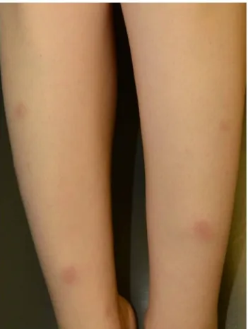
Fig. 1. Erythema nodosum. Tender, bilateral, erythematous nodules and plaques on the anterior aspect of the lower extremities.
나타난다.7 치료법은 국소 또는 전신 항생제, 면역억제제 등이 사용되기도 하나 주로 외과적 치료가 요구된다.8
2) 전이성 크론병(metastatic Crohn’s disease)
전이성 크론병은 드문 질환으로 장과 연속적으로 발생하는 것이 아니며, IBD에 이환된 장으로부터 떨어진 곳에 특이적 인 육아종성 피부 병변이 발생하는 것이다. 즉, 떨어져 발생한 농양, 누공, 궤양 또는 결절에서 크론병과 동일한 병리학적 소견을 보이는 것이 특징이다. 전이성 크론병에서 병변의 위 치와 임상 양상은 연령에 따라 다른 소견을 보인다. 소아의 경우에는 림프 부종의 형태로 생식기 주변에 많이 나타나지 만, 성인의 경우에는 누공, 결절, 궤양의 형태로 하지와 다리 에 주로 나타난다.9-11 전이성 크론병은 IBD의 활동성 정도와 는 관련성이 없는 것으로 보이며,10 치료 방법으로 경구 또는 국소 스테로이드제, 병변 내 스테로이드 주사, 경구 메트로니 다졸, 답손, 인플릭시맵, 탈리도마이드, 미노사이클린, 설파살 라진, 6-메르캅토푸린, 아자치오프린, 수술 등이 제안된다.12
2. 반응성 피부 증상(reactive skin manifestations) 반응성 피부 발현은 IBD의 발병 기전을 공유하는 것으로
생각되지만, 동일한 조직 병리학적 특징을 갖지는 않는다. 비 정상적인 호중구의 기능 또는 손상된 세포성 면역에 의하여 호중구가 자가면역 반응을 일으키는 것이 반응성 피부 발현의 발병 기전으로 생각된다.13
1) 결절 홍반(erythema nodosum, EN)
EN은 IBD의 가장 흔한 피부 증상이다. 유병률은 UC보다 CD에서 더 높게 보고된다. EN의 발병 빈도는 CD에서 3-15%, UC에서 6-10%이며 여성에서 발병률이 더 높다.5,14,15 국내 건강보험 청구 데이터 분석에서도 CD와 여성에서 유병 률이 더 높았으며, IBD 환자에서 EN의 유병률은 일반 인구 대비 CD에서 9.91배(95% CI, 6.47-13.34), UC에서 4.06배 (95% CI, 2.69-5.42)였다.16 EN은 일반적으로 IBD로 확진된 후에 나타나며 IBD 진단 전에는 15% 미만에서 나타난다.17 IBD 환자의 EN 발생 원인에 대해서는 정확히 알려져 있지 않다. 염색체 6번에 있는 인체 백혈구 항원(human leukocyte antigen, HLA)-B의 변이(mutation)와 관련이 있다는 보고와18 지연형 IV형 과민 반응이 EN 발생과 연관이 있다는 보고가 있다.19 대부분의 환자에서 지름 1-5 cm의 압통을 동반하는 다수의 염증성 결절이 하지의 신근부(extensor surfaces)에 주로 발생하며 특히 정강이 부분에서 호발한다(Fig. 1).5 반면, 안면부, 체간부, 상지 등은 거의 영향을 받지 않는다. 진단은 임상 증상을 근거로 하기 때문에 일반적으로 조직 검사는 필요 하지 않지만 비특이적인 임상 증상의 경우에는 조직 검사가 도움이 될 수 있다. EN은 IBD의 범위 및 활동성 정도와 밀접한 관련성이 있다.19 대부분의 경우 EN은 IBD 치료 시 흉터 없이 치유되며 약 25%에서는 자연적으로 호전되는 경과를 보인다.19 보조 치료로는 다리 올리기, 진통제, 요오드칼륨 등이 있다. 질 병 경과가 심한 경우에는 전신 스테로이드(0.5-1 mg/kg/day) 를 사용한다. 스테로이드 치료에 불응성 또는 의존성인 경우 에는 tumor necrosis factor (TNF)-α 억제제를 고려해볼 수 있다.20,21
2) 괴저 고름 피부증(pyoderma gangrenosum, PG) PG는 IBD와 관련된 가장 심한 피부 병변으로, UC의 5-12%에서 관찰되고 CD에서는 1-2%로 드물게 나타난다.18,22 소아에서의 유병률은 0.6%로 낮은 편이다.23 특징적으로 PG 병변을 가지고 있는 환자의 50%는 IBD에 이환되어 있으며,24 대부분의 경우에는 IBD가 PG 발생 이전에 나타나지만 약 15% 정도에서는 IBD 발병 이전에 PG 병변이 먼저 나타난 다.17 아직 병인이 명확히 밝혀지지는 않았지만 대장과 피부의 공통 항원에 대한 교차 반응 항체가 비정상적인 면역 반응을 유발하여 발생하는 것으로 생각되고 있다.25,26 또한 inter- leukin (IL)-8, IL-16, IL-17 및 TNF-α와 같은 염증성 사이토
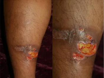
Fig. 2. Pyoderma gangrenosum. Ulcer shows an edematous pale border with granulation tissue on the leg.
Fig. 3. Sweet’s syndrome. Erythematous plaques and patches on the extremities and trunk.
카인의 과발현, 호중구 기능 장애, 비정상적인 T세포 반응들 도 PG의 병인에 대한 기전으로 생각된다.26 PG는 고름 물집 이나 결절 종기로 시작하고 급격히 병변의 중심부가 괴사하면 서 주변부로 잠식해가며 통증성의 만성 궤양을 형성한다. 궤 양의 가장자리는 푸른색 또는 자색을 띠며 주변과의 경계가 명확하고 심부로 궤양이 계속 파고 들어간다(Fig. 2). PG는 피부의 어느 곳에서나 발생 가능하지만, 주로 하지나 체간부 에 발생한다. 약 25%에서는 외상을 받은 부위에서 PG가 발생 하는데, 이러한 현상을 “pathergic phenomenon”이라고 한 다. 일단 발생하면 빠르게 진행하며 수주에서 수개월에 걸쳐 오랜 시간 지속된다.17 진단은 다른 피부 질환을 배제하여 임 상적인 판단으로 한다. 피부 조직 검사는 비특이적이지만 다 른 피부 질환을 배제할 때 도움이 될 수 있다. PG와 장질환의 활성도의 연관성에 대해서는 아직 논쟁의 여지가 있다. 일부 보고에서는 IBD 질환의 경과와 관련성이 있다고 하였지만4 관련성이 없다는 보고도 있다.27 질병 경과는 대개 예측이 힘 들며 완치까지는 장기간이 걸릴 수도 있다. PG는 조기 치료 와 신속한 치료가 필요하다. 기저 IBD 질환을 치료하는 것이 중요하고, 국소 병변의 경우에는 이차 감염을 예방하면서 치 료를 진행하는 것이 필요하다. 국소 치료로써 강력한 국소 스 테로이드 도포나 병변 내 스테로이드 주사 요법 또는 국소 타크로리무스나 4% 크로몰린이 효과적일 수 있다. 그러나 대 부분은 전신적인 약제 투여가 필요하며, 그중 전신 스테로이 드(0.5-1 mg/kg/day)가 가장 효과적이다.25 질환이 조절되기 시작하면 점차 감량해 나간다. 질환이 침습적이고 스테로이드 투여에도 빠르게 회복되지 않는 경우에는 전신 면역조절제(사 이클로스포린)도 사용해볼 수 있다.19 최근 TNF-α 억제제 치 료법이 많이 시도되고 있으며 여러 연구에서 PG 치료에 매우 좋은 결과를 보이고 있다.28 무작위 대조군 임상시험에서 인플
릭시맙군이 위약군 대비 유의하게 더 많은 환자에서 임상적 호전을 보였으며, 치료 6주째에 인플릭시맙군에서 69%의 임 상적 호전과 21%의 관해를 보였다.29 따라서, 스테로이드에 대한 신속한 치료 반응이 이루어지지 않을 경우에는 TNF-α 억제제 치료를 고려해야 한다.30 외과적 치료는 대개 PG를 재 발 또는 악화시키므로 피해야 한다.
3) Sweet’s 증후군(Sweet’s syndrome, acute febrile neutrophilic dermatosis)
급성 발열 호중구 피부병으로도 알려진 Sweet’s 증후군은 매우 드문 피부 질환으로, IBD 환자에서의 유병률은 정확히 알려져 있지 않다. IBD와의 연관성은 Becuwe 등에 의하여 1989년 처음 제시되었으며, 보고된 사례의 87%가 여성으로 여성의 유병률이 더 높다.31 피부와 장의 공통된 항원에 반응 하는 이상 면역 반응에 의하여 발병한다는 가설이 있으나 정 확한 병인은 아직 알려져 있지 않다.32 Sweet’s 증후군은 얼 굴, 목, 팔, 몸통, 다리 등에 갑자기 홍반성의 구진 또는 판이 비대칭적으로 나타나며 통증 또는 압통을 동반한다. 병변은 2-10 mm 정도로 대개 다발성인 경우가 많다(Fig. 3). 발진과 함께 지속적인 발열이 있는 경우가 대부분이고 두통, 관절통, 전신 쇠약 등을 호소하며 결막염 및 상공막염을 동반할 수도 있다. 특징적인 임상 증상 및 진피 상부에 호중구 염증세포 침윤이 있는 병리조직학적 소견과 더불어 혈액 검사상 호중구 의 증가로 진단할 수 있다.17 스테로이드 연고가 효과적이며 스테로이드의 전신 투여에 신속히 반응하고 수일 내에 반흔 없이 병변이 소실된다. 조기에 스테로이드를 중단하면 재발할 수 있으며 치료를 하지 않은 경우에는 대부분 수주 내지 수개 월 후에 자연적으로 소실되는 경과를 보인다.19 그 외에 요오 드칼륨, 콜히친, 인도메타신, 사이클로스포린, 답손을 전신 치 료제로 고려할 수 있다. 또한 TNF-α 억제제도 효과적이라는
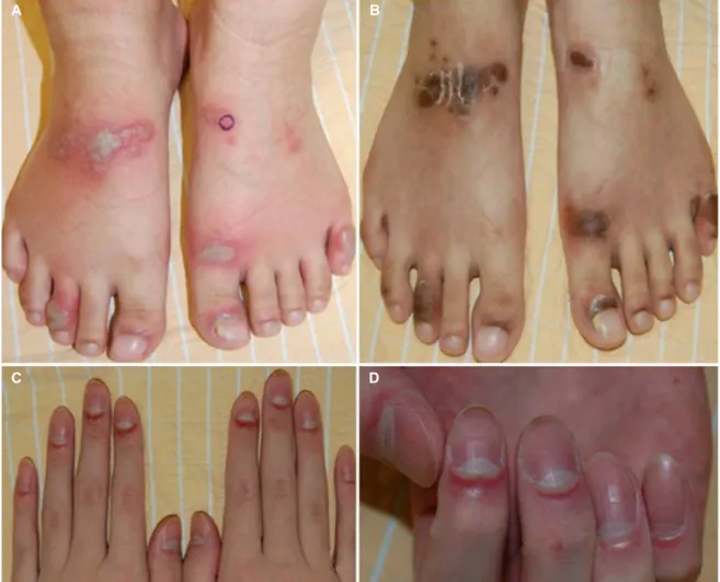
Fig. 4. Acrodermatitis enteropathica. (A) Multiple flaccid bullae and vesicles on dorsum of the feet. (B) Hyperpigmented macules and patches with scale resolved on the dorsum of the feet. (C, D) Paronychia.
보고가 있다.31
4) 구강 병변
구강 병변으로는 치주염(periodontitis), 아프타 입안염 (aphthous stomatitis) 및 가장 불량한 경과를 보이는 증식 화농 구내염(pyostomatitis vegetans) 등이 있다. 구강 병변 의 유병률은 CD에서 10%로 UC의 4%보다 높았고, 환자의 25% 이상에서 IBD 진단을 받기 전에 구강 병변이 나타났다 는 보고가 있다.17 치주염은 세균성 플라그에 대한 만성 염증 성 반응으로 뼈와 연조직의 파괴를 일으키며 발진, 출혈, 부종 등을 일으킨다. 치주염은 IBD에 특이성을 가지지 않는 빈번 한 증상이나 항문 주위 질환과는 일부 관련이 있는 것으로 보고되었다.32 아프타 입안염은 여러 개의 원형 또는 타원형의 통증을 수반하는 궤양으로 볼과 혀 밑 점막에 발생한다.19 증 식 화농 구내염은 IBD와 밀접한 관계를 가지며 아주 드물게 발생하는 구강 내 염증 질환으로, 모든 연령 대에서 발생하고 남녀의 발생 비는 3:1로 알려져 있다.33 특징적으로 구강 내
점막에 부종과 홍반을 형성하며 그 위에 다발성의 농포를 동 반하는 염증성 병변으로 나타난다. 다발성의 농포는 쉽게 터 지고 서로 합쳐져 미란과 궤양을 형성하기도 한다. 주로 구순 과 구강 점막을 침범하고 혀는 잘 침범하지 않는다.33,34 구강 병변의 진단은 임상 증상, 염증성 장질환과의 관련성, 조직 검사 결과 등으로 할 수 있다. 구강 병변의 활성도는 장질환의 활성도와 밀접하게 관련되어 있으므로 IBD의 치료가 중요하 다. 국소 스테로이드, 요오드팅크, 클로헥시딘 가글 등을 사용 한다. 전신 항생제, 설파제, 답손, 아자치오프린, 전신 스테로 이드 투여 등이 치료로 시도되었고, 고용량의 스테로이드 투 여만이 치료에 효과가 있다는 보고도 있으나 스테로이드 용량 을 줄이거나 중단하면 재발하였다.35 전대장 절제술을 시행한 후 완치가 된 경우도 보고되었다.36 TNF-α 억제제 또한 효과 적인 치료 대안이다. Löfberg 등37의 연구에 따르면 20주 간 의 아달리무맙 투여 후 아프타 입안염의 빈도가 5.2%
(49/945)에서 2.1% (20/942)로 감소하였다. 최근의 IBD 집단 연구 결과는 78.1% (25/32)의 반응률을 보였다.38 하지만 모
A B
C D
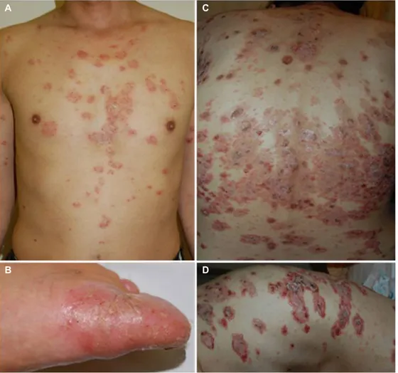
Fig. 5. TNF-α inhibitor treatment-induced skin manifestations. (A, B) Small plaque psoriasiform lesions scattered over the trunk and palmoplantar pustular psoriasiform lesions following TNF-α inhibitor therapy. (C, D) Generalized blistering skin lesions on the trunk of a 40-year-old woman treated for ulcerative colitis with infliximab. TNF-α, tumor necrosis factor-alpha.
든 TNF-α 억제제 치료는 구강 병변보다는 오히려 IBD를 치 료하기 위하여 시작된 것이므로, 구강 병변의 치료보다는 IBD 치료가 우선시되어야 함을 시사한다.
3. IBD와 연관된 피부 질환(dermatosis associated with IBD)
1) 건선(psoriasis)
만성 염증성 피부 질환인 건선은 IBD와의 연관성에 대한 많은 연구 결과가 알려져 있다. 최근 발표된 체계적 문헌고찰 에 따르면 건선과 CD (OR, 1.70; 95% CI, 1.20-2.40) 및 UC (OR, 1.75; 95% CI, 1.49-2.05)와의 연관성이 보고되었다. 또 한 건선 환자에서 IBD 발생 위험도가 상대적으로 증가한다는 보고도 있다.39 국내 건강보험 청구 데이터를 이용하여 IBD와 염증성 피부염 관계를 연구한 자료에 따르면 IBD 환자에서 건선의 유병률은 1.30-1.78배 높았다.16,40두 질환에서 동시에 연관이 있다고 생각되는 병인으로는 피부와 장 점막에
Th1/Th17 세포성 만성 면역 염증 반응이 가설로 제시되었 다.41 또한 CD와 UC 그리고 건선에 서로 일치하는 유전자 변이가 확인되었는데 이를 바탕으로 특정 유전자 변이 또는 HLA가 특정 면역 반응과 염증 반응을 유발하여 두 질환을 일으키는 것으로 생각되고 있다.42 치료는 크게 국소 치료, 광 선 치료, 전신 치료, 생물학적 제제 등이 있다. 중증도에 따라 경증인 경우에는 주로 국소 치료를 시행하고, 중등증인 경우 에는 국소 치료와 광선 치료를 시행하며, 중증인 경우에는 국 소 치료, 전신 치료, 광선 치료를 모두 시행한다. 국소 치료로 는 보습제를 기본으로 국소 스테로이드, 비타민 D3 유도체를 사용한다. 미국 식품의약국(Food and Drug Administration) 에서 승인된 전신 치료제는 레티노이드, 메토트렉세이트, 사 이클로스포린 등이 있다. 그중 메토트렉세이트는 건선과 IBD 모두에서 치료 효과가 있음이 보고되었으며 사이클로스포린 도 치료제로 고려해볼 수 있다.43 모든 치료에도 반응이 없는 경우에는 생물학적 제제 치료를 고려한다. 건선과 IBD 모두 에서 미국 식품의약국 승인을 받은 생물학적 제제로는 인플릭
A C
B D
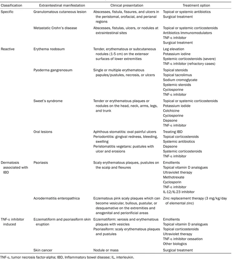
Table 1. Cutaneous Extra-intestinal Manifestations of Inflammatory Bowel Disease
Classification Extraintestinal manifestation Clinical presentation Treatment option
Specific Granulomatous cutaneous lesion Abscesses, fistula, fissures, and ulcers in the peristomal, orofacial, and perianal regions
Topical or systemic antibiotics Surgical treatment
Metastatic Crohn’s disease Abscesses, fistulas, ulcers, or nodules at extraintestinal sites
Topical or systemic corticosteroids Antibiotics Immunomodulators TNF-α inhibitor
Surgical treatment Reactive Erythema nodosum Tender, erythematous or subcutaneous
nodules (1-5 cm) on the extensor surfaces of lower extremities
Leg elevation Potassium iodine
Systemic corticosteroids (severe) TNF-α inhibitor (refractory cases) Pyoderma gangrenosum Single or multiple erythematous
papules/pustules, necrosis, or ulcers
Topical steroids Topical tacrolimus Sodium cromoglycate Systemic steroids Cyclosporine TNF-α inhibitor Sweet’s syndrome Tender or erythematous plaques or
nodules on the head, neck, arms, legs, and trunk
Topical or systemic corticosteroids Potassium iodide
Colchicine Cyclosporine Dapsone TNF-α inhibitor Oral lesions Aphthous stomatitis: oval painful ulcers
Periodontitis: gingival redness, bleeding, swelling
Peristomatitis vegetans: pustules with ulcer and erosions
Treating IBD
Topical corticosteroids Systemic antibiotics Dapsone
Systemic corticosteroids TNF-α inhibitor Dermatosis
associated with IBD
Psoriasis Scaly erythematous plaques, pustules on
the scalp and flexures
Emollients
Topical vitamin D analogues Ultraviolet therapy Methotrexate Cyclosporin TNF-α inhibitor IL-12/IL-23 inhibitor Acrodermatitis enteropathica Eczematous pink scaly plaques which can
become vesicular, bullous, pustular, or desquamative on the extremities and anogenital and periorificial areas
Zinc replacement therapy (3 mg/kg/day of elemental zinc)
TNF-α inhibitor induced
Eczematiform and psoriasiform skin eruption
Eczematiform: xerosis and erythematous plaques with vesicles
Psoriasiform: scaly erythematous plaques and pustules
Emollients
Topical vitamin D analogues Topical corticosteroids Ultraviolet therapy TNF-α inhibitor cessation Other biologics
Skin cancer Nodule or mass Surgical treatment
TNF-α, tumor necrosis factor-alpha; IBD, Inflammatory bowel disease; IL, interleukin.
시맙, 아달리무맙, 우스테키누맙이 있다.43 최근 중등도 이상 의 CD에서 치료제로 승인된 우스테키누맙은 IL-12, IL-23과 같은 pro-inflammatory 사이토카인을 억제하므로,44 건선 등 과 같은 장외 증상이 동반된 환자에서 좋은 치료 방법으로 제시되고 있다. 건선 환자에서 우스테키누맙의 장기간 안정성
에 대한 연구 결과들은 우스테키누맙이 일반적으로 안전하며 심각한 감염과 악성 종양의 발병은 드물다고 보고하였다.45-47
2) 장병 말단 피부염(acrodermatitis enteropathica) 장병 말단 피부염은 생체 기능을 위한 필수적인 원소인 아
연 결핍으로 인하여 신체 개구부 주변(periorificial)과 말단부 의 피부염, 설사, 탈모 등의 증상이 특징적으로 나타나는 질 환이다. IBD로 인한 가장 흔한 영양 결핍성 피부 증상이며 UC보다 CD에서 더 흔하게 나타난다.48 발생 원인은 흡수 장 애, 만성 설사, 골다공증의 예방 목적으로 복용하는 칼슘제 등이 알려져 있다. 피부 병변의 발현은 홍반성 판 또는 반의 형태로 나타나며, 수포, 물집, 농포의 형태로 진행하면서 나 타날 수도 있다. 병변은 입, 항문, 팔다리, 손가락 및 두피 주 변에서 자주 발생한다(Fig. 4).49 국내 장병 말단 피부염 연구 에 따르면 후천적인 장병 말단 피부염의 경우 그 병인으로 장질환에 의한 흡수 장애가 가장 큰 비중을 차지하였다. 병변 의 위치는 항문 주위에 발생한 경우가 53.3% (24/45)로 가장 많았고, 형태는 홍반성 인설이나 미란을 동반한 피부 병변의 발생 빈도가 77.8% (35/45)로 가장 많았다.50 치료는 아연을 2-3 mg/kg/day로 투여하는 것이며 일반적으로 투여 후 수일 내에 피부 병변은 소실된다. 황산아연을 경구 투여할 경우에 는 부작용으로 오심, 구토, 위장관 출혈이 발생할 수 있으니 주의해야 한다.
4. 치료 유도 피부 증상(TNF-α inhibitor induced skin mani- festations)
IBD의 치료제로 TNF-α 억제제가 널리 보급됨에 따라 역설 적(paradoxical) 피부 반응이 자주 보고되고 있다. TNF-α 억 제제로 치료한 IBD 환자 중 5-10%에서 역설적 피부 반응이 보고된다.51 특히,TNF-α 억제제가 건선의 효과적인 치료제로 도 사용됨에도 불구하고, 일부 환자에서는 역설적 피부 반응 에 의하여 건선양 병변을 유발할 수 있다.51,52 대규모 연구에 서 4년의 추적 관찰 기간 동안 TNF-α 억제제로 치료한 환자 중 5%에서 TNF-α 억제제 유발 피부 병변이 발생하였으며, 모든 TNF-α 억제제(인플릭시맙, 아달리무맙 및 세르톨리주 맙)에서 나타났다.51 국내 문헌에 따르면 IBD 치료를 위하여 TNF-α 억제제를 투여받은 환자 500명 중 9.6%에서 피부 병 변이 발생하였다. TNF-α 억제제 투여 횟수의 중앙값은 12.7회 였으며 투약 후 피부 병변 발생까지의 기간은 평균 13개월이 었다. 습진양 병변(38%)이 가장 많았으며, 건선양 병변(28%) 도 흔히 관찰되었다. 건선양 병변은 소판상건선양 병변이 80%로 가장 흔하였으며, 농포성건선양 병변도 관찰되었다 (Fig. 5).53 건성양 병변은 여성(70%)에서 더 흔하고, 손·발바 닥(43%)과 두피(42%)가 가장 흔한 발생 위치이다.52 명확한 병인은 아직 밝혀지지 않았지만 TNF-α 억제제 치료 시 inter- feron-α의 상향 조절에 의하여 역설적 피부 반응이 나타날 것 이라는 가설이 가장 유력하다. TNF-α는 골수에서 형질세포 모양 수지상세포(plasmacytoid dendritic cells)의 성숙을 억 제하는 역할을 한다. 이러한 작용이 TNF-α 억제제에 의하여
차단되면 형질세포 모양 수지상세포에 의하여 증가된 inter- feron-α로 인하여 T세포의 CXCR3 발현을 증가시키는 IL-1β, IL-6, IL-17, IL-21 등과 같은 염증성 사이토카인의 발현이 증 가하여, T세포들이 피부 쪽으로 유입되면서 역설적 피부 병변 이 발생하는 것으로 추정하고 있다.54,55 역설적 피부 병변의 치료는 TNF-α 억제제의 투약을 중단하기보다는 TNF-α 억제 제 치료를 유지하면서 피부 병변에 국소 치료를 병합하는 방 법이 추천된다. 국소 치료 방법에는 국소 스테로이드, 각질 용해제, 피부 연화제, 자외선 치료 등이 있다.56 Fiorino 등56은 국소 치료에도 불구하고 5-35% 정도는 역설적 피부 병변이 심해져서 TNF-α 억제제 치료를 중지해야 한다고 보고하였다.
다시 TNF-α 억제제로 치료하였을 때는 약 절반에서 재발하 였다는 보고도 있다.52하지만 국내 보고에서는 대부분 스테로 이드 도포 및 경구 항히스타민제 복용으로 피부 병변의 호전 을 보였으며, TNF-α 억제제 치료를 중단하였던 경우는 단 1예 만 보고되었다.53 이는 인종이나 기저 질환의 차이에 따라 국 소 치료 반응이 다르게 나타났기 때문이라고 생각된다. 따라 서 국소 치료 후의 치료 반응에 따라 향후 치료 방향을 결정하 는 것을 추천한다.
IBD 치료제로 TNF-α 억제제의 사용이 증가하면서 이로 인 한 피부암 발생도 보고되고 있다. 비흑색종(non-melanoma) 과 흑색종(melanoma) 피부암 모두에서 TNF-α 억제제 사용 으로 인하여 발생 위험도가 증가한다는 연구 결과들이 있
다.57-59 IBD 치료로 TNF-α 억제제를 사용할 경우 피부암 발
생 가능성이 높아지므로 이에 대한 예방책으로 자외선 차단제 사용과 정기적인 피부 병변 검진이 권고된다.
결 론
지금까지 살펴본 내용은 Table 1에 정리하였다. IBD 환자 에서 나타나는 장외 증상으로 피부 병변은 흔히 발생하며 EN 과 같은 치료하기 쉬운 병변부터 PG와 같은 심한 병변의 형 태까지 다양하게 나타날 수 있다. IBD와 관련된 피부 병변의 발현은 IBD의 활성도와 관련이 있으므로 기저 IBD의 치료가 중요하다. 최근에는 IBD와 장외 합병증 치료로 TNF-α 억제 제가 많이 사용되고 있다. 그러나 이러한 치료도 역설적 피부 반응으로 인하여 피부 병변을 유발할 수 있음에 주의해야 한 다. IBD 환자에서 피부 병변이 발생한 경우에는 피부과 전문 의와의 협진을 통하여 환자의 질환 치료 및 삶의 질 향상을 위하여 노력해야 한다.
REFERENCES
1. Podolsky DK. Inflammatory bowel disease. N Engl J Med 2002;
347:417-429.
2. Lee JW, Im JP, Cheon JH, Kim YS, Kim JS, Han DS. Inflammatory bowel disease cohort studies in Korea: present and future. Intest Res 2015;13:213-218.
3. Bernstein CN, Wajda A, Blanchard JF. The clustering of other chronic inflammatory diseases in inflammatory bowel disease:
a population-based study. Gastroenterology 2005;129:827-836.
4. Veloso FT, Carvalho J, Magro F. Immune-related systemic mani- festations of inflammatory bowel disease. A prospective study of 792 patients. J Clin Gastroenterol 1996;23:29-34.
5. Vavricka SR, Brun L, Ballabeni P, et al. Frequency and risk factors for extraintestinal manifestations in the Swiss inflammatory bowel disease cohort. Am J Gastroenterol 2011;106:110-119.
6. Guest GD, Fink RL. Metastatic Crohn's disease: case report of an unusual variant and review of the literature. Dis Colon Rectum 2000;43:1764-1766.
7. Marzano AV, Borghi A, Stadnicki A, Crosti C, Cugno M. Cutaneous manifestations in patients with inflammatory bowel diseases:
pathophysiology, clinical features, and therapy. Inflamm Bowel Dis 2014;20:213-227.
8. Sandborn WJ. Evidence-based treatment algorithm for mild to moderate Crohn's disease. Am J Gastroenterol 2003;98(12 Suppl):S1-S5.
9. Ploysangam T, Heubi JE, Eisen D, Balistreri WF, Lucky AW.
Cutaneous Crohn's disease in children. J Am Acad Dermatol 1997;36(5 Pt 1):697-704.
10. Palamaras I, El-Jabbour J, Pietropaolo N, et al. Metastatic Crohn's disease: a review. J Eur Acad Dermatol Venereol 2008;22:
1033-1043.
11. Lee KG, Koh BT, Kwon ES, Myung KB, Cheong SH. Crohn’s dis- ease initially presenting as vulvar swelling. Korean J Dermatol 2018;56:49-52.
12. Bender-Heine A, Grantham JT, Zaslau S, Jansen R. Metastatic Crohn disease: a review of dermatologic manifestations and treatment. Cutis 2017;99:E33-E40.
13. Huang W, McNeely MC. Neutrophilic tissue reactions. Adv Dermatol 1997;13:33-64.
14. Vavricka SR, Schoepfer A, Scharl M, Lakatos PL, Navarini A, Rogler G. Extraintestinal manifestations of inflammatory bowel disease. Inflamm Bowel Dis 2015;21:1982-1992.
15. Farhi D, Cosnes J, Zizi N, et al. Significance of erythema nodosum and pyoderma gangrenosum in inflammatory bowel diseases: a cohort study of 2402 patients. Medicine (Baltimore) 2008;87:
281-293.
16. Yang BR, Choi NK, Kim MS, et al. Prevalence of extraintestinal manifestations in Korean inflammatory bowel disease patients.
PLoS One 2018;13:e0200363.
17. Vavricka SR, Rogler G, Gantenbein C, et al. Chronological order of appearance of extraintestinal manifestations relative to the time of IBD diagnosis in the Swiss inflammatory bowel disease cohort. Inflamm Bowel Dis 2015;21:1794-1800.
18. Yüksel I, Başar O, Ataseven H, et al. Mucocutaneous manifes- tations in inflammatory bowel disease. Inflamm Bowel Dis 2009;
15:546-550.
19. Timani S, Mutasim DF. Skin manifestations of inflammatory bow- el disease. Clin Dermatol 2008;26:265-273.
20. Clayton TH, Walker BP, Stables GI. Treatment of chronic erythema
nodosum with infliximab. Clin Exp Dermatol 2006;31:823-824.
21. Ortego-Centeno N, Callejas-Rubio JL, Sanchez-Cano D, Caballero- Morales T. Refractory chronic erythema nodosum successfully treated with adalimumab. J Eur Acad Dermatol Venereol 2007;
21:408-410.
22. Trost LB, McDonnell JK. Important cutaneous manifestations of inflammatory bowel disease. Postgrad Med J 2005;81:580-585.
23. Greuter T, Bertoldo F, Rechner R, et al. Extraintestinal manifes- tations of pediatric inflammatory bowel disease: prevalence, presentation, and anti-TNF treatment. J Pediatr Gastroenterol Nutr 2017;65:200-206.
24. Rothfuss KS, Stange EF, Herrlinger KR. Extraintestinal manifes- tations and complications in inflammatory bowel diseases.
World J Gastroenterol 2006;12:4819-4831.
25. Crowson AN, Mihm MC Jr, Magro C. Pyoderma gangrenosum: a review. J Cutan Pathol 2003;30:97-107.
26. Feliciani C, De Simone C, Amerio P. Dermatological signs during inflammatory bowel diseases. Eur Rev Med Pharmacol Sci 2009;13 Suppl 1:15-21.
27. Menachem Y, Gotsman I. Clinical manifestations of pyoderma gangrenosum associated with inflammatory bowel disease. Isr Med Assoc J 2004;6:88-90.
28. Agarwal A, Andrews JM. Systematic review: IBD-associated pyo- derma gangrenosum in the biologic era, the response to therapy.
Aliment Pharmacol Ther 2013;38:563-572.
29. Brooklyn TN, Dunnill MG, Shetty A, et al. Infliximab for the treat- ment of pyoderma gangrenosum: a randomised, double blind, placebo controlled trial. Gut 2006;55:505-509.
30. Lee JH, Chang IK, Lee HE, et al. Treatment of recalcitrant pyo- derma gangrenosum with ulcerative colitis by adalimumab injection. Ann Dermatol 2017;29:260-262.
31. Ali M, Duerksen DR. Ulcerative colitis and Sweet's syndrome: a case report and review of the literature. Can J Gastroenterol 2008;22:296-298.
32. Vavricka SR, Manser CN, Hediger S, et al. Periodontitis and gingi- vitis in inflammatory bowel disease: a case-control study.
Inflamm Bowel Dis 2013;19:2768-2777.
33. Calobrisi SD, Mutasim DF, McDonald JS. Pyostomatitis vegetans associated with ulcerative colitis. Temporary clearance with fluo- cinonide gel and complete remission after colectomy. Oral Surg Oral Med Oral Pathol Oral Radiol Endod 1995;79:452-454.
34. Kim TH, Kim SC. Pyodermatitis-pyostomatitis vegetans asso- ciated with Crohn's disease. Ann Dermatol 2015;27:624-625.
35. Bens G, Laharie D, Beylot-Barry M, et al. Successful treatment with infliximab and methotrexate of pyostomatitis vegetans as- sociated with Crohn's disease. Br J Dermatol 2003;149:181-184.
36. Campisi G, Compilato D, Cirillo N, et al. Oral ulcers: three questions on their physiopathology. Minerva Stomatol 2007;56:293-302.
37. Löfberg R, Louis EV, Reinisch W, et al. Adalimumab produces clin- ical remission and reduces extraintestinal manifestations in Crohn's disease: results from CARE. Inflamm Bowel Dis 2012;
18:1-9.
38. Vavricka SR, Gubler M, Gantenbein C, et al. Anti-TNF treatment for extraintestinal manifestations of inflammatory bowel dis- ease in the Swiss IBD cohort study. Inflamm Bowel Dis 2017;
23:1174-1181.
39. Fu Y, Lee CH, Chi CC. Association of psoriasis with inflammatory bowel disease: a systematic review and meta-analysis. JAMA Dermatol 2018;154:1417-1423.
40. Kim M, Choi KH, Hwang SW, Lee YB, Park HJ, Bae JM.
Inflammatory bowel disease is associated with an increased risk of inflammatory skin diseases: a population-based cross-sec- tional study. J Am Acad Dermatol 2017;76:40-48.
41. Asarch A, Barak O, Loo DS, Gottlieb AB. Th17 cells: a new para- digm for cutaneous inflammation. J Dermatolog Treat 2008;19:
259-266.
42. Cho JH, Brant SR. Recent insights into the genetics of in- flammatory bowel disease. Gastroenterology 2011;140:1704-1712.
43. Whitlock SM, Enos CW, Armstrong AW, et al. Management of psoriasis in patients with inflammatory bowel disease: from the medical board of the national psoriasis foundation. J Am Acad Dermatol 2018;78:383-394.
44. Wittig BM. Drug evaluation: CNTO-1275, a mAb against IL-12/IL-23p40 for the potential treatment of inflammatory diseases. Curr Opin Investig Drugs 2007;8:947-954.
45. Papp KA, Griffiths CE, Gordon K, et al. Long-term safety of usteki- numab in patients with moderate-to-severe psoriasis: final re- sults from 5 years of follow-up. Br J Dermatol 2013;168:
844-854.
46. Menter A, Papp KA, Gooderham M, et al. Drug survival of biologic therapy in a large, disease-based registry of patients with psor- iasis: results from the psoriasis longitudinal assessment and registry (PSOLAR). J Eur Acad Dermatol Venereol 2016;30:
1148-1158.
47. Papp K, Gottlieb AB, Naldi L, et al. Safety surveillance for usteki- numab and other psoriasis treatments from the psoriasis longi- tudinal assessment and registry (PSOLAR). J Drugs Dermatol 2015;14:706-714.
48. Danese S, Semeraro S, Papa A, et al. Extraintestinal manifes- tations in inflammatory bowel disease. World J Gastroenterol 2005;11:7227-7236.
49. Maverakis E, Fung MA, Lynch PJ, et al. Acrodermatitis enter- opathica and an overview of zinc metabolism. J Am Acad Dermatol 2007;56:116-124.
50. Lim YS, Lee MW, Choi JH, Sung KJ. Original articles : the clinical
study of zinc deficiency presented as a skin manifestation of ac- rodermatitis enteropathica. Korean J Dermatol 2000;38:
155-162.
51. Rahier JF, Buche S, Peyrin-Biroulet L, et al. Severe skin lesions cause patients with inflammatory bowel disease to dis- continue anti-tumor necrosis factor therapy. Clin Gastroenterol Hepatol 2010;8:1048-1055.
52. Cullen G, Kroshinsky D, Cheifetz AS, Korzenik JR. Psoriasis asso- ciated with anti-tumour necrosis factor therapy in inflammatory bowel disease: a new series and a review of 120 cases from the literature. Aliment Pharmacol Ther 2011;34:1318-1327.
53. Moon IJ, Moon HR, Noh TK, et al. Multiple cases of psoriasiform skin lesions followinganti-TNF-α therapy: a single center experience. J Korean Soc Psori 2018;13:14-18.
54. Sfikakis PP, Iliopoulos A, Elezoglou A, Kittas C, Stratigos A.
Psoriasis induced by anti-tumor necrosis factor therapy: a para- doxical adverse reaction. Arthritis Rheum 2005;52:2513-2518.
55. Harbord M, Annese V, Vavricka SR, et al. The first European evi- dence-based consensus on extra-intestinal manifestations in in- flammatory bowel disease. J Crohns Colitis 2016;10:239-254.
56. Fiorino G, Danese S, Pariente B, Allez M. Paradoxical im- mune-mediated inflammation in inflammatory bowel disease patients receiving anti-TNF-α agents. Autoimmun Rev 2014;13:
15-19.
57. Long MD, Martin CF, Pipkin CA, Herfarth HH, Sandler RS, Kappelman MD. Risk of melanoma and nonmelanoma skin cancer among patients with inflammatory bowel disease.
Gastroenterology 2012;143:390-399.e1.
58. Askling J, Fahrbach K, Nordstrom B, Ross S, Schmid CH, Symmons D. Cancer risk with tumor necrosis factor alpha (TNF) inhibitors: meta-analysis of randomized controlled trials of adali- mumab, etanercept, and infliximab using patient level data.
Pharmacoepidemiol Drug Saf 2011;20:119-130.
59. McKenna MR, Stobaugh DJ, Deepak P. Melanoma and non-mela- noma skin cancer in inflammatory bowel disease patients follow- ing tumor necrosis factor-α inhibitor monotherapy and in combi- nation with thiopurines: analysis of the Food and Drug Administration adverse event reporting system. J Gastrointestin Liver Dis 2014;23:267-271.