ISSN 0378-6471 (Print)⋅ISSN 2092-9374 (Online)
https://doi.org/10.3341/jkos.2017.58.12.1388
Original Article
열공망막박리 유리체절제술 시 백내장수술 동시 시행 여부에 따른 재발 차이와 위험요인 분석
Comparison of the Recurrence of RRD in PPV Combined Cataract Surgery with PPV Alone, and Risk Factors in PPV Combined Cataract Surgery
박혜인⋅윤명헌⋅진희승
Hye In Park, MD, Myung Hun Yoon, MD, Hee Seung Chin, MD, PhD
인하대학교 의과대학 인하대병원 안과학교실
Department of Ophthalmology, Inha University Hospital, Inha University School of Medicine, Incheon, Korea
Purpose: To compare the recurrence percentage and risk factors of recurrence in rhegmatogenous retinal detachment (RRD) af- ter pars plana vitrectomy (PPV) by in two groups of patients according to combined cataract surgery.
Methods: The recurrence percentage of RRD and risk factors after PPV over 20 years, performed by a single surgeon from January 1997 to September 2016, were retrospectively evaluated by classification into two groups according to combined cata- ract surgery. The risk factors were the patients' factors (age and sex), duration of disease, preoperative visual analyses, posteri- or capsular tears (PCR) during cataract surgery, the tamponade used, retinal detachment ranges, macular status, number and position of retinal tears, proliferative vitreoretinopathy (PVR) grade, and vitreous opacity. A total of 158 cases were included in the study.
Results: The recurrence percentage of RRD was not associated with combined cataract surgery. In the combined cataract sur- gery group, PCR (p = 0.020), inferior retinal tears (p = 0.037), and PVR above grade B (p = 0.037) were associated with the re- currence of RRD using univariate logistic regression analyses, but PCR (odds ratio 1.880, p = 0.021) was the only significant risk factor for RRD using multivariate logistic regression analyses.
Conclusions: Cataract surgery can be performed at any time, but there should be consideration of the recurrence of RRD if there was PCR during combined cataract surgery.
J Korean Ophthalmol Soc 2017;58(12):1388-1395
Keywords: Combined cataract surgery, Recurrence, Rhegmatogenous retinal detachment, Risk factors, Vitrectomy
■Received: 2017. 7. 13 ■ Revised: 2017. 9. 11.
■Accepted: 2017. 11. 23.
■Address reprint requests to Hee Seung Chin, MD, PhD Department of Ophthalmology, Inha University Hospital, #27 Inhang-ro, Jung-gu, Incheon 22332, Korea
Tel: 82-32-890-2400, Fax: 82-32-890-2417 E-mail: [email protected]
*Conflicts of Interest: The authors have no conflicts to disclose.
ⓒ2017 The Korean Ophthalmological Society
This is an Open Access article distributed under the terms of the Creative Commons Attribution Non-Commercial License (http://creativecommons.org/licenses/by-nc/3.0/) which permits unrestricted non-commercial use, distribution, and reproduction in any medium, provided the original work is properly cited.
열공망막박리는 망막의 전층 열공을 통해 액화된 유리체 액이 망막 아래로 유입되면서 망막색소상피와 감각신경망
막 사이가 분리되는 질환이다.1 열공망막박리의 일차적 치 료는 수술이며 이 종류에는 공막돌륭술, 냉동법, 레이저광 응고술, 기체망막유착술, 유리체절제술 등이 있다. 이 중 유 리체절제술은 수술기구와 기술의 발전으로 인해 일차적으 로 가장 많이 선택되는 수술이며 다른 수술들과 병행하기 도 한다. 수술 이후 재발률은 높지 않으나 여전히 큰 과제 이며 이 위험인자에 대한 보고와 이를 기반으로 하여 재발 을 막기 위한 수술 기법이나 환자 상태에 따른 수술 선택의 기준에 관한 연구가 활발히 진행되고 있다.2-7
백내장수술 후의 망막박리는 7-10년 동안 0.16-2.31%의
확률로 보고될 정도로 드물지만 술 후 심각한 시력 저하의 가장 흔한 원인 중 하나이다.8-13 나이를 보정한 수정체안과 인공수정체안의 비교에서 백내장수술 후 8-20년 사이에 인공 수정체안의 경우 4배에서 9배까지 망막박리의 위험성이 높아 진다는 보고도 있으며10,11 이의 위험 요인으로는 성별, 나이, 안축장 길이, 후낭파열, 앞유리체절제술 등이 포함되었다.12-16
망막박리 재발의 위험 요인, 백내장수술 후의 망막박리 등에 관한 연구는 많으나 망막박리로 유리체절제술과 백내 장수술을 동시에 시행한 경우의 재발률과 위험요인을 분석 한 연구는 드물다. 그러나 현재 망막박리의 진단 당시 백내 장 여부에 따라 유리체절제술 시 백내장수술을 병행하는 케이스가 증가하고 있는 추세이며 이에 대한 명확한 기준 은 제시되고 있지 않아 망막박리 진단 당시의 백내장 상태 나 환자의 선택에 따라 결정하는 경우가 일반적이다.
이에 저자들은 열공망막박리를 처음 진단 받고 유리체절 제술을 받은 환자를 대상으로, 유리체절제술만 시행한 환 자군(group I)과 백내장수술을 병행한 환자군(group II) 간 의 망막박리의 재발 빈도를 비교하고 백내장수술을 병행한 군(group II)에서 기존에 알려진 망막박리 재발의 위험요인 을 분석해 보고자 한다.
대상과 방법
1997년 1월부터 2016년 9월까지 20년에 걸쳐 본원에 내 원한 환자 중 처음으로 열공망막박리를 진단 받고 유리체 절제술을 시행 받은 유수정체안(phakic eye) 158명을 대상 으로, 유리체절제술과 동시에 백내장수술을 받지 않은 환 자군(group I) 유리체절제술과 동시에 백내장수술을 받은 환자군(group II)으로 나눈 후 이들의 의무기록을 후향적으 로 분석하였다.
의무기록을 통해 유리체절제술과 동시에 백내장수술을 받지 않은 군과 받은 군 간의 망막박리 재발 빈도를 분석하 고 백내장수술을 시행한 군(group II)에서 성별, 나이, 증상 발생부터 수술까지의 기간, 술 전 시력, 백내장 술 중 후낭 파열 유무, 눈속 충전물(tamponade) 종류, 망막박리의 범위, 황반 침범 여부, 망막열공의 수와 위치, 증식성 유리체망막 병증의 유무와 단계, 유리체 혼탁의 유무, 술 중 망막절개술 유무, 술 후 레이저 후낭절개술(Yttrium-Aluminum-Garnet [YAG] capsulectomy) 등의 각 요인이 망막박리 재발에 미치 는 영향을 분석하였다. 이전에 망막박리나 망막열공으로 유 리체 절제술, 레이저 시술(Barrier)을 받은 과거력이 있는 환자, 처음 내원 시 망막 열공을 발견할 수 없었던 환자는 제외하였다. 본 연구는 본원의 임상시험심사위원회로부터 승인을 받았다(승인번호: INHAUH 2017-05-021).
처음 내원 시 최대교정시력, 전안부 및 수정체 검사, 안 저검사와 스펙트럼영역 빛간섭단층촬영(Spectral domain optical coherence tomography [SD-OCT], Cirrus HD OCT® Model 4000, Carl Zeiss Meditec Inc., Dublin, CA, USA)을 시행하였다. 세극등 현미경 검사를 통해 전안부 및 수정체 를 평가하였으며 간접검안경 및 3-mirror lens를 이용한 안 저 검사를 통해 망막박리의 범위와 위치, 망막열공의 수와 위치, 유리체 혼탁의 유무 등을 평가하였고 빛간섭단층촬 영을 통해 황반의 침범 여부를 확인하였다.
수술은 구후마취 혹은 전신마취하에 시행되었으며, 백 내장이 있는 경우에는 환자의 나이와 백내장 정도에 의한 수술을 위한 매체의 투명도(media clarity)를 고려하여 술자 의 판단하에 수정체초음파유화술과 인공수정체삽입술을 함 께 시행하였다. 유리체절제술 시스템으로 2011년 7월까지 는 Storz PREMIERE Microsurgical System (Storz medical AG, Tagerwilen, Lohstampfestrasse, Switzerland), 이후에는 The CONSTELLATION® Vision System (Alcon Laboratories Inc., Fort Worth, TX, USA)을 이용하였으며 유리체절제술과 동시에 백내장수술을 진행한 경우는 동일한 장비를 이용하였 으나 유리체절제술 이후 백내장수술을 한 경우는 2015년 7월 까지는 LegacyTM Microsurgical System (Alcon Laboratories Inc., Fort Worth, TX, USA), 이후에는 CENTURION® Vision System (Alcon Laboratories Inc., Fort Worth, TX, USA)을 이용하였다. 또한 술자의 판단에 따라 망막 하액 배출과 액 체가스치환술(SF6 또는 C3F8) 및 실리콘기름 삽입술을 시 행하였고, 모든 경우에서 망막 열공 주변부에 안내 레이저 를 시행하였다. 모든 수술은 한 명의 망막 전문의에 의해 시행되었다.
술 후 경과관찰은 입원 기간 동안 매일, 이후 일주일 뒤, 한 달 뒤 이후로는 필요에 따라 관찰기간을 조정하여 최대 교정시력, 세극등 현미경 검사, 골드만 안압검사, 안저검사, 황반부 빛간섭단층촬영을 시행하여 망막박리의 재발 여부 를 검사하였다.
통계학적 분석은 SPSS ver. 19.0 (IBM Corp., Armonk, NY, USA)을 이용하여 유리체절제술과 백내장수술 병행 유무에 따른 망막박리 재발률과 백내장수술을 병행한 케이스들에 한 해 각 요인들이 망막박리 재발에 미치는 영향을 Student T-test, Chi-square test, Fisher’s exact test, univariate/multivariate lo- gistic regression analysis를 시행하였다. p-value가 0.05 미 만인 경우를 통계학적 의의가 있는 것으로 정의하였다.
결 과
전체 158안의 대상안 중 I군이 105안, II군이 53안이었으
Table 1. Demographic characteristics of retinal detachment
Characteristics No combine op (Group I)
(n = 105)
Combine op (Group II)
(n = 53) p-value
Gender (n, %) 0.275
Male 68 (64.8) 31 (58.5)
Female 37 (35.2) 22 (41.5)
Age (years) 49.99 ± 13.40 58.66 ± 15.76 0.002*
Eyes (n, %) 0.251
OD 51 (48.6) 22 (41.5)
OS 54 (51.4) 31 (58.5)
Duration (days) 5.13 ± 10.08 9.62 ± 12.96 0.534
Preop BCVA (logMAR) 0.45 ± 0.34 0.68 ± 0.29 0.856
Postop BCVA (logMAR) 0.26 ± 0.45 0.37 ± 0.58 0.586
Posterior capsular tear (n, %)
Yes 13 (24.5)
No 40 (75.5)
Tamponade (n, %) 0.256
SF6 or C3F8 89 (84.8) 42 (79.2)
Silicone oil 16 (15.2) 11 (20.8)
Range of retinal detachment (n, %) 0.593
1 quadrant 25 (23.8) 15 (28.3)
2 quadrant 60 (57.1) 31 (58.5)
3 quadrant 7 (6.7) 4 (7.5)
4 quadrant 13 (12.4) 3 (5.7)
Macula state (n, %) 0.213
On 45 (42.9) 26 (49.1)
Off 60 (57.1) 27 (50.9)
Number of retinal break (n, %) 0.537
1 70 (66.7) 35 (66.0)
≥2 35 (33.3) 18 (34.0)
Position of retinal break (n, %) 0.027*
Superior 53 (50.5) 36 (67.9)
Inferior 52 (49.5) 17 (32.1)
PVR (n, %) 0.469
No or A 91 (86.7) 45 (84.9)
≥B 14 (13.3) 8 (15.1)
Vitreous opacity (n, %) 0.133
Yes 29 (27.6) 20 (37.7)
No 76 (72.4) 33 (62.3)
Retinotomy (n, %) 0.368
Yes 54 (51.4) 25 (47.2)
No 51 (48.6) 28 (52.8)
Follow up period (week) 0.138
Median 60.00 64.57
Values are presented as mean ± SD unless otherwise indicated. Statistics were analyzed by T-test, chi-square test, Fisher’s exact test.
op = operative; OD = oculus dexter; OS = oculus sinister; BCVA = best corrected visual acuity; PVR = proliferative vitreoretinopathy.
*Statistically significant difference among groups.
며, 남녀의 비율은 I군이 남자 68명(64.8%), 여자 37명 (35.2%), II군이 남자 31명(58.5%), 여자 22명(41.5%)이었 고, 평균 나이는 I군이 49.99 ± 13.40세, II군이 58.66 ± 15.76세였다. 평균 증상 발생 시부터 수술까지의 기간은 I 군이 5.13 ± 10.08일, II군이 9.62 ± 12.96일이었다. 술 전 평균 시력(logMAR)은 I군이 0.45 ± 0.34, II군이 0.68 ± 0.29, 술 후 평균 시력(logMAR)은 I군이 0.26 ± 0.45, II군
이 1.03 ± 0.58이었다. 술 중 눈 속 충전물로는 I군의 경우 가스(SF6나 C3F8)는 89안(84.8%), 실리콘 오일 16안(15.2%) 이었으며 II군의 경우 각각 42안(79.2%), 11안(20.8%)이었 다. 안저검사로 확인한 망막박리의 범위는 I군이 1사분면 25안(23.8%), 2사분면 60안(57.1%), 3사분면 7안(6.7%), 4 사분면 13안(12.4%)이었고 II군이 1사분면 15안(28.3%), 2 사분면 31안(58.5%), 3사분면 4안(7.5%), 4사분면 3안
Figure 1. Cumulative frequency of time of recurrence of retinal de-
tachment after surgery. The median time of recurrence of retinal de- tachment was 60.00 weeks in group I and 64.57weeks in group II.Table 2. Comparison of recurrence of retinal detachment be-
tween 2 groupsGroup No recurrence Recurrence p-value
Group I (No combine op) 79 (75.2) 26 (24.8) 0.363 Group II (Combine op) 42 (79.2) 11 (20.8)
Values are presented as n (%) unless otherwise indicated. Statistics were analyzed by chi-square test.
Table 3. Comparison of recurrence of retinal detachment ac-
cording to vitrectomy equipmentNo recurrence Recurrence p-value Storz medical AG
(~2011.7)
51 (70.8) 21 (29.2) 0.085 Constellation vision
system (2011.8~)
70 (81.4) 16 (18.6)
Values are presented as n (%) unless otherwise indicated. Statistics were analyzed by chi-square test.
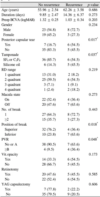
Table 4. Risk factors of recurrence of retinal detachment in
group IINo recurrence Recurrence p-value Age (years) 53.96 ± 2.54 62.26 ± 3.58 0.686 Duration (days) 9.85 ± 2.47 14.36 ± 6.37 0.273 Preop BCVA (logMAR) 1.32 ± 0.25 1.03 ± 0.34 0.203
Gender 0.234
Male 23 (54.8) 8 (72.7)
Female 19 (45.2) 3 (27.3)
Posterior capsular tear 0.017*
Yes 7 (16.7) 6 (54.5)
No 35 (83.3) 5 (45.5)
Tamponade 0.037*
SF6 or C3F8 36 (85.7) 6 (54.5)
Silicone oil 6 (14.3) 5 (45.5)
RD range 0.219
1 quadrant 13 (31.0) 2 18.2)
2 quadrant 25 (59.5) 6 (54.5)
3 quadrant 3 (7.1) 1 (9.1)
4 quadrant 1 (2.4) 2 (18.2)
Macula state 0.273
On 22 (52.4) 4 (36.4)
Off 20 (47.6) 7 (63.6)
No. of break 0.443
1 27 (64.3) 8 (72.7)
≥2 15 (35.7) 3 (27.3)
Position of break 0.018*
Superior 32 (76.2) 4 (36.4)
Inferior 10 (23.8) 7 (63.6)
PVR 0.048*
No or A 38 (90.5) 7 (63.6)
≥B 4 (9.5) 4 (36.4)
Vit.opacity 0.173
Yes 14 (33.3) 6 (54.5)
No 28 (66.7) 5 (45.5)
Retinotomy
0.585
Yes 20 (47.6) 5 (45.5)
No 22 (52.4) 6 (54.5)
YAG capsulectomy 0.606
Yes 7 (77.8) 2 (22.2)
No 35 (79.5) 9 (20.5)
Values are presented as mean ± SD or n (%) unless otherwise indicated. Statistics were analyzed by T-test, chi-square test, and Fisher’s exact test.
Preop = preoperative; BCVA = best corrected visual acuity; RD = reti- nal detachment; PVR = proliferative vitreoretinopathy; Vit.opacity
= vitreous opacity; YAG = Yttrium-Aluminum-Garnet.
*Statistically significant difference among groups.
(5.7%)이었다. 또한 황반을 침범한 경우가 I군은 전체 105 안 중 60안(57.1%), II군은 전체 53안 중 28안(49.1%)이었 다. I군에서 망막열공의 수가 2개 이상인 경우가 35안 (33.3%)이었던 것에 비해 II군은 18안(34.0%)이었다. 망막 열공의 위치가 위쪽인 경우는 I군에서 53안(50.5%), II군에 서는 36안(61.9%)이었다. 증식성 유리체망막병증 grade B 이상은 I군 14안(13.3%), II군 8안(15.1%)이었고 유리체 혼 탁은 I군 29안(27.6%), II군 20안(37.7%)에서 관찰되었다.
술 중 망막절개술을 시행한 경우는 I군 54안(51.4%), II군 25안(47.2%)이었다. 각 요인 중 백내장수술을 병행한 군의 평균 나이(p=0.002)와, 망막열공이 위쪽인 경우(p=0.027)가
통계학적으로 유의하게 더 많았으며 그 이외의 요인은 두 군 간에 유의한 상관관계를 보이지 않았다(Table 1). 두 군 간의 나이 차는 나이에 따라 진행한 백내장 정도에 따라 수 술 병행 여부를 결정했기 때문이라고 추측할 수 있다. 수술 이후 경과관찰 기간 중간값은 I군 60.00주, II군 64.57주로 두 군 간에 유의한 차이는 없었다(p=0.138) (Table 1, Fig. 1).
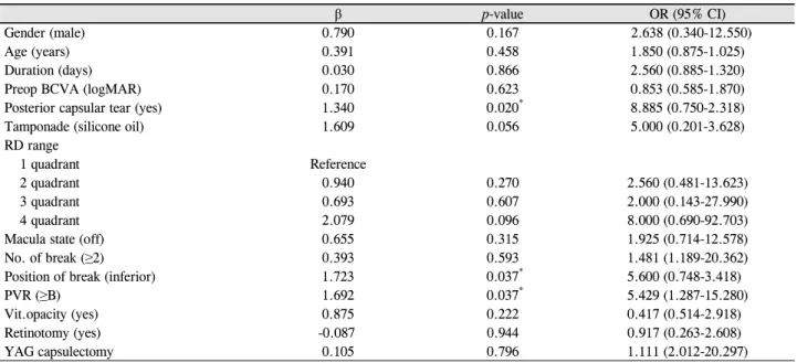
Table 5. Univariate analysis about risk factors of recurrence of retinal detachment in group II
β p-value OR (95% CI)
Gender (male) 0.790 0.167 2.638 (0.340-12.550)
Age (years) 0.391 0.458 1.850 (0.875-1.025)
Duration (days) 0.030 0.866 2.560 (0.885-1.320)
Preop BCVA (logMAR) 0.170 0.623 0.853 (0.585-1.870)
Posterior capsular tear (yes) 1.340 0.020* 8.885 (0.750-2.318)
Tamponade (silicone oil) 1.609 0.056 5.000 (0.201-3.628)
RD range
1 quadrant Reference
2 quadrant 0.940 0.270 2.560 (0.481-13.623)
3 quadrant 0.693 0.607 2.000 (0.143-27.990)
4 quadrant 2.079 0.096 8.000 (0.690-92.703)
Macula state (off) 0.655 0.315 1.925 (0.714-12.578)
No. of break (≥2) 0.393 0.593 1.481 (1.189-20.362)
Position of break (inferior) 1.723 0.037* 5.600 (0.748-3.418)
PVR (≥B) 1.692 0.037* 5.429 (1.287-15.280)
Vit.opacity (yes) 0.875 0.222 0.417 (0.514-2.918)
Retinotomy (yes) -0.087 0.944 0.917 (0.263-2.608)
YAG capsulectomy 0.105 0.796 1.111 (2.012-20.297)
Statistics were analyzed by backward stepwise method in logistic regression analysis.
β = regression coefficient; OR = odds ratio; CI = confidence interval; Preop = preoperative; BCVA = best corrected visual acuity; RD = retinal detachment; PVR = proliferative vitreoretinopathy; Vit.opacity = vitreous opacity; YAG = Yttrium-Aluminum-Garnet.
*Statistically significant difference among groups.
Table 6. Multivariate analysis about risk factors of recurrence of retinal detachment in group II
β p-value OR (95% CI)
Gender 7.357 0.286 1.567 (0.290-0.765)
Age (years) 0.595 0.220 3.919 (0.443-1.430)
Posterior capsular tear 58.196 0.021* 1.880 (39.733-64.776)
Tamponade (silicone oil) 42.156 0.087 2.032 (14.881-48.204)
Position of break 3.338 0.348 2.035 (13.404-25.302)
PVR 36.082 0.109 5.281 (1.15-25.148)
Statistics were analyzed by backward stepwise method in logistic regression analysis.
β = regression coefficient; OR= odds ratio; CI= confidence interval; PVR = proliferative vitreoretinopathy.
*Statistically significant difference among groups.
유리체절제술과 백내장수술을 병행하지 않은 환자군 (group I)과 유리체절제술과 동시에 백내장수술을 받은 환 자군(group II)의 망막박리 재발률을 비교 분석한 결과, Group I에서는 105명 중 26명(24.8%), Group II는 53명 중 11명(20.8%)에서 망막박리 재발을 보였으며 백내장수술을 병행하는 것이 망막박리의 재발을 높인다는 근거를 발견하 지 못했다(p=0.363) (Table 2). 유리체절제술 장비의 발달로 인한 영향이 혼란변수로 작용할 수 있음을 감안하여, con- stellation system 장비의 도입 시점을 기준으로 망막박리 재 발률을 비교한 결과 유의한 차이를 보이지 않았다(p=0.085) (Table 3).
또한 유리체절제술과 동시에 백내장수술을 한 군(Group II)에서 망막박리 재발의 위험인자를 분석하였다. 백내장수 술 중 후낭파열이 망막박리 재발이 없는 경우에서 16.7%, 재발이 있는 경우에서 54.5%로 유의한 빈도 차이를 보였고
(p=0.017) 눈 속 충전물로 silicone oil이 다른 충전물(SF6, C3F8)에 비해 망막박리 재발의 빈도가 높았다(14.3% vs.
45.5%, p=0.037). 또한 아래쪽의 망막열공이 망막박리 재발 이 없는 경우에서 23.8%, 재발이 있는 경우에서 63.6%로 유의한 빈도 차이를 보였으며(p=0.018). 증식성 유리체망막 병증 grade B 이상이 재발이 없는 경우에서 9.5%, 재발이 있는 경우에서 36.4%로 유의한 차이를 보였다(p=0.048) (Table 4).
이분형 로지스틱 회귀분석을 이용한 단변량 분석 결과, 백 내장수술 중 후낭파열이 있었던 경우(odds ratio [OR] 8.885, p=0.020), 망막열공의 위치가 아래쪽인 경우(OR 5.600, p=0.037), 증식성 유리체망막병증 grade B 이상인 경우 (OR 5.429, p=0.037)에 망막박리의 재발과 유의하게 연관 이 있는 것으로 나타났지만, 다변량 분석 결과에서는 백내 장수술 중 후낭파열 여부(OR 1.880, p=0.021)만이 망막박
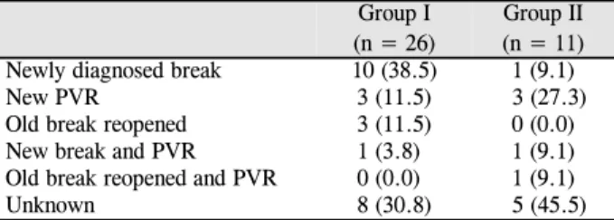
Table 7. Causes of recurrence of retinal detachment
Group I (n = 26)Group II (n = 11) Newly diagnosed break 10 (38.5) 1 (9.1)
New PVR 3 (11.5) 3 (27.3)
Old break reopened 3 (11.5) 0 (0.0)
New break and PVR 1 (3.8) 1 (9.1)
Old break reopened and PVR 0 (0.0) 1 (9.1)
Unknown 8 (30.8) 5 (45.5)
Unknown indicated that no new break, a reopened old break or sig- nificant PVR was detected during second surgery.
PVR = proliferative vitreoretinopathy.
리 재발과의 상관관계가 증명되었다(Table 5, 6).
두 군에서 망막박리가 재발했을 당시의 안저 검사를 통 해 재발의 원인을 분석해 보았을 때, Group I에서는 새롭게 발견된 망막 열공이 10안(38.5%), PVR이 3안(11.5%), 이전 의 열공이 다시 열린 경우가 3안(11.5%), 새롭게 발견된 열 공과 PVR이 같이 보인 눈이 1안(3.8%), 이전의 망막열공과 PVR이 재발 원인이라고 추측되는 경우는 없었고, 원인을 모르는 경우 9안(34.6%)이었으며, Group II에서는 각각 1 안(9.1%), 3안(27.3%), 0안(0.0%), 1안(9.1%), 1안(9.1%), 5 안(45.5%)이었다(Table 7).
고 찰
열공망막박리의 일차적 치료로서 유리체 절제술은 많은 비중을 차지하고 있으며 성공률은 대략 72-94%로 보고되 고 있다.2 유리체절제술은 여전히 대략 15%의 적지 않은 수의 망막박리 재발과 관련이 있다고 알려져 있으며 5%에 서 여러 번의 수술에도 불구하고 망막의 재박리가 생겼다 는 연구 결과가 있다.3 또한 망막박리 재발의 위험 인자에 대해서는 망막열공 혹은 망막박리의 위치가 아래였던 경우, 증식성 유리체망막병증 grade B 이상이었던 경우 등이었다.
Heimann et al4은 512명의 열공망막박리 환자를 대상으 로 한 연구에서 하부 망막박리인 경우, 망막열공의 위치가 아래쪽인 경우에 유리체절제술 후 재발 위험이 높다고 보 고하였고 이는 상대적으로 오랜 유병기간과 술 전 유리체 섬유세포 증식의 높은 비율과 관련이 있고 유리체절제술 중 넣은 충전물이 충분히 아래쪽 박리 부분을 지지해 주지 못하기 때문이라고 하였다. 따라서 긴 작용기간을 가진 충 전물이나 실리콘 기름을 넣는 등 유리체 절제술 기법의 변 형이나 공막돌륭술을 선택하는 것을 첫 번째 치료 대안으 로 제시하였다.
증식성 유리체 망막병증이 망막박리 재발의 큰 위험인자 라는 것은 이미 증명되었다. 증식성 유리체 망막병증의 grade는 크게 A, B, C 세 단계로 나뉜다. A단계는 유리체
흐림과 세포가 존재하는 경우로 제한되며 B단계는 망막열 공 가장자리가 불규칙하거나 말려 있고 내망막표면 주름이 있는 경우로 무증상의 수축을 의미한다. C단계는 망막앞 또는 망막하 증식막이 존재하는 단계로 적도부보다 앞부분 은 Ca, 뒷부분은 Cb로 세분한다. 이는 망막박리 수술 환자 의 5.1-11.7%에서 나타나며 열공망막박리 치료의 가장 흔 한 실패요인이다. 망막박리 후 유리체절제술 시행 환자의 1/4 이상이 이후 유리체 망막 견인으로 인해 결국 망막박리 의 재발을 경험한다. 망막 색소 상피세포가 가장 큰 역할을 하며 수용성 매개 물질과 세포외기질 구성요소도 보조 역 할을 한다. 유리체절제술 시 증식성 유리체망막병증을 예방 하기 위한 연구는 지속되고 있으며 최근 연구에서 세포의 증식과 막 수축을 억제하는 daunorubicin, 5-fluorouracil, heparin 등의 약물학적 치료 방법도 제시하고 있다.5
또한 수정체안과 가성수정체안에서 유리체절제술 후 망 막박리 재발률을 분석했을 때 3사분면 범위까지의 망막박 리에서는 비슷하였으나 그 이상에서는 가성수정체안에서 더 망막박리의 재발률이 높았다는 보고도 있다.6 물론 술자 의 수술 경험에 따른 술 후 재발률의 차이도 크다.7
후낭파열은 백내장수술 중 대략 1.92-2.09%에서 발생하 며 이는 가성수정체안의 망막박리와 연관성이 있다.9,16-21 후낭파열 후 3년 이내 망막박리의 위험률(odd ratio)이 15 배, 1년 이내는 18배, 3개월 이내는 무려 42배라고 보고된 바도 있다.16,19 Western Australia entire population study9에 서는 후낭파열 후 5년 이내 망막박리가 발생한 위험비 (hazard ratio)가 27.6이었고 France21에서는 4년 이내 4.4로 보고되었다.
Kon et al22은 백내장수술 후 후낭결손(defective posterior capsule)이 있거나 유리체 단백질 단계(vitreous protein lev- el)가 높은 경우, 또는 술 전 증식성 유리체망막병증의 유무 가 술 후 유리체망막병증과 관련이 있으며 술 후 유리체망 막병증은 해부학적, 기능적으로 평가하는 유리체절제술 성 공 여부에 큰 영향을 미친다고 보고하였다.
본 연구는 후향적 연구임과 동시에 20년 동안의 데이터 를 분석한 것으로, 술자의 경험 축적에 의해 좋은 수술 결 과를 보일 수 있다는 측면을 고려하지 못했다는 한계가 있 지만 열공망막박리로 유리체절제술과 백내장수술 병행여 부에 따른 망막박리 재발 빈도를 분석함으로써 유리체절제 술 시행 예정인 환자에서 백내장수술의 병행에 대해 생각 해 볼 수 있고, 백내장수술을 시행한 경우에 한해 망막박리 재발의 위험요인을 좀 더 정밀하게 분석하여 하부 망막열 공, 증식성 유리체망막병증 등 기존 연구에서 증명된 망막 박리 재발의 위험인자를 재확인하였다. 특히 후낭파열은 기존의 연구에서 망막박리의 위험인자로 밝혀졌지만 망막
박리의 재발에 영향을 미치는지에 대한 연구는 없었다는 점에서 본 연구가 큰 의미를 가진다. 또한 20년 동안 한 명 의 안과 망막전문의에 의해 시행된 수술 환자 데이터를 분 석한 것으로 이는 망막박리의 상황에 따른 결정과 수술적 기법의 다양성을 배제한 결과라 그 신뢰성이 높으며, 발달 한 수술장비의 도입으로 인한 영향력을 배제할 수 있는 결 과를 도출하였다.
백내장수술과 유리체 망막수술을 동시에 시행한 군과 백 내장수술만 단독으로 시행한 군의 후낭혼탁에 미치는 영향 에 대한 기존의 비교 연구에서는 유리체 망막수술을 시행 한 경우 상대적으로 높은 빈도로 후낭혼탁이 일어났으며 통계적으로 유의하였다. 이는 술 후 염증반응과 관련이 있 으며 염증반응은 아마도 긴 수술시간과 안내에서 이루어지 는 많은 수술기구들의 조작과 관련이 있을 것이라는 추측 이다.23 본 연구에서는 유리체절제술과 백내장수술을 병행 한 군에 한해서 후낭혼탁으로 인해 레이저 후낭절개술 시 행 유무에 따른 망막박리 재발률을 분석한 결과 유의한 차 이는 발견하지 못했다. 따라서 추후 백내장수술 시행에 대 한 환자의 부담이나 유리체절제술 당시 백내장 상태 등을 고려하여 수술 병행 여부를 결정하고, 병행 수술 이후 혹여 후낭 혼탁이 발생하면 적절한 시기에 레이저 후낭절개술을 시행하는 것이 합당할 것이다.
다른 관점에서 보면, 유리체절제술 시행 이후 망막이 안 정된 뒤 백내장수술을 시행할 경우 인공수정체 도수 측정 의 정확도에 따른 백내장수술의 성공률을 높일 수 있으며 유리체절제술과 백내장수술 병행 시에 긴 수술 시간과 수 술 기구로 인해 발생할 수 있는 염증, 자세 유지 때문에 발 생하는 침착물로 인한 부작용 등을 걱정하지 않아도 된다.
결국 백내장수술을 병행할 경우와 그렇지 않은 경우의 망 막박리 재발의 차이를 증명할 수 있는 근거를 발견하지 못한 본 연구 결과로 볼 때, 망막박리 진단 당시 매체 투명성 (media clarity)을 고려한 백내장의 상태를 파악하여 유리체 절제술에 지장이 있을 정도의 백내장이거나 환자가 동시 수 술을 원할 때 백내장수술을 병행하고, 그렇지 않을 경우에는 망막이 안정된 뒤 백내장수술을 시행할 경우의 장점을 고려 하여 백내장수술을 유리체절제술 이후에 시행하는 방법을 생각해 보는 것이 합리적이라고 판단된다. 또한 망막박리 수 술과 동시에 혹은 2년 이내 백내장수술을 시행했을 시 백내 장수술 중 후낭파열이 있었다면, 특히 망막박리 재발의 위험 이 높으므로 술 후 주의 깊게 망막의 상태를 살펴야 하겠다.
REFERENCES
1) Machemer R. The importance of fluid absorption, traction, intra- ocular currents, and chorioretinal scars in the therapy of rhegma-
togenous retinal detachments. XLI Edward Jackson memorial lecture. Am J Ophthalmol 1984;98:681-93.
2) Lee E, Housseini ZE, Steel DH, Williamson TH. An analysis of the outcomes for patients with failed primary vitrectomy for rhegma- togenous retinal detachment. Graefes Arch Clin Exp Ophthalmol 2014;252:1711-6.
3) SPR Study group. View 2: the case for primary vitrectomy. Br J Ophthalmol 2003;87:784-7.
4) Heimann H, Zou X, Jandeck C, et al. Primary vitrectomy for rheg- matogenous retinal detachment: an analysis of 512 cases. Graefe's Arch Clin Exp Opathalmol 2006;244:69-78.
5) Kwon OW, Song JH, Roh MI. Retinal detachment and proliferative vitreoretinopathy. Dev Ophthalmol 2016;55:154-62.
6) Halberstadt M, Brandenburg L, Sans N, et al. Analysis of risk fac- tors for the outcome of primary retinal reattachment surgery in phakic and pseudophakic eyes. Klin Monatsbl Augenheikd 2003;
220:116-21.
7) Miki D, Hida T, Hotta K et al. Comparison of scleral buckling and vitrectomy for retinal detachment resulting from flap tears in supe- rior quadrants. Jpn J Ophthalmol 2001;45:187-91.
8) Quek DT, Lee SY, Htoon HM, Ang CL. Pseudopharic rhegmatoge- nous retinal detachment in large Asian tertiary eye centre: a cohort study. Clin Exp Ophthalmol 2012;40:e1-7.
9) Clark A, Morlet N, Ng JQ, et al. Risk for retinal detachment after phacoemulsification: a whole-population study of cataract surgery outcomes. Arch Ophthalmol 2012;130:882-8.
10) Boberg-Ans G, Henning V, Villumsen J, la Cour M. Longterm in- cidence of rhegmatogenous retinal detachment and survival in a defined population undergoing standardized phacoemulsification surgery. Acta Ophthalmol Scand 2006;84:613-8.
11) Erie JC, Raecker MA, Baratz KH, et al. Risk of retinal detachment after cataract extraction, 1980-2004: a population-based study.
Ophthalmology 2006;113:2026-32.
12) Lin JY, Ho WL, Ger LP, Sheu SJ. Analysis of factors correlated with the development of pseudophakic retinal detachment—a long-term study in a single medical center. Graefes Arch Clin Exp Ophthalmol 2013;251:459-65.
13) Sheu SJ, Ger LP, Ho WL. Late increased risk of retinal detachment after cataract extraction. Am J Ophthalmol 2010;149:113-9.
14) Clark A, Morlet N, Ng JQ, et al. Whole population trends in com- plications of cataract surgery over 22 years in Western Australia.
Ophthalmology 2011;118:1055-61.
15) Sheu SJ, Ger LP, Chen JF. Axial myopia is an extremely significant risk factor for young-aged pseudophakic retinal detachment in Taiwan. Retina 2006;26:322-7.
16) Jakobsson G, Montan P, Zetterberg M, et al. Capsule complication during cataract surgery: retinal detachment after cataract surgery with capsule complication: Swedish Capsule Rupture Study Group report 4. J Cataract Refract Surg 2009;35:1699-705.
17) Jaycock P, Johnson RL, Taylor H, et al. The Cataract National Dataset electronic multi-centre audit of 55,567 operations: updat- ing benchmark standards of care in the United Kingdom and internationally. Eye (Lond) 2009;23:38-49.
18) Lundström M, Behndig A, Kugelberg M, et al. Decreasing rate of capsule complications in cataract surgery: eight-year study of in- cidence, risk factors, and data validity by the Swedish National Cataract Register. J Cataract Refract Surg 2011;37:1762-7.
19) Day AC, Donachie PH, Sparrow JM, et al. The Royal College of
= 국문초록 =
열공망막박리 유리체절제술 시 백내장수술 동시 시행 여부에 따른 재발 차이와 위험요인 분석
목적: 열공망막박리에서 유리체절제술 시 백내장수술 병행 여부에 따른 망막박리의 재발 빈도의 차이와 기존에 알려진 망막박리 재발 의 위험요인을 분석해 보고자 한다.
대상과 방법: 1997년 1월부터 2016년 9월까지 20년에 걸쳐 내원 당시 처음으로 열공망막박리를 진단 받고 유리체절제술을 시행 받은 환자 중 진단 당시 유수정체안 158안을 대상으로, 유리체절제술과 동시에 본원에서 백내장수술을 받은 환자군과 그렇지 않은 환자군 으로 나누어 이들의 의무기록을 후향적으로 분석하였다.
결과: 유리체절제술과 동시에 백내장수술을 받은 군과 받지 않은 군 간 비교에서는 백내장수술을 병행한 군이 평균 나이가 더 많았고 (p=0.002) 두 군 간 망막박리 재발률은 유의한 차이를 보이지 않았다. 백내장수술을 받은 군에서 망막박리 재발의 위험 요인으로는, 단변량 분석에서 백내장수술 중 후낭파열이 있었던 경우(p=0.020), 아래쪽의 망막 열공(p=0.037), 증식성 유리체망막병증 grade B 이상(p=0.037)이었으나 다변량 분석에서는 백내장수술 중 후낭파열이 있는 경우에만 망막박리의 재발 위험이 높은 것으로 나타났다 (odds ratio 1.880, p=0.021).
결론: 본 연구에서는 열공망막박리로 인한 유리제절제술 시 백내장수술 동시 시행 여부가 망막박리의 재발을 높인다는 근거를 발견하 지 못했다. 백내장수술 동시 시행 여부는 여러 상황을 고려하여 탄력적으로 결정할 수 있으며, 백내장수술을 동시에 시행 시 후낭파열 이 있었던 경우에 망막박리의 재발이 있는지를 추후 잘 살펴야 한다.
<대한안과학회지 2017;58(12):1388-1395>
Ophthalmologists’ National Ophthalmology Database study of cataract surgery: report 1, visual outcomes and complications. Eye (Lond) 2015;29:552-60.
20) Tuft SJ, Minassian D, Sullivan P. Risk factors for retinal detach- ment after cataract surgery: a case-control study. Ophthalmology 2006;113:650-6.
21) Daien V, Le Pape A, Heve D, et al. Incidence, risk factors, and im- pact of age on retinal detachment after cataract surgery in France: a
national population study. Ophthalmology 2015;122:2179-85.
22) Kon CH, Asaria RH, Occleston NL, et al. Risk factors for pro- liferative vitreoretinopathy after primary vitrectomy: a prospective study. Br J Ophthalmol 2000;84;506-11.
23) Ariki G, Ogino N. Postoperative anterior chamber inflammation af- ter posterior chamber intraocular lens implantation concurrent with pars plana vitrectomy and lensectomy. Nippon Ganka Gakkai Zasshi 1992;96:1300-5.