ISSN 0378-6471 (Print)⋅ISSN 2092-9374 (Online)
https://doi.org/10.3341/jkos.2018.59.8.797
Case Report
곰팡이나비굴염에 의한 가돌림신경마비와 시신경주위염 1예
Abducens Nerve Palsy and Optic Perineuritis Caused by Fungal Sphenoidal Sinusitis
서영범1⋅김경주2⋅김원제3
Youngbeom Seo, MD1, Kyung-Ju Kim, MD, PhD2, Won Jae Kim, MD3
영남대학교 의과대학 신경외과학교실1, 영남대학교 의과대학 병리학교실2, 영남대학교 의과대학 안과학교실3 Department of Neurosurgery, Yeungnam University College of Medicine1, Daegu, Korea
Department of Pathology, Yeungnam University College of Medicine2, Daegu, Korea Department of Ophthalmology, Yeungnam University College of Medicine3, Daegu, Korea
Purpose: To report a case of abducens nerve palsy and optic perineuritis caused by fungal sphenoidal sinusitis.
Case summary: A 48-year-old male visited emergency department for retrobulbar pain, decreased vision, and horizontal dip- lopia for 3 days. He reported that previous medical history was non-specific, however, blood glucose level was 328 mg/dL (70-110). He had experienced severe headache for 7 days. The best corrected visual acuity was 20/20 at right eye and 20/25 at left eye. The pupil of left eye did not have relative afferent pupillary defect. Left mild proptosis was noted. The extraocular exami- nation showed 30 prism diopters left esotropia at primary gaze and –4 abduction limitation of left eye. The left eye showed mild optic disc swelling and inferior field defect by field test. Brain magnetic resonance imaging showed enhancement of sphenoidal sinus, ethmoidal sinus, and around optic nerve at left eye. Three days after antibiotics treatment, the vision of left eye deterio- rated to 20/40 and periorbital pain developed. The drainage and biopsy of sphenoidal sinus were performed. The histopathologic examination showed hyphae consistent with aspergillosis. The ocular symptoms were improved with anti-fungal treatment.
Follow-up magnetic resonance imaging performed 1 month after treatment showed improvement of lesion at left orbit. Five months after surgery, the visual acuity of left eye was improved to 20/25. The patient showed orthotropia at primary gaze without limitation.
Conclusions: The abducens nerve palsy and optic perineuritis can be caused by fungal sphenoidal sinusitis. The early diagnosis and appropriate treatment can lead to favorable outcome.
J Korean Ophthalmol Soc 2018;59(8):797-801
Keywords: Abducens nerve palsy, Fungus, Optic perineuritis, Sphenoidal sinusitis
■Received: 2018. 4. 26. ■ Revised: 2018. 5. 23.
■Accepted: 2018. 7. 24
■Address reprint requests to Won Jae Kim, MD
Department of Ophthalmology, Yeungnam University Hospital,
#170 Hyeonchung-ro, Nam-gu, Daegu 42415, Korea Tel: 82-53-620-4191, Fax: 82-53-626-5936 E-mail: [email protected]
* Conflicts of Interest: The authors have no conflicts to disclose.
ⓒ2018 The Korean Ophthalmological Society
This is an Open Access article distributed under the terms of the Creative Commons Attribution Non-Commercial License (http://creativecommons.org/licenses/by-nc/3.0/) which permits unrestricted non-commercial use, distribution, and reproduction in any medium, provided the original work is properly cited.
나비굴(sphenoidal sinus)은 안구, 시신경, 해면정맥굴 (cavernous sinus), 속목동맥(internal carotid artery) 등의 구 조물과 인접해 있다.1,2 따라서 나비굴 염증 병변의 진행은 시신경과 해면정맥굴 내의 눈돌림신경(oculomotor nerve), 활차신경(trochlear nerve), 삼차신경(trigeminal nerve), 가돌 림신경(abducens nerve)에 영향을 줄 수 있고, 경우에 따라 심각한 머리속(intracranial) 또는 눈 합병증이 발생할 수 있 다.1,3 하지만 나비굴 염증 병변의 초기 증상은 흔히 비특이 적이기 때문에, 적절한 진단과 치료가 늦어질 수 있다.3 저 자들은 당뇨 환자에서 발생한 곰팡이나비굴염(fungal sphe-
A
B C
Figure 1. Findings of ocular examination at initial visit. (A) Image of the patient in nine diagnostic position of gaze demonstrating esotropia at primary gaze and abduction limitation of the left eye. (B) Fundus examination shows mild swelling of optic disc at the left eye. (C) The visual field test reveals inferior visual field defect at the left eye (Humphrey Field Analyzer, Carl Zeiss M editec, Inc., Dublin, CA, USA).
noidal sinusitis)에 의한 가돌림신경마비와 시신경주위염 (optic perineuritis)을 경험하였기에 이를 보고하고자 한다.
증례보고
48세 남자가 3일 전부터 갑자기 시작된 좌안 눈뒤통증과 시력저하, 수평복시로 응급실에 내원하였다. 7일 전부터 이 전에 없었던 머리 전체의 심한 두통이 발생하였으며, 3일 전부터 두통이 좌측으로 이동하면서 눈뒤통증이 심해졌다 고 하였다. 두통은 지속적이었고, 특별한 악화나 완화요인 은 없었다. 이전에 전신 질환, 종양, 자가면역 질환, 외상, 약복용, 수술 등의 과거력은 없다고 하였다. 그러나 혈당이 328 mg/dL (70-110)였다. 시력은 우안이 20/20, 좌안이 20/25였고, 색각검사에서는 정상 소견을 보였다. 동공반응 검사에서 상대구심동공운동 장애는 없었다. 안구돌출계검 사에서 좌안이 우안보다 2 mm 돌출되어 있었다. 안구운동
검사에서 30프리즘디옵터(prism diopters, PD)의 내사시와 좌안 -4의 가쪽운동장애를 보였다(scale of -4 to 0, Fig. 1A).
안저검사에서 좌안 시신경유두의 경도 부종 소견을 보였으 며, 시야검사에서는 하측의 시야장애 소견을 보였다(Fig.
1B, C). 혈액검사에서 백혈구수치(white blood cell)는 8,350 /μL (4,000–10,000)로 정상이었고, 적혈구침강속도(erythrocyte sedimentation rate, ESR)는 81 mm/H (0-20), 고감도C반응 단백(high-sensitivity C-reactive protein, hsCRP)이 3.448 mg/dL (0-0.06)로 증가되어 있었다. 뇌 자기공명영상(magnetic res- onance imaging, MRI)에서 좌측 나비굴과 벌집굴(ethmoid sinus) 점막을 따라서 벽(wall)에 국한된 조영증강 소견이 관찰되었다. 시신경관(optic canal) 및 안구 내의 시신경집 (optic nerve sheath)에 조영증강 소견이 관찰되었다(Fig.
2A-C). 나비굴염에 의한 가돌림신경마비와 시신경주위염 가 능성을 고려하여 정맥 항생제 치료(ceftriaxone 4 g/day, ne- tilmicin 300 mg/day, clindamycin 1,200 mg/day)를 시작하
A B C
D E F
Figure 2. Findings of brain magnetic resonance (MR) imaging during follow-ups. (A-C) The initial magnetic resonance (MR) imaging. (A) Axial T1-weighted gadolinium-enhanced MR image showing an enhancement of the circumferential mucosa in the sphenoid sinus and the ethmoidal sinus (arrowhead). (B, C) Axial and coronal T1-weighted gadolinium-enhanced MR images dem- onstrate an enhancement of the left optic nerve sheath (arrow). (D-F) The follow-up MR imaging at 1 month after the anti-fungal treatment. The patient underwent an endoscopic endonasal surgery for sinusitis. (E, F) Axial and coronal T1-weighted gadoli- nium-enhanced MR images showing a normalization of the left optic nerve.
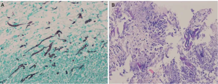
였다. 3일 뒤 좌안 시력이 20/40으로 저하되고, 눈주위 통 증이 발생하였다. 나비굴절개술과 조직검사를 시행하였고, 코곁굴염(paranasal sinusitis)으로 인한 안구 및 머릿속 침 범으로 시신경주위염 및 해면정맥굴혈전정맥염(cavernous sinus thrombophlebitis)이 의심되어 신경외과로 전과되어 치료를 지속하였다. 조직에 대한 hematoxylin and eosin stain (H&E stain)과 GMS 특수염색(Grocott’s methenamine sliver stain)에서 나뭇가지 모양의 진균의 균사가 관찰되었 다. 이것은 예각(acute angle)으로 분지하였고, 두께는 약 3 µm로 측정되었다(Fig. 3). 이러한 조직 소견은 aspergillosis에 합당하여, 정맥 항진균치료(amphotericin B 70 mg/day)를 시 작하였다. 항진균치료 1개월째 MRI에서 초기 MRI와 비교 하여 시신경주위 조영증강의 호전 양상을 관찰할 수 있었 다(Fig. 2D-F). 정맥 주사치료를 경구 항진균제(fluconazole 250 mg/day)로 변경하여 5주간 지속하였다. 수술 5개월째
좌안 시력은 20/25로 호전되었다. 안구운동검사에서도 정 면주시 시 정위를 보였으며, 좌안 가쪽운동 장애 소견도 소 실되었다(Fig. 4).
고 찰
곰팡이나비굴염은 성인에서 가돌림신경마비와 시신경주 위염의 흔한 원인은 아니다.2,4 해면정맥굴 내에서 가돌림신 경은 뇌막 가쪽에 부착되어 있는 다른 신경들과 달리 나비 굴과 인접해 있기 때문에, 나비굴염의 침범에 더 쉽게 영향 을 받을 수 있다.4 이전에 국내에서 곰팡이나비굴염에 의한 가돌림신경마비 또는 시신경염이 몇 예 보고되었으나,5-7 곰 팡이나비굴염에 의한 가돌림신경마비와 시신경주위염의 발생과 회복에 대한 보고는 없었다.
나비굴 염증 병변의 초기 증상은 대개 비특이적이기 때
A B
Figure 3. Histopathology of the sphenoid sinus tissue showed hyphae, consistent with the aspergillosis. (A) Grocott’s methenamine silver stain ×200. (B) Haematoxylin and eosin stain ×200.
Figure 4. Finding of ocular motility at 5 months after the anti-fungal treatment. Images of the patient in nine diagnostic position of gaze at 5 months after the anti-fungal treatment, he showed stable ocular alignment without abduction limitation.
문에, 빠른 진단과 적절한 치료가 늦어질 수 있다. Chen et al3의 연구에서 총 23명의 나비굴 염증 병변에 의한 시력저 하 또는 복시 환자에서 가장 흔한 눈 외 증상은 두통(18/23 명)이었으며, 단 5명의 환자에서 콧물, 코막힘, 혈성콧물 등 의 코증상이 있었다. 따라서 정확한 감별 진단을 위해서는 뇌 MRI, 코곁굴전산화단층촬영술(computed tomography, CT) 등의 영상검사가 필수적이다.3 성인에서 가돌림신경마 비의 가장 흔한 원인은 허혈이며, 이 경우에도 환자는 눈주 위 통증을 동반할 수 있다. 허혈 원인은 대부분 3-6개월 내 에 완전 회복을 보인다.2,4,8 따라서 가돌림신경마비가 허혈 에 의한 것으로 생각된다면 3개월까지 회복 여부를 확인 후 뇌영상검사를 시행하는 것이 제시되고 있다.2,8,9 그러나 허혈로 생각되었던 가돌림신경마비 환자의 약 4.7%에서 다른 원인이 밝혀졌다는 연구가 있어 허혈 가돌림신경마비 가 의심되는 경우의 뇌영상검사 시기에 대해서는 논란의 여지가 있다.10 따라서 이 증례와 같이 지속적인 심한 두통 과 눈통증을 동반하는 경우는 비허혈 원인에 의한 가능성
을 고려하여 빨리 뇌영상검사를 시행하는 것이 필요하다.9 시신경주위염은 눈확 내의 시신경수초와 주위 눈확 조직 의 염증으로 특발성으로 발생하는 경우가 대부분이며, 감 염이나 자가면역 질환에 동반되어도 발생할 수 있다.11,12 시 신경주위염은 스테로이드 치료에서 비교적 빠른 시력 회복 을 보이지만, 이 증례와 같이 감염에 의한 경우는 스테로이 드 치료 이전에 감염원에 대한 치료가 선행되어야 할 것이 다.3,11
이 환자에서 심한 가쪽운동 장애를 동반한 가돌림신경마 비와 시신경주위염이 항진균 치료 5개월째에 완전 회복을 보인 것이 흥미롭다. Miller et al13은 코곁굴 병변에 의한 가돌림신경마비와 회복에 대한 연구에서 압박성(compressive) 병변에 의한 경우 좋은 경과를 보였으나, 침습적 곰팡이감 염과 같이 파괴성(destructive) 병변에 의해 가돌림신경이나 주위 혈관에 직접 손상을 주는 경우는 나쁜 결과로 이어질 수 있다고 하였다. Chen et al3의 보고에서는 9명의 곰팡이 나비굴염에 의한 시력저하 환자에서 5명의 환자가 치료 후
= 국문초록 =
곰팡이나비굴염에 의한 가돌림신경마비와 시신경주위염 1예
목적: 곰팡이나비굴염(fungal sphenoidal sinusitis)에 의한 가돌림신경마비(abducens nerve palsy)와 시신경주위염(optic perineuritis) 을 경험하였기에 이를 보고하고자 한다.
증례요약: 48세 남자가 3일 전부터 시작된 좌안 눈뒤통증과 시력저하, 수평복시로 내원하였다. 이전에 전신 질환의 과거력은 없다고 하였으나, 혈당이 328 mg/dL (70-110)였다. 7일 전부터 심한 두통이 있었다. 시력은 우안이 20/20, 좌안이 20/25였고, 동공반응검사 에서 상대구심동공운동 장애는 없었다. 좌안 안구돌출을 동반하였고, 안구운동검사에서 30프리즘디옵터(prism diopters, PD)의 내사 시와 좌안 -4의 가쪽운동 장애를 보였다. 안저검사에서 좌안 시신경유두의 경도의 부종 소견과 시야검사에서 하측 시야결손을 보였 다. 뇌 자기공명영상(magnetic resonance imaging, MRI)에서 나비굴 및 벌집굴(ethmoid sinus), 시신경주위의 조영증가 소견을 보였 다. 항생제 치료 3일째 좌안 시력이 20/40으로 저하되고, 눈주위 통증이 발생하였다. 부비동염에 대해 내시경적 부비동 수술을 통해 배액술 및 조직검사를 시행하였고, aspergillosis를 확인하였다. 수술 후 항진균치료를 시작하였고, 환자의 증상은 호전되었다. 치료 후 1개월 뒤 시행한 MRI에서 안구 내 병변의 호전을 확인하였다. 수술 5개월 뒤, 좌안 시력은 20/25로 호전되었다. 안구운동검사에서 는 정위를 보였고, 좌안 가쪽운동 장애 소견은 없었다.
결론: 가돌림신경마비와 시신경주위염의 원인으로 곰팡이나비굴염이 고려될 수 있고, 빠른 진단과 적절한 치료로 좋은 경과를 얻을 수 있음을 확인하였다.
<대한안과학회지 2018;59(8):797-801>
에도 시력호전이 없었는데, 이들은 모두 눈 증상 발생 후 치료까지 1개월 이상의 시간이 걸린 경우였다. 이 환자의 경우, 눈 증상 발생 후 뇌영상검사를 통한 빠른 진단과 항 진균 치료를 통해 감염원이 뇌신경과 주위 혈관으로 더 침 범하는 것을 막을 수 있어서 상대적으로 좋은 예후를 보일 수 있었다고 생각된다.
결론적으로, 당뇨 환자에서 곰팡이나비굴염에 의한 가돌 림신경마비와 시신경 주위염의 발생을 확인하였고 나비굴 절개술과 항진균 치료를 통하여 좋은 결과를 얻을 수 있음 을 확인하였다. 심한 두통과 눈주위통증을 동반한 가돌림 신경마비의 경우는 정확한 원인 감별을 위해서 적극적인 영상검사의 시행이 필요하다.
REFERENCES
1) Witterick IJ, Vescan AD. Complications of rhinosinusitis. In:
Kennedy DW, Hwang PH, eds. Rhinology, 1st ed. New York:
Thieme Medical Publishers, 2012; chap. 21.
2) Miller NR, Subramanian PS, Patel VR. Walsh and Hoyt's Clinical Neuro-Ophthalmology: the Essentials, 3rd ed. Philadelphia:
Wolters Kluwer, 2016; 341-71.
3) Chen L, Jiang L, Yang B, Subramanian PS. Clinical features of visu-
al disturbances secondary to isolated sphenoid sinus inflammatory diseases. BMC Ophthalmol 2017;17:237.
4) Kline LB, Foroozan R. Neuro-Ophthalmology Review Manual, 7th ed. Thorofare: SLACK, 2013; 83-92.
5) Jang JH, Kim YC, Chang SD, et al. A case of optic neuritis in acute sphenoid sinusitis. J Korean Ophthalmol Soc 2007;48:1742-6.
6) Kim DH, Kim HK, Nam JK, Park JG. A case of isolated aspergillus sphenoid sinusitis. J Korean Neurol Assoc 2005;23:402-4.
7) Lee HR, Kim HJ, Seong SY, Chang JH. A case of orbital apex syndrome related to sphenoid fungal sinusitis. Korean J Otorhinolaryngol-Head Neck Surg 2010;53:644-7.
8) Biousse V, Newman NJ. Neuro-ophthalmology Illustrated, 2nd ed.
New York: Thieme, 2016; 321-465.
9) Pane A, Miller NR, Burdon M. The neuro-ophthalmology survival guide, 2nd ed. Beijing: Elsevier, 2017; 170-239.
10) Tamhankar MA, Biousse V, Ying GS, et al. Isolated third, fourth, and sixth cranial nerve palsies from presumed microvascular versus other causes: a prospective study. Ophthalmology 2013;120:2264-9.
11) Purvin V, Kawasaki A, Jacobson DM. Optic perineuritis: clinical and radiographic features. Arch Ophthalmol 2001;119:1299-306.
12) Lim HC, Choi HY, Choi JH, Jung JH. Clinical manifestations and treatment of idiopathic optic perineuritis. J Korean Ophthalmol Soc 2014;55:891-7.
13) Miller C, Suh JD, Henriquez OA, et al. Prognosis for sixth nerve palsy arising from paranasal sinus disease. Am J Rhinol Allergy 2013;27:432-5.