© 2020 Neurology India, Neurological Society of India | Published by Wolters Kluwer - Medknow 1509
Linear Trigeminal Pontine Lesion in Multiple Sclerosis‑related Trigeminal Neuropathy
A 31‑year‑old man presented with ten days of numbness in his left tongue and lips as if he just had dental anesthesia.
The numbness subsequently extended to the left cheek and chin. There were no associated pain, weakness, or any other neurological symptoms. Nine months previously, he had been admitted for short transverse myelitis at the C2 level, which was his first clinical event [Figure 1].
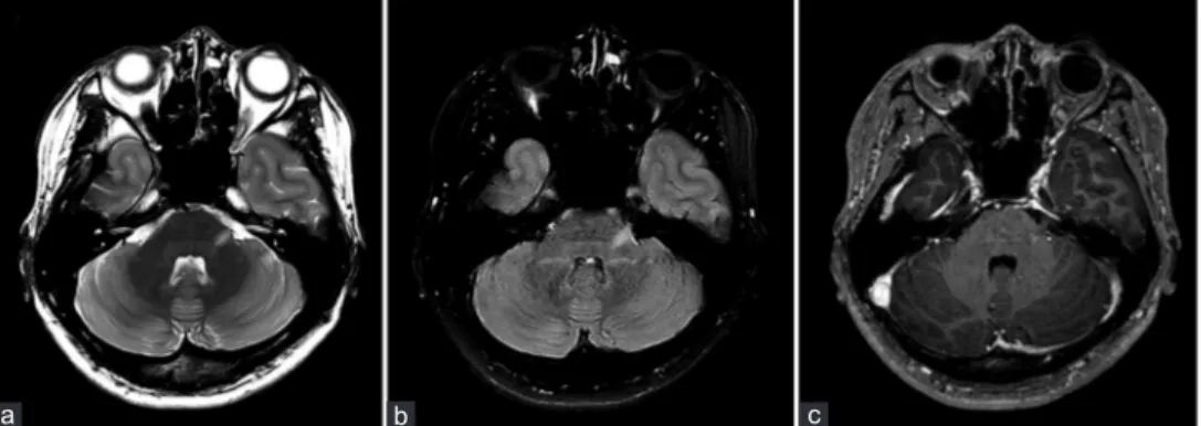
On examination, facial sensation was intact to pinprick, light touch, and vibration. Brain magnetic resonance imaging (MRI) revealed a linear hyperintense lesion in the left pontine trigeminal root entry zone (REZ) with gadolinium enhancement [Figure 2]. Immunologic serum markers
including anti‑aquaporin‑4 antibody and cerebrospinal fluid analysis were normal. We diagnosed him with clinically definite relapsing‑remitting multiple sclerosis (MS) based on the McDonald criteria.[1] He was treated with five days of intravenous methylprednisolone (1g/day) and was then started subcutaneous interferon beta‑1b to prevent further attacks. Two weeks later, his symptoms were almost improved. In MS, the trigeminal REZ lesion can appear to be either symptomatic or asymptomatic.[2,3] In symptomatic cases, clinical phenotypes are diverse, ranging from trigeminal neuralgia, which is characterized by facial pain, to trigeminal neuropathy, which is characterized by facial numbness.[4,5] Pontine trigeminal REZ lesion can also be seen in pontine infarction.[6] Distinguishing MS from pontine infarction in patients with trigeminal REZ lesion is difficult by imaging features alone and must be considered together with clinical findings. MS usually has a trigeminal REZ lesion with a linear appearance and no other ischemic lesions on MRI.[7] On the other hand, the trigeminal REZ lesion in the pontine infarction is not only linear but also wedge‑shaped, and its size is relatively larger than that of MS.
In addition, pontine infarction is relatively older at symptom onset and has more history of vascular risk factors than MS.[7]
Although the pontine trigeminal REZ lesion is not specific to MS, it may provide further clues to the diagnosis of MS when combined with other MS typical clinical events.
Financial support and sponsorship Nil.
Conflicts of interest
There are no conflicts of interest.
Neuroimage
Figure 1: Spinal MRI. (a) Sagittal T2-weighted image shows a short T2 hyperintense lesion at the C2 level (arrow). (b) Axial T2-weighted image shows
partial cord involvement, with a triangular lesion involving the dorsal columns a b
Figure 2: Brain MRI. (a) Axial T2-weighted and (b) fluid-attenuated inversion recovery images show a linear hyperintense lesion in the left pontine trigeminal root entry zone.
(c) The lesion is enhanced in axial T1-weighted gadolinium-enhanced image b c
a
Neuroimage
1510 Neurology India | Volume 68 | Issue 6 | November-December 2020
References
1. Polman CH, Reingold SC, Banwell B, Clanet M, Cohen JA, Filippi M, et al. Diagnostic criteria for multiple sclerosis: 2010 revisions to the McDonald criteria. Ann Neurol 2011;69:292‑302.
2. da Silva CJ, da Rocha AJ, Mendes MF, Maia AC, Jr., Braga FT, Tilbery CP. Trigeminal involvement in multiple sclerosis: Magnetic resonance imaging findings with clinical correlation in a series of patients. Mult Scler 2005;11:282‑5.
3. Mills RJ, Young CA, Smith ET. Central trigeminal involvement in multiple sclerosis using high‑resolution MRI at 3 T. Br J Radiol 2010;83:493‑8.
4. Gass A, Kitchen N, MacManus DG, Moseley IF, Hennerici MG, Miller DH. Trigeminal neuralgia in patients with multiple sclerosis:
Lesion localization with magnetic resonance imaging. Neurology 1997;49:1142‑4.
5. Nakashima I, Fujihara K, Kimpara T, Okita N, Takase S, Itoyama Y.
Linear pontine trigeminal root lesions in multiple sclerosis: Clinical and magnetic resonance imaging studies in 5 cases. Arch Neurol 2001;58:101‑4.
6. Kim JB, Yu S. Neurological picture. Trigeminal neuralgia after pontine infarction affecting the ipsilateral trigeminal nerve. J Neurol Neurosurg Psychiatry 2013;84:881‑2.
7. Renard D, Le Floch A, Aerts C, Freitag C, Pereira F. Intrapontine root entry zone FLAIR hyperintensity in classical trigeminal neuralgia.
Headache 2014;54:1543‑4.
Hung Youl Seok, Mi-Yeon Eun
1 Department of Neurology, Dongsan Medical Center,Keimyung University School of Medicine, Daegu,
1Department of Neurology, School of Medicine, Kyungpook National University, Kyungpook National University Chilgok Hospital, Daegu, Republic of Korea
Address for correspondence:
Dr. Mi-Yeon Eun, Department of Neurology, School of Medicine, Kyungpook National University, Kyungpook National University Chilgok Hospital, 807 Hoguk-ro, Buk-gu, Daegu 41404, Republic of Korea.
E-mail: [email protected]
Access this article online Website:
www.neurologyindia.com
Quick Response Code
DOI:10.4103/0028-3886.304117
This is an open access journal, and articles are distributed under the terms of the Creative Commons Attribution‑NonCommercial‑ShareAlike 4.0 License, which allows others to remix, tweak, and build upon the work non‑commercially, as long as appropriate credit is given and the new creations are licensed under the identical terms.
How to cite this article: Seok H, Eun M. Linear Trigeminal Pontine Lesion in Multiple Sclerosis-related Trigeminal Neuropathy. Neurol India 2020;68:1509-10.
© 2020 Neurology India, Neurological Society of India | Published by Wolters Kluwer - Medknow