180 Original Article
Korean Circulation J 2005;35:180-182
ISSN 1738-5520
ⓒ 2005, The Korean Society of Circulation CASE REPORT
Floating Thrombus in the Aortic Arch: A Case Report
Sung Hee John, M.D1., Nam-Ho Kim, M.D2,4., Ji Hyun Lim, M.D1., Jay Young Rhew, M.D1., Nam Jin Yoo, M.D2., Seok Kyu Oh, M.D2,4., Sung Hee Shin, M.D2., Eun Mi Lee, M.D2., Yong Moon, M.D1., Jong Bum Choi, M.D3,4. and Jin-Won Jeong, M.D1,4.
1Depratment of Internal Medicine, Presbyterian Medical Center, Jeonju, 2Department of Internal Medicine,
3Thoracic and Cardiovascular Surgery, Wonkwang University School of Medicine, 4The Institute of Medical Sciences, Iksan, Korea
ABSTRACT
Floating thrombi in the aortic arch are very rare, and often go under-diagnosed. Herein, a case of an 8-cm long thrombus in the aortic arch is reported. It was a floating, highly mobile thrombus attached to the atherosclerotic plaque in the proximal aortic arch. The patient was a 59-year-old woman with a history of hypertension. The thrombus was operatively removed, with a favorable outcome. (Korean Circulation J 2005;35:180-182)
KEY WORDS:Arteriosclerosis;Thoracic aorta;Thrombus.
Introduction
Thrombosis and embolism are important causes of morbi- dity and mortality in a variety of disease. An arterial embolism usually reflects heart disease, which accounts for approxima- tely 80% of all cases.1) Ulcerated atherosclerotic plaques in the aorta and carotid artery are an important noncardiac source of emboli. Embolization of thrombi in the aorta can also be associated with cancer, pregnancy and hypercoagulable states, and in rare cases, with the insertion site of ductus arteriosus.2)3) In 10% of patients, the source of a peripheral embolism can- not be identified. Herein, a case of a large, floating thrombus attached to the atherosclerotic plaque in the aortic arch is re- ported, with review of some the literature.
Case
A 59-year-old woman presented with continuous numbness in the left arm and hand of 3 days duration, which was follo- wed by mild epigastric pain, vomiting, diarrhea and chills. She had a 10-year history of hypertension. However, she had no
history of cigarette smoking or medical history of arrhythmias, ischemic heart disease, diabetes mellitus or stroke. On admis- sion, she was hemodynamically stable, with blood pressure of 110/70 mmHg in the right arm, but 90/60 mmHg in the left, with a pulse rate of 78 beats per minute.
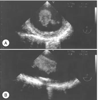
Her chest X-ray and electrocardiogram were normal. The diagnostic work-up to determine the source of the embolism included a transthoracic echocardiogram, which showed the possibility of vegetation on the aortic valve. On examination of the transesophageal echocardiogram, myxomatous changes of the aortic valve were verified. However, at that time, anot- her important lesion was also detected, which was a long floa- ting echogenic mass, with an irregular margin in the aortic arch(Fig. 1). This appeared to be attached to the posterior wall of the proximal aortic arch, extending to the left subcla- vian artery and descending aorta. Chest computed tomography showed a filling defect in the aortic arch(Fig. 2).
The laboratory data included: a white blood cell count of 6600×103/uL, hemoglobin 12.3 g/dL, platelets 235,000/uL, negative ANCA, negative ANA, antithrombin III 90%(nor- mal value: 75-125%), cardiolipin IgM antibody 0.4 MPL U/
mL(normal value <4 MPL U/mL), protein C 100%(normal value: 80-120%) and protein S 70%(normal value: 70- 123%).
She underwent surgery due to the risk of peripheral embo- lization. During the operation, a large thrombus, measuring more than 8 cm in length, was removed from the aortic arch,
Received:August 13, 2004 Revision Received:October 28, 2004 Accepted:December 1, 2004
Correspondence:Nam-Ho Kim, M.D.,Department of Internal Medicine, Wonkwang University School of Medicine, 344-2 Shinyong-dong, Iksan 570-711, Korea
Tel:82-63-850-1071, Fax:82-63-852-8480 E-mail:[email protected]
Sung Hee John, et al: Floating Thrombus in the Aorta·181
left subclavian artery and descending aorta(Fig. 3). A small amount of residual thrombus was removed piecemeal, super- ficially, from the roughened base of the posterior wall of the proximal aortic arch. The base of thrombus was dimpled, like an atheromatous lesion. An organized thrombus, without evi- dence of malignancy, was found on histopathological exami- nation. Two weeks later, after an uneventful recovery, she was discharged, with warfarin anticoagulation therapy.
Discussion
Most systemic embolisms are caused by thrombi in the left
side of the heart. Aortic thrombi, however, are another impor- tant cause of arterial thromboembolism.1) Some hypercoagu- lable states, e.g., primary polycythemia vera, antiphospholipid antibody syndrome, protein C deficiency, depressed activation of protein C and factor V Leiden deficiency, have been asso- ciated with aortic thrombi. Laperche et al.4) reported that 17%
of patients with a thrombosis of the aortic arch had evidence of a hematostatic disorder.
The presence of pedunculated thrombi in the thoracic aorta is a rare entity. These thrombi move freely in the aortic lumen with each cardiac cycle, and their fragmentation can lead to acute ischemic episodes due to peripheral, visceral or cerebral arterial embolization.5) Pathologic studies of the aortic wall in these patients have shown atheromatic lesions, often with minimal atherosclerotic plaques.6) The diagnosis of a hyper- coagulable disorder in our patient was excluded due to the negative coagulation profile and lack of previously unexplai- ned arterial or venous thromboses. The pathologic mechanism of the lesion in our case appeared to be due to the progressive development of an apposition thrombosis on an ulcerated pla- que.
Treatment was considered necessary due to the risk of a massive systemic embolization. Karalis et al.7) reported the incidence of embolic events in 73% of highly mobile aortic thrombi, compared with 12% of immobile ones. The optimal treatment of these pedunculated lesions remains undefined.
Thrombolysis of clots has been reported in the literature.8) Reber et al.9) however, discussed the potential danger of thrombolytic agents selectively lysing the stalk of peduncu- lated lesions, releasing the bulk of the lesion into the blood- stream, thus causing a massive embolization. Thrombolysis for pedunculated thrombi may be contraindicated. In selected A
B
Fig. 1. Transesophageal echocardiography demonstrates a large free-floating thrombus in the aortic arch in transverse (A) and longitudinal (B) views.
Fig. 2. Chest CT scan demonstrates the thrombus in the aortic arch. CT: computed tomographic.
Fig. 3. Organized thrombus. The base of the thrombus (arrow) and the floating end are shown.
182·Korean Circulation J 2005;35:180-182
patients, surgical treatment has been successful. Sadony et al.5) described the successful removal of thrombi using hypo- thermic circulatory arrest. In the present case, the thoracic aortic floating thrombus was located in the aortic arch, so a surgical removal of the thrombus was performed using hypo- thermic circulatory arrest. The exact surgical risk and long- term results are not known, although no embolization has recurred during the 13 months of follow-up.
REFERENCES
1) Panetta T, Thompson JE, Talkington CM, Garrett WV, Smith BL.
Arterial embolectomy: a 34-year experience with 400 cases. Surg Clin North Am 1986;66:339-53.
2) Rhee MY, Myong NH, Park YB. Primary intimal sarcoma of the aorta: role of transesophageal echocardiography. Circ J 2002;
66:111-3.
3) Hirata Y, Ono M, Morota T, Takamoto S. Recurrent embolism caused by floating thrombus originating from the ligamentum ar- teriosum. Eur J Cardiothorac Surg 2003;24:452-4.
4) Laperche T, Laurian C, Roudaut R, Steg PG. Mobile thromboses of the aortic arch without aortic debris: a transesophageal echo- cardiographic finding associated with unexplained arterial embo- lism. Circulation 1997;96:288-94.
5) Sadony V, Walz M, Lohr E, Rimpel J, Richter HJ. Unusual cause of recurrent arterial embolism: floating thrombus in the aortic arch surgically removed under hypothermic cardiocirculatory ar- rest. Eur J Cardiothorac Surg 1988;2:469-71.
6) Rubin BG, Allen BT, Anderson CB, Barzilai B, Sicard GA. An embolizing lesion in a minimally diseased aorta. Surgery 1992;
112:607-10.
7) Karalis DG, Chandrasekaran K, Victor MF, Ross JJ Jr, Mintz GS.
Recognition and embolic potential of intraaortic atherosclerotic debris. J Am Coll Cardiol 1991;17:73-8.
8) Hausmann D, Gulba D, Bargheer K, Niedermeyer J, Comess KA, Daniel WG. Successful thrombolysis of an aortic-arch thrombus in a patient after mesenteric embolism. N Engl J Med 1992;327:
500-1.
9) Reber PU, Patel AG, Stauffer E, Muller MF, Do DD, Kniemeyer HW. Mural aortic thrombi: an important cause of peripheral embolization. J Vasc Surg 1999;30:1084-9.