Update on Acute Aortic Syndrome
9
0
0
전체 글
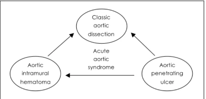
(2) 708·Korean Circulation J 2005;35:707-715. easily detected by MRI; the signal intensity of the thickened aortic wall might vary depending on the amount of methemoglobin formation within the hematoma. TEE is unique as it is the only technique directly used by attending cardiologists familiar with a patient’s medical history or condition needed for clinical decisions at the bedside. In 1994, Dr. R. Erbel’s group in Mainz University first reported the clinical usefulness of TEE for diagnosis of AIH.3) Since then, AIH and AAS have emerged as interesting topics of clinical investigators, especially cardiologists.4)5) TEE is very useful to demonstrate characteristic findings of AIH including circumferential or crescentic aortic wall thickening without an intimal tear as well as displacement of intimal calcification caused by the hemorrhage within the aortic media(Fig. 4). According to our data, the mean thickness(±SD) of type A AIH(n=48 patients) and type B AIH(n=103 patients) was 12.7±6.9 mm(range 540 mm) and 10.5±3.5 mm(range 5-23 mm), respec-. Classic aortic dissection Acute aortic syndrome. Aortic intramural. Aortic penetrating. hematoma. ulcer. Fig. 1. Schematic diagram showing the relationship among 3 differ-. ent entities of acute aortic syndrome. Arrows represent a potential progression from one entity to the other during clinical course.. A. B. tively. As the normal thickness of the aorta is less than 3 mm by any imaging modality, aortic wall thickness greater or equal to 5 mm seems to be adequate for diagnosis of AIH in patients with typical clinical symptoms suggesting AAS. TEE is superior to any imaging modality as it allows direct observation of the entire aortic wall. One characteristic TEE finding of AIH is the presence of an ‘echo-lucent area’ or ‘echo-free space’ within the thickened aortic wall(Fig. 5); the prevalence of this finding is reported to be higher than 60%. Demonstration of an ‘echo-lucent area’ or ‘echo-free space’ is quite unique or pathognomonic for AIH, and we believe it represents different patterns of liquefaction after the initial hemorrhage, which is useful for differential diagnosis from nonspecific aortic wall thickening due to other pathologies. Although some patients with a large ‘echo-free space’ detected in TEE also show contrast enhancement during CT, suggesting flow communication with a true lumen, the presence of an ‘echo-free space’ itself is not a poor prognostic sign and is not associated with future development of classic AD.6) It is generally believed that AIH is caused by rupture of the vasa vasorum,1) which then separates the medial wall layers eventually leading to a secondary tear or to a communication with the adventitial space. However, some sporadic case reports showing accidental development of typical AIH by percutaneous catheter manipulations, such as insertion of a balloon pump,7) coronary angioplasty,8) or catheter ablation of a left sided bypass tract(Fig. 6), support the presence of a ‘primary intimomedial tear’ in AIH. This hypothesis is also supported by the demonstration of a small intimal communicaD. E. C. F. Fig. 2. Representative images of classic dissection (A, B, C) and intramural hematoma (D, E, F). LA: left atrium, LV: left ventricle, Ao: aorta.. A. B. C. D. Fig. 3. Representative pre- and post-contrast enhancement computerized tomographic images of acute aortic intramural hematoma. In precontrast computed tomography (A, C) high attenuation areas along the aortic wall represent intramural hematoma, and no contrast enhancement is characteristic after injection of the contrast agent (B, D)..
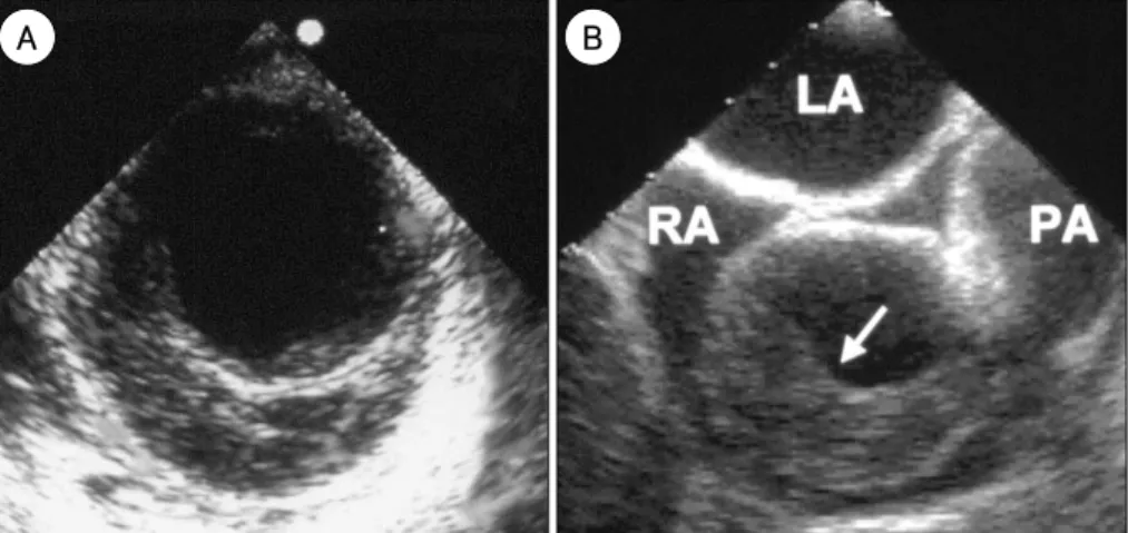
(3) Jae-Kwan Song: Update on Acute Aortic Syndrome·709. tion at the time of surgery.9)10) These observations raise the issue of the diagnostic ability of various noninvasive imaging modalities to identify an ‘intimal tear’, which is considered a critical criterion to differentiate AD from AIH. The only difference between AD and AIH might be whether the intimal tear is big enough to be detected by contemporary noninvasive imaging techniques. Further investigation is necessary to verify if the ‘micro-tear’ is an initiating event of AIH or just a decompression vent. AIH occurs predominantly in the elderly population and the incidence of AIH shows a single peak in the 7th decade. As the population increasingly ages and survival is prolonged despite hypertension, a major risk factor for the development of AAS, the incidence of AIH is expected to increase significantly. In earlier studies, it was reported that AIH accounts for 10 to 20% of patients with a clinical presentation of AAS. However, with the wide application of noninvasive imaging modalities in clinical practice and the resulting increased A. recognition of AIH, the relative incidence of AIH might increase. One interesting point is that there might be a geographical difference in the relative incidence of AIH. Table 1 summarizes the incidence of AIH versus AD reported in the literature. In western countries, the relative incidence of AIH ranges from 5.1 to 22.5%. In Japan and Korea, the incidence is higher than 25%. Table 1. Incidence of aortic dissection versus aortic intramural hematoma reported in the literature. Author. Country 11). Nienaber Germany Vaccari12) Italy USA Ganaha13) Spain Evangelista14). Duration AD (n) AIH (n) AIH (%) ’83-’93 ’91-’99 ’90-’00 ’90-’00. 170 166 663 234. 025 009 065 068. Sohn15) Korea ’95-’99 048 016 Japan ’88-’00 158 083 Kaji16)17) Japan ’90-’02 139 094 Moizumi18) Song Korea ’93-’03 212 159 AD: aortic dissection, AIH: aortic intramural hematoma. 12.8% 05.1% 09.1% 22.5% 25.0% 34.4% 40.3% 42.9%. B. Fig. 4. Representative transesophageal echocardiographic images of aortic intramural hematoma demonstrating crescentic aortic wall thickening. without intimal flap or flow communication in the descending (A) and ascending aorta (B). LA: left atrium, RA: right atrium, PA: pulmonary artery.. Fig. 5. Transesophageal echocardiographic images showing various patterns of an ‘echo-free space’ or ‘echo-lucent area’ in patients with aortic intramural hematoma, which do not show any evidence of flow communication in color Doppler mapping or contrast echocardiography..
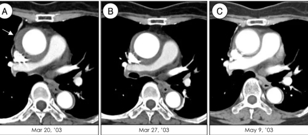
(4) 710·Korean Circulation J 2005;35:707-715. A. B. C. Mar 20, ’03. Mar 27, ’03. May 9, ’03. Fig. 6. A case of aortic intramural hematoma which developed during percutaneous catheter manipulation. A 72-year-old female underwent. radiofrequency catheter ablation of the left -sided bypass tract: during manipulation of the catheter to enter the left ventricle, she complained of sudden chest pain, and developed cardiac tamponade. After successful pericardiocentesis, computed tomography showed typical type A aortic intramural hematoma (A). A follow-up study revealed complete resorption of the hematoma with medical treatment (B & C). 30. Number of events. Number of events. 30. 20. 10. 20. 10. 0. A. 0 1. 4. 7. 10 13 16 Time (clock hour). 19. 22. B. 1. 4. 7. 10 13 16 Time (clock hour). 19. 22. Fig. 7. Circadian variation in onset of acute aortic syndrome (A) and acute myocardial infarction (B).. The relatively larger population of patients with poorly controlled or uncontrolled hypertension in the East might be one of the factors explaining this difference. Further study is necessary to determine whether this difference represents any racial or genetic heterogeneity. Contrary to AD, which more frequently affects the ascending aorta, AIH has a strong tendency to involve the distal aorta. According to recently reported data of the International Registry of Acute Aortic Dissection (IRAD), the total number of patients with AAS involving the proximal(type A) and distal(type B) aorta were 547 and 328, respectively. Among the 547 patients with type A AAS, 8.0%(44/547) were due to AIH.19)20) The prevalence of AIH in type B AAS was 15.6%(60/ 384). Based on our data of AAS from January 1993 to March 2003, AIH accounted for 27.4% of type A AAS (51/186) and 58.3% of type B AAS(108/185). Thus, the relative incidence of AIH affecting the distal aorta is about two times higher than that affecting the proximal aorta; there seems to be no geographical or racial difference of this tendency. Characteristic chronobiological patterns can be found in the occurrence of AAS. According to the analysis of 310 patients with AAS treated in our hospital, a significantly higher frequency of AAS occurred from 6:00. AM to 12:00 MD with a peak at 9:00 AM. The same pattern was also found in patients with acute myocardial infarction(Fig. 7).21) Another small peak was demonstrated at 9:00 PM, which was not found in patients with acute myocardial infarction and had a significant association with patients less than sixty years old with a history of hypertension. This finding may suggest the importance of more aggressive hypertension control in groups with a relatively more active social life style. Although no significant variation was found among the days of the week, the frequency of AIH was significantly higher during the winter months and contrary to acute myocardial infarction, AAS showed significantly lower frequency during the summer months(Fig. 8).21) There was no significant chronobiological difference between AIH and classic AD. These significant circadian and seasonal variations observed may have important implications for the prevention of AIH by tailoring treatment strategies to ensure maximal benefits during these vulnerable periods.. Recent Progress in the Treatment of AAS Aggressive medical treatment for proximal AIH In the past, when classic AD was considered as the.
(5) 50. 50. 40. 40 Number of events. Number of events. Jae-Kwan Song: Update on Acute Aortic Syndrome·711. 30 20 10. 20 10. 0. A. 30. 0 Jan. Mar. May Jul Sep Time (month). Nov. B. Jan. Mar. May Jul Sep Time (month). Nov. Fig. 8. Seasonal variation in onset of acute aortic syndrome (A) and acute myocardial infarction (B). Table 2. Natural history of aortic intramural hematoma with medical. treatment: follow-up imaging data21). Fig. 9. A case showing complete resorption of aortic intramural hem-. atoma involving the ascending aorta with medical treatment.. only disease entity of AAS, the most important factor determining the treatment strategy was the location of the affected aorta; if the proximal ascending aorta was involved, emergent surgery was recommended to prevent sudden death mostly due to aortic rupture and medical treatment was recommended for patients with distal AD, as the risk of aortic rupture was relatively low in this group. There has been considerable debate to determine whether the same principle should be applied for patients with AIH; for patients with distal AIH, the same medical treatment done for classic distal AD seems to be acceptable. However, the optimal treatment option for patients with proximal(type A) AIH still remains a challenging debate. As clinical symptomatology is very similar between AD and AIH, and rapid progression from AIH to classic AD or aortic rupture had been reported in earlier clinical studies, the same therapeutic strategies were recommended for patients with AIH and those with classic. Type A (n=38). Type B (n=82). No change Resorption. 00 (00%)0 24 (67%)0. 02 (03%) 54 (78%). Complete Partial Aggravation Development of AD. 16 (44%)0 08 (22%)0 03 (08%)0 09 (25%)0. 46 (67%) 08 (12%) 02 (03%) 11 (16%). Classic (type I/II/III) Localized AD: aortic dissection. 06 (2/3/1) 3. 10 11. AD.3)11) However, from a pathological viewpoint, AIH is different from AD in terms of the absence of an intimal tear or a continuous flow communication between the true and false lumen; this difference may have a different impact on the clinical course of patients with AIH. This hypothesis was proved in part by a report showing that, with the same medical treatment, AIH involving the distal descending thoracic aorta shows a much higher rate of complete resorption of the aortic pathology than typical AD.22) Table 2 summarizes the results of follow-up imaging studies in patients with AIH who received medical treatment only.23) Development of or progression to classic AD occurred in less than 25%, and more than 70% of patients showed near complete resorption or marked regression of the initial aortic wall thickening with medical treatment only, regardless of location of the affected aorta. These observations may suggest that aggressive medical treatment with surgical back-up and timed surgery in patients showing complications in follow-up imaging studies can be a rational option for patients with proximal or type 1 AIH. It is now widely accepted that AIH is not just a precursor of AD but has diverse remodeling processes after the initial event.24) With serial imaging studies, AIH was proved to have four different possible courses: persistent hematoma; reabsorbed hematoma, so that the appearance of the aortic wall returns to normal(Fig. 9);.
(6) 712·Korean Circulation J 2005;35:707-715. gery, and with medical treatment only, early mortality was very high. This is very similar to recently published IRAD data on patients with classic type A AD undergoing medical treatment only.32) However, other institutions in Spain, Japan, and Korea reported significantly lower mortality in a relatively high prevalence of patients without early surgical intervention undergoing medical treatment only. When aggressive medical treatment is selected for type A AIH, frequent follow-up imaging with surgical ‘back up’ is absolutely necessary, as wall configuration of AIH can change very rapidly, especially in patients with a very thick hematoma in the initial imaging study(Fig. 11).33) Development of an ulcer-like projection at follow-up imaging studies is an excellent predictor for progression to classic AD and close monitoring is warranted despite a stable clinical condition.. progression to aortic aneurysm; or, conversion to classic AD, with the development of a typical intimal flap and flow in a false lumen. Although development of classic AD from AIH with medical treatment is the most disastrous remodeling process, its frequency is less than 30%. Thus, risk stratification based on imaging, including identification of predictors for clinical events such as development of AD or hospital mortality, has been a hot issue. Hitherto, the two most important variables reported as useful predictors for development of adverse clinical events are aortic diameter and hematoma thickness(Fig. 10).25-27) These observations are important as they demonstrate that noninvasive imaging modalities including TEE is useful not only for correct differential diagnosis but for risk stratification and selection of treatment strategy. Although controversial, initial medical treatment for patients with type A AIH has been reported to have acceptable results that are not so significantly different from that of initial surgical intervention.25)28)29) A selective treatment strategy of initial medical treatment and timed surgery with frequent follow-up imaging studies has been proposed28) and is still in debate. Selection of treatment options and results with medical treatment in these patients vary significantly among institutions (Table 3). In Germany, most patients underwent sur-. Endovascular treatment Percutaneous stent-graft placement has been increasing recognized as an important and effective method for patients with acute and chronic AAS.34) Initially it was selectively applied for patients with chronic distal AD who developed local complications such as progressive aneurysmal changes resulting in hypoperfusion including claudication(Fig. 12). Poorly controlled hypertension due to a markedly collapsed true lumen with. 100. 100. B Hematoma thickness<11 mm. Event-free survival (%). Event-free survival (%). A. 75 p<0.05 50. 25. Hematoma thickness≥11 mm. 0. 5. 10. Maximal aortic diameter<48 mm 75 p<0.05 50. 25. Maximal aortic diameter≥48 mm. 0. 15. 5. Months Hematoma thickness <11 mm ≥11 mm. Patients at risk 12 13. 5 4. 10. 15. Months. 3 3. 2 3. Maximal aortic diameter. Patients at risk. <48 mm ≥48 mm. 6 3. 12 13. 4 2. 3 2. Fig. 10. Event-free survival curves of patients with type A aortic intramural hematoma who received initial medical treatment; maximal hematoma. thickness (cut off value of 11 mm, A) and aortic diameter (cut off value of 48 mm, B) were 2 important variables determining the event-free survival in this selected group. Table 3. Comparison of treatment option and results in patients with type A aortic intramural hematoma reported in the literature. Author. 12 38 13 22. Patients with medical treatment only 50.0% (16/12) 28.9% (11/38) 62.5% (18/13) 59.1% (13/22). In-hospital mortality without surgery 16.7% (1/16). 13 41 51. 84.6% (11/13) 51.2% (21/41) 68.6% (35/51). 27.3% (3/11) 14.8% (1/21) 18.6% (3/35). Study duration. Year published. Study design. Number of cases. Evangelista14) von Kodolitsch30) Sueyoshi29) Kaji25). ’90-’00 ’94-’00 ’84-’95 ’91-’97. 2003 2003 1997 1999. Single center Multicenter (4) Multicenter (2) Multicenter (2). Shimizu31) Moizumi18) Song. ’95-’99 ’90-’02 ’93-’03. 2000 2004 Submission. Single center Single center Single center. 54.5% (6/11) 12.5% (1/18) 17.7% (1/13).
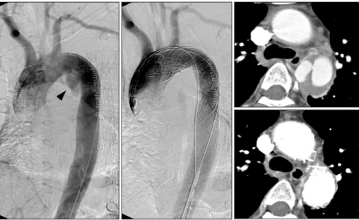
(7) Jae-Kwan Song: Update on Acute Aortic Syndrome·713. dilated false lumen was another good indication for this endovascular treatment option.35) After confirmation of feasibility and safety of this procedure, a more aggressive application to prevent adverse remodeling due to persistent intimal tear has been tried; some investigators applied this promising technique in the acute A. B. stage of distal AD for direct closure of intimal tear, which is believed to be helpful to prevent not only early but also late morbidity and mortality in these patients.36) However, there remain several unresolved issues including persistent leak after stent-graft insertion, ideal stent designs for patients with intimal tear very close to the C. D. Fig. 11. Two cases of aortic intramural hematoma showing very rapid changes of aortic wall configuration at the hyper acute stage. The case. presented in panels A and B shows a dramatic decrease of hematoma within 7 hours, whereas the case presented in C and D shows development of a typical aortic dissection within 12 hours after pain onset.. Fig. 12. A diagram showing percutaneous insertion of a stent-graft for distal aortic dissection to close the intimal tear.. Fig. 13. A case showing evolution of type B aortic intramural hematoma to localized dissection, in which endovascular treatment was performed. Aortogram and computed tomography showed complete normalization of descending thoracic aorta after successful stent-graft deployment and medical treatment..
(8) 714·Korean Circulation J 2005;35:707-715. origin of the subclavian artery, and long-term safety or durability. This treatment option seems to be especially useful for a localized ulcer or dissection(Fig. 13), which can develop during medical treatment of type B distal AIH. Stent-graft management for localized complications of type B AIH seems to be very beneficial for natural healing or remodeling process, and has almost replaced surgical management. For ideal candidate selection for stent-graft management, noninvasive imaging modalities, including TEE, provide invaluable information, such as the location, extent, and size of the localized ulcer or dissection. Monitoring of the descending thoracic lesion during the procedure is another advantage of TEE.. Conclusions For the past decade, we have witnessed dramatic changes in the traditional concept of AAS. These changes include diagnostic identification of AIH, risk stratification, and selection of treatment strategies based on information provided by imaging studies. In the therapeutic aspects, less invasive percutaneous stent-graft insertion has been more widely applied in this syndrome, and clinical application of this treatment option in relatively early stages of AAS to prevent adverse remodeling of the aorta which increases the risk of late mortality and morbidity seems to be promising. REFERENCES 1) Vilacosta I, Roman JA. Acute aortic syndrome. Heart 2001;85:. 365-8. 2) Yamada T, Tada S, Harada J. Aortic dissection without intimal. rupture: diagnosis with MR imaging and CT. Radiology 1988; 168:347-52. 3) Mohr-Kahaly S, Erbel R, Kearney P, Puth M, Meyer J. Aortic intramural hemorrhage visualized by transesophageal echocardiography: findings and prognostic implications. J Am Coll Cardiol 1994;23:658-64. 4) Kim HS, Yoon JH, Park SH, et al. Efficacy of transesophageal echocardiography in detecting aortic dissection. Korean Circ J 1992;22:105-12. 5) Kang DH, Song JK, Lim TH, et al. Clinical usefulness of transesophageal echocardiography in diagnosis of aortic dissection. Korean Circ J 1995;25:787-93. 6) Song JM, Kang DH, Song JK, et al. Clinical significance of echo-free space detected by transesophageal echocardiography in patients with type B aortic intramural hematoma. Am J Cardiol 2002;89:548-51. 7) Vilacosta I, Castillo JA, Peral V, et al. Intramural aortic hematoma following intra-aortic balloon counterpulsation: documentation by transesophageal echocardiography. Eur Heart J 1995; 16:2015-6. 8) Vilacosta I, de Dios RM, Pinto AG. Aortic intramural hematoma during coronary angioplasty: insights into the pathogenesis of intramedial hemorrhage. J Am Soc Echocardiogr 2000;13:403-6. 9) Harris KM, Braverman AC, Guitierrez FR, Barzilai B, DavilaRoman VG. Transesophageal echocardiographic and clinical. features of aortic intramural hematoma. J Thorac Cardiovasc Surg 1997;114:619-26. 10) Berdat PA, Carrel T. Aortic dissection limited to the ascending aorta mimicking intramural hematoma. Eur J Cardiothoracic Surg 1999;15:108-9. 11) Nienaber CA, von Kodolitsch Y, Petersen B, et al. Intramural hemorrhage of the aorta: diagnostic and therapeutic implications. Circulation 1995;92:1465-72. 12) Vaccari G, Caciolli S, Calamai G, et al. Intramural hematoma of the aorta: diagnosis and treatment. Eur J Cardiothoracic Surg 2001;19:170-3. 13) Ganaha F, Miller DC, Sugimoto K, et al. Prognosis of aortic intramural hematoma with and without penetrating ulcer: a clinical and radiological analysis. Circulation 2002;106:342-8. 14) Evangelista A, Dominguez R, Sebastia C, et al. Long-term followup of aortic intramural hematoma: predictors of outcome. Circulation 2003;108:583-9. 15) Sohn DW, Jung JW, Lee MM, et al. Should ascending aortic intramural hematoma be treated surgically? Am J Cardiol 2001; 87:1024-6. 16) Kaji S, Akasaka T, Horibata Y, et al. Long-term prognosis of patients with type A aortic intramural hematoma. Circulation 2002;106(Suppl I):I248-52. 17) Kaji S, Akasaka T, Katayama M, et al. Long-term prognosis of patients with type B aortic intramural hematoma. Circulation 2003;108(Suppl II):II307-11. 18) Moizumi Y, Komatsu T, Motoyoshi N, Tabayashi K. Clinical features and long-term outcome of type A and type B intramural hematoma of the aorta. J Thorac Cardiovasc Surg 2004; 127:421-7. 19) Mehta RH, Suzuki T, Hagan P, et al. Predicting death in patients with acute type A aortic dissection. Circulation 2002;105:200-6. 20) Suzuki T, Mehta RH, Ince H, et al. Clinical profiles and outcomes of acute type B aortic dissection in the current era: lessons from the international registry of aortic dissection (IRAD). Circulation 2003;108(Suppl II):II312-7. 21) Kim SD, Song JK, Park CB, et al. Chronobiological patterns of acute aortic syndrome: comparison with those with acute myocardial infarction. Korean Circ J 2004;34:970-7. 22) Song JK, Kang DH, Lim TH, et al. Different remodeling of descending thoracic aorta after acute event in aortic intramural hemorrhage versus aortic dissection. Am J Cardiol 1999;83: 937-41. 23) Song JK, Kim HS, Song JM, et al. Outcomes of medically treated patients with aortic intramural hematoma. Am J Med 2002;113: 181-7. 24) Song JK, Kim HS, Song JM, et al. Multicenter longitudinal follow-up clinical study comparing the natural course of medically treated patients with aortic dissection and aortic intramural hematoma. Korean Circ J 2001;31:592-601. 25) Kaji S, Nishigami K, Akasaka T, et al. Prediction of progression or regression of type A aortic intramural hematoma by computed tomography. Circulation 1999;100(Suppl II):II281-6. 26) Song JM, Kim HS, Song JK, et al. Usefulness of the initial noninvasive imaging study to predict adverse outcomes in the medical treatment of acute type A aortic intramural hematoma. Circulation 2003;108(Suppl II):II324-8. 27) Sueyoshi E, Imada T, Sakamoto I, Matsuoka Y, Hayashi K. Analysis of predictive factors for progression of type B aortic intramural hematoma with computed tomography. J Vasc Surg 2002;35:1179-83. 28) Song JK, Kim HS, Kang DH, et al. Different clinical features of aortic intramural hematoma versus dissection involving the.
(9) Jae-Kwan Song: Update on Acute Aortic Syndrome·715 ascending aorta. J Am Coll Cardiol 2001;37:1604-10. 29) Sueyoshi E, Matsuoka Y, Sakamoto I, Uetani M, Hayashi K,. Narimatsu M. Fate of intramural hematoma of the aorta: CT evaluation. J Comput Assist Tomogr 1997;21:931-8. 30) von Kodolitsch Y, Csosz SK, Koschyk DH, et al. Intramural hematoma of the aorta: predictors of progression to dissection and rupture. Circulation 2003;107:1158-63. 31) Shimizu H, Yoshino H, Udagawa H, et al. Prognosis of aortic intramural hemorrhage compared with classic aortic dissection. Am J Cardiol 2000;85:792-5. 32) Evangelista A, Mukherjee D, Mehta RH, et al. Acute intramural hematoma of the aorta: a mystery in evolution. Circulation 2005;. 111:1063-70. 33) Song JK. Diagnosis of aortic intramural hematoma. Heart 2004;. 90:368-71. 34) Dake MD. Aortic intramural hematoma: current therapeutic. strategy. Heart 2004;90:375-8. 35) Nienaber CA, Fattori R, Dieckmann C, et al. Nonsurgical recon-. struction of thoracic aortic dissection by stent-graft placement. N Engl J Med 1999;340:1539-45. 36) Dake MD, Kato N, Mitchell RS, et al. Endovascular stent-graft placement for the treatment of acute aortic dissection. N Engl J Med 1999;340:1546-52..
(10)
수치

+3

관련 문서
In addition to the problem of this bias, the problem caused by using the time variable with a weighted wage rate may be a multicollinearity between time value (=time x
Based on the results, a study model was developed and a hypothesis that transformational and transactional leadership would significantly affect organizational
In addition, Wallace and Froum 20) reported that the bone formation rate was superior in the cases using a barrier membrane in comparison with the
Fifth, the result of the verification of the hypothesis were hypothesis1;, hair that had used treatment had better setting capabilities and preservation rate than
This study also shows that real exchange rate misalignment could have a positive impact on export performance in China.. With its implications on
This study was conducted under the hypothesis that a group art therapy program may increase emotional expression and decrease life event stress in patients with
The result of this study on the intervention of function words in children with language development delay shows that intervention of function words by grammatically
At the end of the study, a reevaluation of each study case was performed with the same questionnaire. The result shows there has been a meaningful result in the group
