Cytoprotective Effects of Schisandrin A against Hydrogen Peroxide-induced Oxidative Stress in SW1353 Human Chondrocytes
Jin-Woo Jeong1,2, Eun Ok Choi1,2, Da Hye Kwon1,2, Bum Hoi Kim3, Dong Il Park4, Hye Jin Hwang2,5, Byung Woo Kim2,6 and Yung Hyun Choi1,2*
1Open Lab. for Muscular & Skeletal Disease Control and Department of Biochemistry and Dongeui University College of Korean Medicine, Busan 47227, Korea
2Anti-Aging Research Center and Blue Bio Industry RIC, Dongeui University, Busan 47340, Korea
3Department of Anatomy, Dongeui University College of Korean Medicine, Busan 47227, Korea
4Department of Internal Medicine, Dongeui University College of Korean Medicine, Busan 47227, Korea
5Department of Food and Nutrition, College of Nursing, Healthcare Sciences & Human Ecology, Dongeui University, Busan 47340, Korea
6Biopharmaceutical Engineering Major, Division of Applied Bioengineering, College of Engineering, Dongeui University, Busan 47340, Korea Received July 12, 2017 /Revised September 8, 2017 /Accepted September 8, 2017
Chondrocyte apoptosis induced by reactive oxygen species (ROS) plays an important role in the pathogenesis of osteoarthritis. Schisandrin A, a bioactive compound found in fruits of the Schisandra genus, has been reported to possess multiple pharmacological and therapeutic properties. Although several studies have described the antioxidant effects of analogues of schisandrin A, the underlying molecular mechanisms of this bioactive compound remain largely unresolved. The present study in- vestigated the cytoprotective effect of schisandrin A against oxidative stress (hydrogen peroxide [H2O2]) in SW1353 human chondrocyte cells. The results showed that schisandrin A preconditioning significantly inhibited H2O2-induced growth inhibition and apoptotic cell death by blocking the degra- dation of poly (ADP-ribose) polymerase proteins and down-regulating pro-caspase-3. These anti- apoptotic effects of schisandrin A were associated with attenuation of mitochondrial dysfunction and normalization of expression changes of proapoptotic Bax and antiapoptotic Bcl-2 in H2O2-stimulated SW1353 chondrocytes. Furthermore, schisandrin A effectively abrogated H2O2-induced intracellular ROS accumulation and phosphorylation of histone H2AX at serine 139, a widely used marker of DNA damage. Thus, the present study demonstrates that schisandrin A provides protection against H2O2-induced apoptosis and DNA damage in SW1353 chondrocytes, possibly by prevention of ROS generation. Collectively, our data indicate that schisandrin A has therapeutic potential in the treatment of oxidative disorders caused by overproduction of ROS.
Key words : Antioxidant, apoptosis, DNA damage, ROS, schisandrin A
*Corresponding author
*Tel : +82-51-850-7413, Fax : +82-51-853-4036
*E-mail : choiyh@deu.ac.kr
This is an Open-Access article distributed under the terms of the Creative Commons Attribution Non-Commercial License (http://creativecommons.org/licenses/by-nc/3.0) which permits unrestricted non-commercial use, distribution, and reproduction in any medium, provided the original work is properly cited.
Journal of Life Science 2017 Vol. 27. No. 9. 1070~1077 DOI : https://doi.org/10.5352/JLS.2017.27.9.1070
서 론
Schisandra 속 식물에 속하는 열매인 오미자는 오랫동안 우리나라를 포함한 아시아 지역에서 다양한 질병의 예방 및 치료 목적으로 널리 사용되어왔다. 오미자에 포함되어 있는 생리 활성물질로서 다양한 lignan 성분들이 보고되어 왔으며, schisandrin A는 dibenzocyclooctadiene 유도체 중의 하나이 다[4, 27]. 최근 연구에 의하면 schisandrin A는 대표적인 염증 매개인자인 prostaglandin E2의 생성을 억제함이 보고되었는
데, 이는 cyclooxygenase-2의 발현 차단과 연계성이 있었다 [13]. 또한 schisandrin A는 포도당 결핍 손상에 의한 중추 신 경계 질환 발생과 L-glutamate 유도 신경 독성에 예방 효능이 있으며[15, 19], 산소-포도당 결핍 reoxygenation 모델에서 대 뇌피질 보호 효과가 있음이 알려졌다[26]. 아울러 schisandrin A는 소교세포에서 lipopolysaccharide (LPS)에 의해 유도된 nitric oxide와 염증성 cytokine인 tumor necrosis factor-α 및 interleukin-6의 생산을 감소시킴으로써 염증 유발성 신경 손 상을 억제하는 것으로 보고되었고[24], 이와 유사하게 이 화합 물은 LPS가 자극된 대식세포에서도 항염증 활성을 나타내었 으며, 특히 glutathione S-transferase 활성을 증가시키고 부종 을 억제하며 항산화 효능도 있음이 알려졌다[17]. 그리고 schi- sandrin A와 유사한 구조를 가지는 lignan 계열 물질들이 활성 산소종(reactive oxygen species, ROS)의 생성을 억제하고, 대 표적인 항산화 유전자인 nuclear factor (erythroid-derived 2)-like 2의 발현을 증대시킴으로서 항산화 효능을 가질 것이
- Note -
라는 보고들이 있어왔으나[2, 3, 7, 10, 18, 28], schisandrin A의 항산화 활성에 대한 연구는 여전히 미비한 실정이다.
한편 퇴행성 관절염(골관절염, osteoarthritis)은 세계에서 가장 흔한 합병증 중 하나이며, 고령 인구에서 삶의 질을 저하 시키는 주요 원인이다[20]. 퇴행성 관절염은 연골세포(chon- drocytes), aggrecan 및 II 형 collagen을 비롯한 관절 연골 성분 의 파괴로 인한 관절 질환의 한 유형이다[11]. 특히 퇴행성 관 절염은 연골세포의 손상에 따른 세포 외 기질의 anabolism과 catabolism 사이의 균형 파괴가 주요한 원인으로 인식되고 있 다[11]. 이러한 연골세포의 손상은 과도한 염증 반응을 포함한 다양한 원인에 의하여 발생하지만, 산화적 스트레스에 의한 연골세포의 세포사멸(apoptosis) 유도 역시 주요한 요인으로 작용한다[1, 14]. 특히 과도하게 생성된 ROS 또는 자유 라디칼 에 의해 유도된 산화적 스트레스는 퇴행성 관절염의 병인 발 생과 관련된 주요 원인이다[14, 25]. 또한, 연골세포의 고사와 관절 연골 기능의 저하는 ROS 생산의 증가에 의해 유발 될 수 있으며 연골의 노화와 퇴행성 관절염의 병인에 기여할 수 있다[12, 25]. 따라서 산화적 스트레스에 대한 연골세포의 보호 작용을 가지는 물질들은 퇴행성 관절염의 예방 및 치료에 유 효한 효과를 줄 것으로 예측할 수 있다. 본 연구에서는 퇴행성 관절염 제어에 효과적인 약물의 발굴을 위한 연구의 일환으로 산화적 스트레스(hydrogen peroxide, H2O2)에 노출된 연골세 포(SW1353 human chondrocytes)에서 schisandrin A의 보호 효과와 항산화 효능을 평가하였다.
재료 및 방법
세포배양 및 schisandrin A 처리
본 연구에서 사용된 SW1353 연골세포는 American Type Culture Collection (Manassas, VA, USA)에서 구입하였으며, 10% fetal bovine serum (FBS), 100 U/ml의 penicillin 및 100 mg/ml의 streptomycin이 함유된 Dulbecco-modified Eagle medium (DMEM, Gibco-BRL, Grand Island, NY, USA)에서 37℃ 및 5% CO2의 조건에서 배양하였다. Schisandrin A는 Sigma-Aldrich Chemical Co. (St. Louis, MO, USA)에서 구입 하였으며, dimethyl sulfoxide (DMSO, Sigma-Aldrich Chemical Co.)에 100 mM로 stock solution을 만든 후 DMEM에 적정 농도로 희석하여 처리하였다
세포 생존률 측정
산화적 스트레스에 의한 세포독성에 미치는 schisandrin A 의 영향을 조사하기 위하여 SW1353 세포에 적정 농도의 H2O2
(Sigma-Aldrich Chemical Co.)를 24시간 처리하거나, schisan- drin A를 1시간 전처리 후 H2O2를 24시간 처리하였다. 준비된 세포에 3시간 동안 0.5 mg/ml의 3-(4,5-dimethylthiazol-2- yl)-2,5-diphenyltetrazolium bromide (MTT, Sigma-Aldrich
Chemical Co.)를 처리하여, 형성된 formazan을 DMSO에 용 해시킨 후, enzyme-linked immunosorbent assay (ELISA) reader (Molecular Devices, Sunnyvale, CA, USA)를 이용하여 540 nm에서 흡광도 변화를 측정하여 대조군에 대한 세포 생존 율을 백분율로 표시하였다.
세포 핵의 형태 관찰
Apoptosis가 유발된 세포에서 특이적으로 나타나는 핵의 형태적 변화를 관찰하기 위하여 다양한 조건에서 배양된 세포 를 phosphate-buffered saline (PBS)로 2회 수세 후 0.1 M phosphate buffer (pH 7.2)에 희석된 3.7% paraformaldehyde 고정액(Sigma-Aldrich Chemical Co.)을 1시간 처리하였다. 고 정된 세포를 수회 수세한 후 0.05 M phosphate buffer (pH 7.2)에 2.5 μg/ml의 농도로 희석된 4,6-diamidino-2-phenyl- indole (DAPI, Sigma-Aldrich Chemical Co.) 용액으로 상온 암하에서 20분 염색하였다. 이를 다시 PBS로 수회 수세하여 건조시킨 후 형광현미경(fluorescene microscope, Carl Zeiss) 하에서 핵의 형태를 비교하였다.
단백질의 분리 및 Western blot analysis
Schisandrin A의 산화적 스트레스 보호에 따른 특정 유전자 들의 발현 변화 관찰을 위하여 준비된 세포에 적당량의 lysis buffer [25 mM Tris-Cl (pH 7.5), 250 mM NaCl, 5 mM ethyl- enediaminetetraacetic acid, 1% Nonidet-P40, 1 mM pheny- methylsulfonyl fluoride, 5 mM dithiothreitol]를 첨가하여 4℃에서 1시간 이상 반응시켰다. 상층액에 있는 단백질을 분 리한 후 동량의 단백질을 sdium dodecyl sulfate (SDS)-poly- acrylamide gel을 이용하여 전기영동을 실시하였다. Gel에 함 유된 단백질을 polyvinylidene difluoride membrane (Schleicher and Schuell, Keene, NH, USA)으로 전이시키고 membrane을 5% skim milk 용액에 1시간 처리하여 비특이적인 단백질들에 대한 blocking을 실시하였다. 그리고 Santa Cruz Biotechnol- ogy, Inc. (Dallas, TX, USA) 및 Cell Signaling Technology, Inc. (Danvers, MA, USA)에서 구입한 적정 1차 항체를 처리하 여 상온에서 2시간 이상 반응시킨 후, PBS-T 용액(PBS with Tween 20)으로 세척하고 2차 항체를 상온에서 1시간 정도 반 응시켰다. 반응이 끝난 후 암실에서 enhanced chemilumine- sence (ECL) solution (Santa Cruz Biotechnology Inc.)을 적용 시킨 다음 X-ray film에 감광시켜 특정 단백질의 발현 변화를 분석하였다.
DNA flow cytometry에 의한 apoptosis 유도의 정량적 분석
Apoptosis가 유발된 세포의 정량적 분석을 위해 준비된 세 포를 고정하고 염색하기 위하여 Cycle TEST PLUS DNA REAGENT Kit (Becton Dickinson, San Jose, CA, USA)를 이
A
B
C
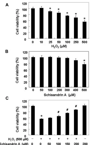
Fig. 1. Effect of schisandrin A on cell viability in H2O2-treated SW1353 cells. The cells were incubated with the in- dicated concentrations of H2O2 and schisandrin A for 24 hr (A and B) or 50-200 μM schisandrin A in the presence or absence of 500 μM H2O2 for 24 hr (C). The cell viability was assessed by the MTT reduction assay, and the re- sults are expressed as the percentage of surviving cells over the control cells (no addition of schisandrin A and H2O2). Values represent the means ± SD of three in- dependent experiments (*p<0.05 compared to the un- treated control. #p<0.05 compared to the to the H2O2
treated group).
용하였다. 이들 세포를 고정 후 4oC, 암실에서 30분 동안 pro- pidium iodide (PI) 용액에 반응을 시킨 후, 실험군당 최소 10,000개 이상의 세포를 flow cytometry (Becton Dickinson)에 적용시켜 세포 내 DNA 함량에 따른 histogram을 대상으로 sub-G1기에 속하는 세포를 apoptosis가 유발된 세포로 산출하 였다.
Mitochondrial membrane potential (MMP, Δψm) 변 화의 분석
세포 내 미토콘드리아 기능 손상의 여부를 확인하기 위하여 MMP 값 변화 정도를 측정하였다. 이를 위하여 다양한 조건에 서 배양된 세포들에 10 μM의 5,5‘,6,6’-tetrachloro-1,1‘,3,3’-tet- raethyl-imidacarbocyanine iodide (JC-1, Sigma-Aldrich Chemical Co.) 용액을 처리하여 암하, 상온에서 20분 동안 반응시켰다.
반응이 끝난 후 상층액을 제거하고 PBS를 첨가하여 세포를 부유시킨 다음 flow cytometer에 적용시켜 MMP의 변화를 측 정하였다.
ROS 생성변화 측정
세포 내 ROS의 생성 변화 정도를 확인하기 위하여 준비된 세포들을 fluorescent probe 인 10 μM의 2‘,7’-di-chlorodihy- drofluorescein diacetate (DCF-DA, Molecular Probes, Leiden, Netherlands)로 20분간 염색 후 flow cytometer를 사용하여 분석을 하였다.
통계 분석
실험 결과들의 유의성을 검정하기 위하여 분산분석(ANOVA) 을 실시한 후 p<0.05 수준에서 Duncan's multiple range tests 를 실시하였으며, 그 결과는 평균(mean) ± 표준편차(standard deviation, SD)로 표시하였다.
결과 및 고찰
산화적 스트레스에 의한 SW1353 연골세포의 증식억제에 미치는 schisandrin A의 영향
SW1353 연골세포 모델에서 산화적 스트레스에 미치는 schisandrin A의 영향을 조사하기 위한 실험 조건의 설정을 위하여 H2O2 및 schisandrin A가 SW1353 연골세포의 생존에 미치는 영향을 먼저 조사하였다. 이를 위하여 다양한 농도의 H2O2 (10~500 μM) 및 schisandrin A (50~500 μM)가 처리된 조건에서 24시간 배양 후 MTT assay를 실시한 결과, Fig. 1A 에 나타낸 바와 같이 H2O2 처리 농도의 증가에 따라 SW1353 연골세포의 생존율이 유의적으로 감소되어, 생존 세포의 백분 율은 250 μM 및 500 μM 처리군에서 각각 70% 및 56% 정도를 보였다. Schisandrin A의 24시간 처리 조건에서는 400 μM 이 하의 농도에서 유의적인 변화를 보이지는 않았지만, 500 μM
처리군에서 약 76% 정도의 생존율을 나타내었다(Fig. 1B). 따 라서 500 μM의 H2O2를 산화적 스트레스 조건으로 설정하였 으며, 이에 따른 schisandrin A의 세포독성 보호 효과를 조사 하였다. Fig. 1C에 나타낸 바와 같이 H2O2를 처리하기 1시간 전에 schisandrin A를 노출시켰을 경우, H2O2 단독 처리군에 비하여 높은 세포 생존률(100 μM 및 200 μM의 schisandrin A 동시 처리군에서 약 76% 및 87%)을 보여 schisandrin A는 H2O2 처리에 의한 세포 생존율 저하를 향상시킬 수 있었음을 알 수 있었다. 따라서 모든 후속 실험은 200 μM의 schisandrin A로 1시간 전처리 한 후 500 μM의 H2O2에 24시간 노출시킨 세포를 대상으로 수행했다.
A
B
C
D
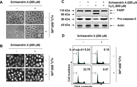
Fig. 2. Protection against H2O2-induced apoptosis by schisandrin A in SW1353 cells. The cells were pre-treated with 200 μM schisandrin A for 1 hr and then incubated without or with 500 μM H2O2 for 24 hr. (A) Cellular morphological changes were monitored by obtaining photomicrographs under an inverted phase contrast microscope (x200 original magnification). (B) The cells were fixed and stained with DAPI solution. The stained nuclei were observed under a fluorescence microscope (x400 original magnification). (B) Cellular proteins were separated on SDS-polyacrylamide gels and were subsequently transferred onto membranes. The membranes were probed with an antibodies against PARP and caspase-3. Proteins were visualized using an ECL detection system. Actin was used as an internal control. (D) To quantify the degree of apoptosis, the cell cycle analysis was performed using flow cytometry, and the cell percentages of the sub-G1 peaks in the cell cycle were analyzed.
Data are presented as mean of two independent experiments.
산화적 스트레스에 의한 SW1353 연골세포의 apoptosis 유도에 미치는 schisandrin A의 영향
이상에서 관찰된 schisandrin A의 산화적 스트레스에 대한 SW1353 연골세포의 보호 효과가 apoptosis 유도 억제와 연관 성이 있는지의 여부를 조사하였다. 이를 위하여 apoptosis가 일어난 세포의 핵에서 특이적으로 관찰되는 염색질의 응축 (chromatin condensation)에 따른 apoptotic body의 형성에 미치는 영향을 조사하였다. Fig. 2B에 나타낸 바와 같이, H2O2
만이 처리된 SW1353 연골세포에서는 DAPI 염색에 의한 전형 적인 핵의 형태 변화가 관찰되었으나, schisandrin A 전처리군 에서는 이러한 현상을 거의 관찰할 수 없었다. 이는 또한 H2O2
처리에 의한 세포의 전체적인 형태 변화가 schisandrin A 전처 리에 의하여 거의 대조군 수준으로 유지되었던 결과와 잘 일 치되는 현상이다(Fig. 2A).
한편 산화적 스트레스에 의한 연골세포의 사멸은 caspase 의 활성과 연관되어 있으며, caspase 활성 의존적인 apoptosis 유발에서는 effector caspase의 활성에 따른 poly (ADP-ri- bose) polymerase (PARP)와 같은 기질 단백질의 분해가 동반 된다[23]. 따라서 schisandrin A의 산화적 스트레스 억제 효능 이 apoptosis 유발의 억제에 의한 것인지를 확인하기 위하여
PARP의 발현 변화를 관찰한 결과, H2O2만이 단독 처리된 조 건에서 배양된 SW1353 연골세포에서는 전형적인 PARP 단편 화 현상이 관찰되었으며, 이러한 현상은 schisandrin A가 전처 리된 조건에서는 대조군 수준으로 억제되었다(Fig. 2C). 아울 러, PARP의 단편화와 연관된 대표적인 effector caspase인 caspase-3 [22]의 불활성형 발현도 H2O2 단독 처리군에서는 뚜렷한 감소 경향을 보였으나, schisandrin A의 전처리군에서 는 대조군 수준으로 유지되었다(Fig. 2C). 이러한 schisandrin A의 산화적 스트레스에 의한 SW1353 연골세포의 apoptosis 유도 억제 여부를 정량적으로 분석하기 위하여 flow cytome- try 분석을 수행한 결과, H2O2가 단독 처리된 세포에서는 apoptosis 유발군에 해당하는 sub-G1기에 속하는 세포의 빈도 가 약 22.75% 정도임에 비하여 schisandrin A의 전처리군에서 는 약 8.47% 정도로 나타났다. 그리고 이러한 전반적인 실험 과정에서 schisandrin A 단독 처리군에서는 대조군과 유사하 게 유의적인 특별한 차이점들은 관찰할 수 없었다. 따라서 이 상의 결과는 산화적 스트레스에 의하여 유도되는 SW1353 연 골세포의 생존율 저하는 apoptosis 유도에 의한 것이며, schi- sandrin A는 apoptosis 유도에 핵심이 되는 caspase 활성 경로 를 차단함으로서 이를 효과적으로 억제하였음을 알 수 있었다.
A
B
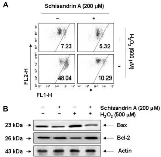
Fig. 3. Effects of schisandrin A on the MMP values and the expression in H2O2-treated SW1353 cells. The cells were pre-treated with 500 μM schisandrin A for 1 hr and then stimulated with and without 500 μM H2O2 for 24 hr.
(A) The cells were collected and incubated with 10 μM JC-1 for 20 min at 37oC in the dark. The cells were then washed with PBS and analyzed using a flow cytometer.
Data are presented as mean of two independent experi- ments. (B) The cells were lysed, and the aliquots con- taining the total proteins were subjected to SDS-poly- acrylamide gels followed by immunoblot analysis with anti-Bax and anti-Bcl-2 antibodies. Actin was used as an internal control.
산화적 스트레스에 의한 SW1353 연골세포의 미토콘드리 아 기능 손상에 미치는 schisandrin A의 영향
선행 연구들에 의하면 산화적 스트레스에 따른 연골세포의 apoptosis 유도는 미토콘드리아의 기능적 손상에 따른 in- trinsic apoptosis 경로의 활성과 직접적인 관련이 있는 것으로 알려져 있다[5, 23]. 이러한 미토콘드리아 기능 손상과 연계된 apoptosis 유도 조절에 가장 중요하게 작용하는 유전자군이 Bcl-2 family이며, Bcl-2 family에 속하는 단백질은 apoptosis 를 유도하거나 억제하는 인자들로 구성되어 있다[9]. 현재 가 장 대표적인 미토콘드리아의 기능 측정 방법은 MMP 값의 변화 여부를 조사하는 것이며, MMP 값의 저하는 미토콘드리 아의 기능이 손상되었음을 나타내는 지표이다[16]. 따라서 산 화적 스트레스에 의한 SW1353 연골세포의 apoptosis 유도에 미토콘드리아 기능 손상이 연계되어 있는지, 그리고 schisan- drin A가 이를 차단할 수 있는지의 여부를 조사하였다. Fig.
3A에 나타낸 바와 같이, 대조군과 schisandrin A 단독 처리군 에 비하여 H2O2가 함유된 배지에서 배양된 SW1353 연골세포 에서의 MMP 손상 정도는 48% 정도 나타나 대조군에 비하여 약 7배 증가되었지만, schisandrin A 전처리 조건군에서는
10% 정도를 보였다. 아울러 H2O2의 처리에 의하여 증가되었 던 미토콘드리아 기능 손상을 유발하는 pro-apoptotic Bax의 발현이 증가된 반면에, anti-apoptotic Bcl-2의 발현은 감소되 었으나 schisandrin A의 전처리에 의해서는 두 Bcl-2 family 단백질들의 발현이 대조군 수준으로 유지되었다(Fig. 3B). 이 러한 결과들은 schisandrin A가 H2O2에 의한 Bcl-2 family 인 자들의 발현 변화를 억제함으로서 미토콘드리아 기능 손상을 차단시켜 apoptosis 유도를 억제하였음을 보여 주는 것으로 생각된다.
산화적 스트레스에 의한 SW1353 연골세포의 DNA 손상 에 미치는 schisandrin A의 영향
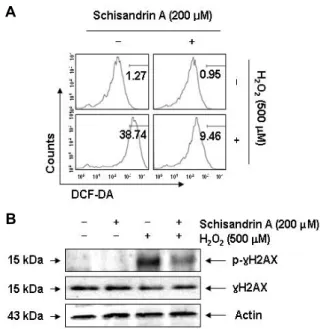
다음은 이상에서 관찰된 schisandrin A의 산화적 스트레스 방어 효과가 ROS의 생성 및 DNA 손상 억제와 직접적인 연관 성이 있는지의 여부를 조사하였다. 이는 세포 내에서 생산되 는 ROS의 대부분은 미토콘드리아 기능 손상과 연관되며, 이 로 인한 apoptosis 유발은 또한 DNA 손상과 연계되기 때문이 다[6, 8]. 이를 위하여 H2O2가 처리된 SW1353 연골세포를 대상 으로 DCF-DA 염색을 통하여 ROS의 생성 여부를 조사한 결 과, H2O2 처리 30분 이내에 ROS의 생성이 증가하기 시작하여, 6시간 경과 후 최고치를 나타낸 후 다시 감소되었다(data not shown). 그러나 schisandrin A를 1시간 전처리 후 H2O2를 6시 간 처리하였을 경우 대조군 수준까지는 아니지만 현저하게 감소되어 schisandrin A가 강력한 항산화 효능을 가지고 있음 을 확인할 수 있었다(Fig. 4A). 따라서 schisandrin A의 항산화 효능이 산화적 스트레스에 의한 DNA 손상 차단과의 연관성 이 있는지를 확인하기 위하여 DNA 이중 나선 손상 지표인 γH2AX 단백질의 인산화(serine 139) [21]에 미치는 schisan- drin A의 영향을 조사하였다. Fig. 4B의 immunoblotting 결과 에 의하면, H2O2에 노출된 SW1353 연골세포에서 γH2AX 단 백질의 전체적인 발현에는 큰 변화 없이 인산화가 매우 증가 되었으나, schisandrin A 전처리된 세포에서는 이러한 현상을 관찰할 수 없었다. 따라서 schisandrin A에 의한 DNA 손상의 방어 효과는 ROS 생성의 차단과 직접적인 연관성이 있음을 알 수 있었다.
본 연구에서는 퇴행성 관절염의 예방과 치료에 효과적인 약물의 발굴을 위하여 SW1353 연골세포 모델을 대상으로 오 미자에서 유래된 lignan 계열 물질인 schisandrin A의 산화적 손상 방어 효능을 검증하였다. Schisandrin A는 H2O2에 의한 SW1353 세포의 생존율 저하를 유의적으로 차단하였으며, 이 는 apoptosis 유도 억제에 의한 것임을 알 수 있었다. 이러한 결과는 오미자 추출물 또는 schisandrin A와 동일 lignan 계열 물질들이 다양한 실험모델에서 세포 보호 효과가 있어왔다는 결과들을 잘 뒷받침하여 주는 결과이다. 예를 들어 schisan- drin A는 쥐의 피질 뉴런의 일차 배양에서 산소 및 포도당 결핍과 재관류에 의해 유발된 손상에 대한 보호 효과가 있었
A
B
Fig. 4. Protection of H2O2-induced ROS generation and DNA damage in SW1353 cells. The cells were pre-treated with 500 μM schisandrin A for 1 hr and then stimulated with and without 500 μM H2O2 for 6 hr (A) or 24 hr (B). (A) In order to monitor ROS production, the cells were in- cubated at 37˚C in the dark for 20 min with culture me- dium containing 10 μM DCF-DA. ROS generation was evaluated using a flow cytometer. Data are the mean of two different experiments. (B) The cells were lysed and then equal amounts of cell lysates were separated on SDS-polyacrylamide gels and transferred to mem- branes. The membranes were probed with specific anti- bodies against p-γH2AX, γH2AX, and actin as an in- ternal control, and the proteins were visualized using an ECL detection system.
으며[26], glucose 결핍에 의한 신경세포의 보호 효과 효능도 보고된 바 있다[19]. 또한 schisandrin A와 유사한 lignan 계열 물질들에서도 Parkinson 병과 연관된 신경 손상[2], 심혈관 질 환 유발 관련 세포[4] 및 UV-B에 의한 피부세포[10] 보호 효과 등이 보고된 바 있어, 오미자 구성 물질들이 광범위한 세포독 성의 차단효과가 있음을 알 수 있다. 아울러 schisandrin A는 미토콘드리아 기능 손상을 보호하였으며, 이로 인한 ROS의 생성을 억제하였다. 다양한 인체 질환의 발병과 악화가 산화 적 스트레스 및 염증성 반응을 동반한다는 측면에서 이러한 schisandrin A의 항산화 효능은 항염증 효능[17, 24]과 함께 인체 항상성 유지를 위한 기능성 소재로서의 가치가 매우 높 을 수 있음을 의미한다. 비록 향후 다양한 모델에서 schisan- drin A의 효능에 대한 추가적인 연구가 필요하지만, 본 연구의 결과는 schisandrin A의 연골세포의 보호 기능은 항산화 효능 에 의한 것임을 알 수 있었다.
감사의 글
본 결과물은 농림축산식품부의 재원으로 농림수산식품기 술기획평가원의 고부가가치식품 기술개발사업(No. 314043-3) 및 산업통상자원부ㆍ부산광역시 지원 지역혁신센터사업(RIC08- 06-07) 동의대학교 블루바이오 소재개발 및 실용화 지원센터 의 지원으로 이루어졌음.
References
1. Asada, S., Fukuda, K., Nishisaka, F., Matsukawa, M. and Hamanisi, C. 2001. Hydrogen peroxide induces apoptosis of chondrocytes; involvement of calcium ion and ex- tracellular signal-regulated protein kinase. Inflamm. Res. 50, 19-23.
2. Ba, Q., Cui, C., Wen, L., Feng, S., Zhou, J. and Yang, K.
2015. Schisandrin B shows neuroprotective effect in 6-OHDA-induced Parkinson's disease via inhibiting the negative modulation of miR-34a on Nrf2 pathway. Biomed.
Pharmacother. 75, 165-172.
3. Checker, R., Patwardhan, R. S., Sharma, D., Menon, J., Thoh, M., Bhilwade, H. N., Konishi, T. and Sandur, S. K. 2012.
Schisandrin B exhibits anti-inflammatory activity through modulation of the redox-sensitive transcription factors Nrf2 and NF-κB. Free Radic. Biol. Med. 53, 1421-130.
4. Chun, J. N., Cho, M., So, I. and Jeon, J. H. 2014. The pro- tective effects of Schisandra chinensis fruit extract and its li- gnans against cardiovascular disease: a review of the molec- ular mechanisms. Fitoterapia 97, 224-233.
5. Dave, M., Attur, M., Palmer, G., Al-Mussawir, H. E., Kennish, L., Patel, J. and Abramson, S. B. 2008. The anti- oxidant resveratrol protects against chondrocyte apoptosis via effects on mitochondrial polarization and ATP pro- duction. Arthritis Rheum. 58, 2786-2797.
6. Decker, P. and Muller, S. 2002. Modulating poly (ADP-ri- bose) polymerase activity: potential for the prevention and therapy of pathogenic situations involving DNA damage and oxidative stress. Curr. Pharm. Biotechnol. 3, 275-283.
7. Dong, Q., Hou, H., Wu, J. and Chen, Y. 2016. The Nrf2-ARE pathway is associated with schisandrin b attenuating ben- zo(a)pyrene-induced HTR cells damages in vitro. Environ.
Toxicol. 31, 1439-1449.
8. Fleury, C., Mignotte, B. and Vayssière, J. L. 2002. Mitochon- drial reactive oxygen species in cell death signaling.
Biochimie 84, 131-141.
9. Galluzzi, L., Morselli, E., Kepp, O. and Kroemer, G. 2009.
Targeting post-mitochondrial effectors of apoptosis for neuroprotection. Biochim. Biophys. Acta. 1787, 402-413.
10. Gao, C., Chen, H., Niu, C., Hu, J. and Cao, B. 2017. Protective effect of Schizandrin B against damage of UVB irradiated skin cells depend on inhibition of inflammatory pathways.
Bioengineered 8, 36-44.
11. Goldring, M. B. and Goldring, S. R. 2007. Osteoarthritis. J.
Cell. Physiol. 213, 626-634.
12. Hadjigogos, K. 2003. The role of free radicals in the patho- genesis of rheumatoid arthritis. Panminerva. Med. 45, 7-13.
13. Huyke, C., Engel, K., Simon-Haarhaus, B., Quirin, K. W. and Schempp, C. M. 2007. Composition and biological activity of different extracts from Schisandra sphenanthera and Schis- andra chinensis. Planta Med. 73, 1116-1126.
14. Hwang, H. S. and Kim, H. A. 2015. Chondrocyte apoptosis in the pathogenesis of ssteoarthritis. Int. J. Mol. Sci. 16, 26035-26054.
15. Kim, S. R., Lee, M. K., Koo, K. A., Kim, S. H., Sung, S. H., Lee, N. G., Markelonis, G. J., Oh, T. H., Yang, J. H. and Kim, Y. C. 2004. Dibenzocyclooctadiene lignans from Schisandra chinensis protect primary cultures of rat cortical cells from glutamate-induced toxicity. J. Neurosci. Res. 76, 397-405.
16. Kroemer, G., Galluzzi, L. and Brenner, C. 2007. Mitochondrial membrane permeabilization in cell death. Physiol. Rev. 87, 99-163.
17. Leong, P. K., Wong, H. S., Chen, J., Chan, W. M., Leung, H. Y. and Ko, K. M. 2016: Differential action between schi- sandrin A and schisandrin B in eliciting an anti-inflamma- tory action: The depletion of reduced glutathione and the induction of an antioxidant response. PLoS One 11, e0155879.
18. Park, S. Y., Park, S. J., Park, T. G., Rajasekar, S., Lee, S. J.
and Choi, Y. W. 2013. Schizandrin C exerts anti-neuro- inflammatory effects by upregulating phase II detoxify- ing/antioxidant enzymes in microglia. Int. Immunopharmacol.
17, 415-426.
19. Qun, E., Tang, M., Zhang, X., Shi, Y., Wang, D., Gu, Y., Li, S., Liang, X., Wang, Z. and Wang, C. 2015. Protection of seven dibenzocyclooctadiene lignans from Schisandra chi- nensis against serum and glucose deprivation injury in SH-SY5Y cells. Cell Biol. Int. 39, 1418-1424.
20. Rahmati, M., Mobasheri, A. and Mozafari, M. 2016. Inflam- matory mediators in osteoarthritis: A critical review of the state-of-the-art, current prospects, and future challenges.
Bone 85, 81-90.
21. Rogakou, E. P., Pilch, D. R., Orr, A. H., Ivanova, V. S. and Bonner, W. M. 1998. DNA double-stranded breaks induce histone H2AX phosphorylation on serine 139. J. Biol. Chem.
273, 5858-5868.
22. Rosen, A. and Casciola-Rosen, L. 1997. Macromolecular sub- strates for the ICE-like proteases during apoptosis. J. Cell.
Biochem. 64, 50-54.
23. Sakata, S., Hayashi, S., Fujishiro, T., Kawakita, K., Kanzaki, N., Hashimoto, S., Iwasa, K., Chinzei, N., Kihara, S., Haneda, M., Ueha, T., Nishiyama, T., Kuroda, R. and Kurosaka, M. 2015. Oxidative stress-induced apoptosis and matrix loss of chondrocytes is inhibited by eicosapentaenoic acid. J. Orthop. Res. 33, 359-365.
24. Song, F., Zeng, K., Liao, L., Yu, Q., Tu, P. and Wang, X.
2016. Schizandrin A inhibits microglia-mediated neuro- ninflammation through inhibiting TRAF6-NF-κB and Jak2- Stat3 signaling pathways. PLoS One 11, e0149991.
25. Sutipornpalangkul, W., Morales, N. P. and Harnroongroj, T. 2009. Free radicals in primary knee osteoarthritis. J. Med.
Assoc. Thai 92, S268-S274.
26. Wang, C. P., Li, G. C., Shi, Y. W., Zhang, X. C., Li, J. L., Wang, Z. W., Ding, F. and Liang, X. M. 2014. Neuroprotec- tive effect of schizandrin A on oxygen and glucose depriva- tion/reperfusion-induced cell injury in primary culture of rat cortical neurons. J. Physiol. Biochem. 70, 735-747.
27. Xiao, W. L., Huang, S. X., Wang, R. R., Zhong, J. L., Gao, X. M., He, F., Pu, J. X., Lu, Y., Zheng, Y. T., Zheng, Q. T.
and Sun, H. D. 2008. Nortriterpenoids and lignans from Schisandra sphenanthera. Phytochemistry 69, 2862-2866.
28. Xie, Y., Hao, H., Wang, H., Guo, C., Kang, A. and Wang, G. 2014. Reversing effects of lignans on CCl4-induced hep- atic CYP450 down regulation by attenuating oxidative stress. J. Ethnopharmacol. 155, 213-221.
초록:SW1353 인간 연골세포에서 산화적 스트레스에 대한 schisandrin A의 세포 보호 효과
정진우1,2․최은옥1,2․권다혜1,2․김범회3․박동일4․황혜진2,5․김병우2,6․최영현1,2*
(1동의대학교 근·골격계 질환제어 융합연구실 및 한의과대학 생화학교실, 2동의대학교 항노화연구소 및 블루바이
오소재개발 및 실용화지원센터, 3동의대학교 한의과대학 해부학교실, 4동의대학교 한의과대학 내과학교실, 5동의
대학교 의료ˑ보건ˑ생활대학 식품영양학과, 6동의대학교 바이오응용공학부 바이오의약공학전공)
활성산소종으로 유도되는 연골 세포의 apoptosis는 퇴행성 관절염의 발병 기전에 중요한 역할을 한다. Schisan- drin 속의 과일에서 발견되는 생체 활성 화합물인 schisandrin A는 여러 가지 약리학적 작용을 하는 것으로 보고 되고 있다. 현재까지 schisandrin A의 유도체들의 항산화 효과에 대해서는 여러 연구가 보고되었지만, schisandrin A의 항산화 효능의 분자 기전은 아직 미해결 상태로 남아 있다. 본 연구는 SW1353 인간 연골세포에서 산화적 스트레스(H2O2)에 대한 schisandrin A의 세포 보호 여부를 조사하였다. 본 연구의 결과에 의하면 schisandrin A는 PARP 단백질의 분해와 caspase-3의 활성 차단을 통해 H2O2에 의해 유도된 성장 억제와 apoptosis를 유의적으로 억제하였다. 이러한 schisandrin A의 anti-apoptotic 효과는 미토콘드리아 기능 손상의 억제와 pro-apoptotic Bax 의 발현 증가 및 anti-apoptotic Bcl-2의 발현 감소의 차단과도 관련이 있었다. 또한, schisandrin A는 ROS의 생성 과 DNA 손상 마커인 H2AX의 인산화도 효과적으로 저해하였다. 따라서 SW1353 연골세포에서 schisandrin A는 산화적 스트레스에 의한 ROS 생성의 억제를 통하여 apoptosis와 DNA 손상을 보호하였음을 알 수 있었다. 결론 적으로 본 연구의 결과는 schisandrin A가 ROS의 과잉 생산으로 인한 산화적 장애에 치료적 잠재력이 있음을 보여준다.