INTRODUCTION
Previous studies have demonstrated that myocardial injuries, such as myocardial infarction or radiofrequency catheter ab- lation (RFCA), lead to cardiac nerve sprouting and sympa-
thetic hyper innervation in animal models.1-4 Such cardiac nerve sprouting and cardiac sympathetic hyper innervation have been documented to be related to sudden cardiac death5,6 or atrial fibrillation (AF).7 The nerve regeneration is triggered by the expression of nerve growth factor-β (NGF-β) gene in the non-neuronal cells around the injury site, conse- quently raising plasma levels of NGF-β.8 Therefore, the in- crease of the plasma level of NGF-β may indicate active nerve sprouting and generation. However, the relationship between the plasma level of NGF-β and cardiac autonomic nerve activ- ity or arrhythmia in human heart has not yet been evaluated.
Heart rate variability (HRV) is a measurement of the cyclic variation of the time intervals between consecutive normal heart beats, and has been widely used to assess cardiac auto- nomic activity, and may be considered as a marker of sympa- thetic and parasympathetic influence on the modulation of heart rate (HR). Therefore, HRV is one of the integral compo-
Catheter Ablation of Atrial Fibrillation Raises the Plasma Level of NGF- β Which Is Associated with Sympathetic Nerve Activity
Jae Hyung Park, Sung Yu Hong, Jin Wi, Da Lyung Lee, Boyoung Joung, Moon Hyoung Lee, and Hui-Nam Pak
Department of Cardiology, Yonsei University Health System, Seoul, Korea.
Purpose: The expression of nerve growth factor-β (NGF-β) is related to cardiac nerve sprouting and sympathetic hyper innerva- tion. We investigated the changes of plasma levels of NGF-β and the relationship to follow-up heart rate variability (HRV) after ra- diofrequency catheter ablation (RFCA) of atrial fibrillation (AF).
Materials and Methods: This study included 147 patients with AF (117 men, 55.8±11.5 years, 106 paroxysmal AF) who underwent RFCA. The plasma levels of NGF-β were quantified using double sandwich enzyme linked immunosorbent assay method before (NGF-βpre) and 1 hour after RFCA (NGF-βpost-1hr). HRV at pre-procedure (HRVpre), 3 months (HRVpost-3mo), and 1 year post-procedure (HRVpost-1yr) were analyzed and compared with plasma levels of NGF-β.
Results: 1) The plasma levels of NGF-β significantly increased after RFCA (20.05±11.09 pg/mL vs. 29.60±19.43 pg/mL, p<0.001).
The patients who did not show increased NGF-βpost-1hr were older (p=0.023) and had greater left atrial volume index (p=0.028) than those with increased NGF-βpost-1hr. 2) In patients with NGF-βpre >18 pg/mL, low frequency components (LF)/high-frequency com- ponents (HF) (p=0.003) and the number of atrial premature contractions (APCs, p=0.045) in HRVpost-3mo were significantly higher than those with ≤18 pg/mL. 3) The LF/HF at HRVpost-3mo was linearly associated with the NGF-βpre (B=4.240, 95% CI 1.114–7.336, p=0.008) and the NGF-βpost-1hr (B=7.617, 95% CI 2.106–13.127, p=0.007). 4) Both NGF-βpre (OR=1.159, 95% CI 1.045–1.286, p=0.005) and NGF-βpost-1hr (OR=1.098, 95% CI 1.030–1.170, p=0.004) were independent predictors for the increase of LF/HF at HRVpost-3mo. Conclusion: AF catheter ablation increases plasma level of NGF-β, and high plasma levels of NGF-βpre was associated with higher sympathetic nerve activity and higher frequency of APCs in HRVpost-3mo.
Key Words: Atrial fibrillation, catheter ablation, nerve growth factor, sympathetic nerve Yonsei Med J 2015 Nov;56(6):1530-1537
http://dx.doi.org/10.3349/ymj.2015.56.6.1530 pISSN: 0513-5796 · eISSN: 1976-2437
Received: August 29, 2014 Revised: December 23, 2014 Accepted: February 2, 2015
Co-corresponding authors: Dr. Hui-Nam Pak, Department of Cardiology, Yonsei University Health System, 50-1 Yonsei-ro, Seodaemun-gu, Seoul 03722, Korea.
Tel: 82-2-2228-8459, Fax: 82-2-393-2041, E-mail: [email protected] and
Dr. Sung Yu Hong, Department of Cardiology, Yonsei University Health System, 50-1 Yonsei-ro, Seodaemun-gu, Seoul 03722, Korea.
Tel: 82-2-2228-0322, Fax: 82-2-393-2041, E-mail: [email protected]
•The authors have no financial conflicts of interest.
© Copyright: Yonsei University College of Medicine 2015
This is an Open Access article distributed under the terms of the Creative Com- mons Attribution Non-Commercial License (http://creativecommons.org/ licenses/
by-nc/3.0) which permits unrestricted non-commercial use, distribution, and repro- duction in any medium, provided the original work is properly cited.
nents of autonomic nervous system assessment.9 Extensive but titrated atrial tissue damage during catheter ablation of AF was known to increase trans-cardiac NGF concentration in patients with AF.10 However, it has not been elucidated whether the plasma level of NGF increases after RFCA and it may reflect post-procedural cardiac autonomic activity in pa- tients who underwent AF catheter ablation. Therefore, we hy- pothesized that RFCA for AF changes the plasma concentra- tion of NGF-β, which is associated with 3rd month HRV or frequency of arrhythmias. We also tested the feasibility of double sandwich enzyme linked immunosorbent assay (ELI- SA) technique for detection of the minimal change of NGF-β in the peripheral blood of patients with AF.
MATERIALS AND METHODS
Study population
The study protocol adhered to the Declaration of Helsinki and was approved by the Institutional Review Board of Yonsei University Health System. All patients provided written in- formed consent. This study initially included consecutive 229 patients with AF who underwent RFCA guided by computed tomography (CT) merged 3D NavX electroanatomical map.
Exclusion criteria were as follows: 1) permanent AF refractory to the electrical cardioversion, 2) left atrial (LA) anterior pos- terior dimension >55 mm measured on echocardiogram, 3) uncontrolled thyroid disease, 4) aortic aneurysm or dissec- tion, 5) intracardiac thrombi detected by transesophageal echocardiography, 6) significant rheumatic valvular disease, or 7) previous AF ablation or maze surgery. Among 229 pa- tients, 82 patients were excluded because one of HRV data (pre-RFCA, post-RFCA 3rd months, and post-RFCA 1 year) was not available due to frequent AF or other arrhythmias. We did not include the patients with cardiac implantable elec- tronic device in this study. Finally, 147 patients with accept- able 3 times of HRV data were included for data analysis. The baseline characteristics of the patients are summarized in Ta- ble 1. All patients maintained optimal anticoagulation (target international normalized ratio 2.0–3.0) before the procedure and antiarrhythmic drugs (AADs) were discontinued for at least five half-lives of each drug and for at least 4 weeks espe- cially in amiodarone. We examined all patients with 3D-spiral CT (64 Channel, Light Speed Volume CT, Philips, Brilliance 63, the Netherlands) in order to visually define the anatomy of LA.
Table 1. Patient Characteristics and Comparisons Based on the Median Value of NGF-βpre and Whether Increase of NGF-βpost-1hr
Overall (n=147)
NGF-βpre
>18 pg/mL (n=73)
NGF-βpre
≤18 pg/mL (n=74)
p value
Increase of NGF-βpost-1hr
(n=127)
No increase of NGF-βpost-1hr
(n=20)
p value
Male (%) 117 (79.6) 57 (78.1) 60 (81.1) 0.652 101 (79.5) 16 (80.0) 0.961
Age, yrs 55.8±11.5 54.2±12.2 57.4±10.6 0.099 54.9±11.4 61.2±10.8 0.023
PAF (%) 106 (72.1) 52 (71.2) 54 (73.0) 0.814 90 (70.9) 16 (80.0) 0.397
AF duration, months 27.2±6.1 26.4±6.2 28.1±6.0 0.119 27.1±6.4 28.2±4.8 0.457
CHADS2 score 0.80±1.03 0.82±1.00 0.77±1.07 0.763 0.76±1.04 1.00±0.97 0.344
Heart failure (%) 0 (0.0) 0 (0.0) 0 (0.0) 1.000 0 (0.0) 0 (0.0) 1.000
Hypertension (%) 62 (42.2) 31 (42.5) 31 (41.9) 0.944 52 (40.9) 10 (50.0) 0.446
Age >75 yrs (%) 4 (2.7) 3 (4.1) 1 (1.4) 0.304 3 (2.4) 1 (5.0) 0.500
Diabetes mellitus (%) 20 (13.6) 9 (12.3) 11 (14.9) 0.654 17 (13.4) 3 (15.0) 0.845
Prior stroke or TIA (%) 15 (10.2) 8 (11.0) 7 (9.5) 0.764 12 (9.5) 3 (15.0) 0.446
Body mass index (kg/m2) 25.0±2.5 24.9±2.7 25.0±2.4 0.887 25.1±2.5 23.9±2.6 0.048
Echocardiography
LA size, mm 41.3±5.4 41.6±5.9 41.1±5.0 0.646 41.0±5.3 43.4±5.8 0.067
LVEF, % 63.8±6.9 63.5±7.3 64.2±6.6 0.559 63.8±7.1 64.0±5.8 0.926
E/Em 10.1±3.6 10.0±4.0 10.3±3.2 0.604 10.0±3.6 10.6±4.1 0.506
CT & NavX
LA volume index, mL/m2 64.0±19.2 66.6±19.5 61.3±18.8 0.114 62.5±18.6 73.2±21.2 0.028
LA voltage, mV 1.23±0.56 1.23±0.58 1.22±0.53 0.912 1.21±0.56 1.30±0.56 0.534
Clinical outcome
Ablation time, min 88.0±26.7 88.3±26.6 87.7±26.9 0.891 89.0±26.6 81.9±27.1 0.272
Early recurrence (%) 39 (26.5) 19 (26.0) 20 (27.0) 0.891 35 (27.6) 4 (20.0) 0.477
Clinical recurrence (%) 43 (29.3) 20 (27.4) 23 (31.1) 0.624 38 (29.9) 5 (25.0) 0.653
Post-RFCA antiarrhythmic drugs (%) 42 (28.6) 24 (32.9) 18 (24.3) 0.251 35 (27.6) 7 (35.0) 0.494 NGF-β, nerve growth factor-β; AF, atrial fibrillation; LA, left atrial; RFCA, radiofrequency catheter ablation.
Electrophysiological mapping and 3D voltage mapping
Intracardiac electrograms were recorded using a Prucka Car- dioLabTM Electrophysiology system (General Electric Health Care System Inc., Milwaukee, WI, USA). Double trans-septal punctures were performed and multi-view pulmonary veno- grams were obtained. After obtaining trans-septal access, sys- temic anticoagulation was achieved with intravenous heparin to maintain an activated clotting time of 350–400 sec. We gen- erated 3D-spiral CT merged 3D electroanatomical mapping (NavX system, St. Jude Medical Inc., Minneapolis, MN, USA).
We generated a LA 3D voltage map by obtaining contact bipo- lar electrograms from 350–500 points of the LA endocardium during high right atrial pacing (pacing cycle length: 500 ms) using a multi-polar ring catheter (Lasso, Johnson & Johnson Inc., Diamond Bar, CA, USA). The bipolar electrograms were filtered from 32 to 300 Hz. Color-coded voltage maps were generated by recording bipolar electrograms and measuring peak-to-peak voltage. The percentage of color-coded areas of voltage maps was analyzed by customized software (Image Pro software 6.0, Media Cybernetics Inc., Silver Spring, MD, USA), referenced to the color scale bars, and utilized for the calculation of the mean and regional endocardial voltages.11
AF ablation techniques
We used an open irrigated-tip catheter (Celsius, Johnson &
Johnson Inc., Diamond Bar, CA, USA; irrigation flow rate 20 to 30 mL/min; 30 W; 47°C) to deliver RF energy for ablation (Stockert generator, Biosense Webster Inc., Diamond Bar, CA, USA). Patients with both PAF and PeAF initially underwent circumferential pulmonary vein isolation (CPVI) and cavotri- cuspid isthmus block. Following CPVI in PeAF patients, we generated an LA roof line, a posterior inferior line, and an LA anterior line, and confirmed bidirectional blocks by differen- tial pacing.12 Depending on the operator’s decision, additional ablations for superior vena cava, non-PV foci or complex frac- tionated electrogram were conducted. If AF persisted beyond the aforementioned ablation protocols for PAF or PeAF, we stopped the procedure after internal cardioversion. The end point of our procedure was the point of no immediate recur- rence of AF after cardioversion with isoproterenol infusion (5 µg/min). If there were non-PV foci under isoproterenol infu- sion, we ablated them all.
Post-ablation management and follow-up schedule
Patients were asked to visit the outpatient clinic 1 week, 1 month, 3 months, 6 months, and 12 months after RFCA. AADs were stopped in all patients after procedure, but AAD was prescribed for patients with AF recurrence or highly symp- tomatic ECG-documented frequent atrial premature beats.Warfarin was maintained for at least 2 months after RFCA.
The Holter monitoring (24 hr or 48 hr) were evaluated at pre- RFCA and 3, 6, 12, 18, and 24 months after RFCA following the
HRS/EHRA/ECAS Expert Consensus Statement guidelines.13 Patients were also advised to call a clinician or visit the outpa- tient clinic if they experienced symptoms suggestive of an ar- rhythmia and Holter (24- or 48-h) or event recorder was per- formed to document ECG in those symptomatic patients.
Patients with any documented AF episode lasting longer than 30 sec after the 3-month follow-up were deemed as having clinical recurrence and AADs were prescribed.
HRV analyses
All patients had analyzable HRV data in Holter monitoring taken at 3 different periods (pre-RFCA, post-RFCA 3rd month, and post-RFCA 1 year) by utilizing a GE Marquette MARS 8000 Holter analyzer (GE Medical System, Milwaukee, WI, USA).
We excluded the patients whose HRV was not analyzable due to sinus node dysfunction, high number of AF or other ar- rhythmia episodes. Premature ventricular contractions (PVCs), atrial premature contractions (APCs), and electrical artifacts were also excluded from the analysis. Only high- quality recordings were considered for analysis. All recordings were converted to a digitized format and reviewed by an ex- perienced operator. HRV parameters were obtained and used as an indicator of autonomic activity according to the guide- lines previously published.14 The mean HR, time-domain HRV parameters [mean RR interval (mean NN interval), the standard deviation of NN intervals (SDNN), the standard de- viation of 5-minute means of NN intervals (SDANN), the root- mean square of differences between successive NN intervals (rMSSD), the proportion of adjacent NN intervals differing by
>50 ms (%) (pNN50)], frequency domain parameters [very- low-frequency components (<0.04 Hz), low frequency com- ponents (LF; 0.04–0.15 Hz), high-frequency components (HF;
0.15–0.40 Hz), and the ratio of LF/HF] were analyzed, respec- tively. The HF and rMSSD served as an indicator of parasym- pathetic nervous activity, and the LF and LF/HF ratio reflect- ed sympathetic nervous activity.
Statistical analysis
Continuous data were expressed as mean±SD and normality tests were performed for each variable to determine whether or not a data set was well-modeled by normal distribution.
The baseline characteristics of the two groups were compared using the Student t-test for continuous variables and the chi- square test and Fisher’s exact test for categorical variables. We analyzed HRV parameters in Holter monitoring according to the plasma levels of NGF-β before and after RFCA. Baseline characteristics and clinical variables associated with RFCA were also compared according to the plasma levels of NGF-β.
Continuous variables were divided and assessed using the median value as the cut-off points. Statistical significance was established at a value of p<0.05. Statistical analysis was per- formed by SPSS version 20.0 (SPSS Inc., Chicago, IL, USA).
RESULTS
AF ablation raises the NGF-β
post-1hr, but not in old patients with significant LA remodeling
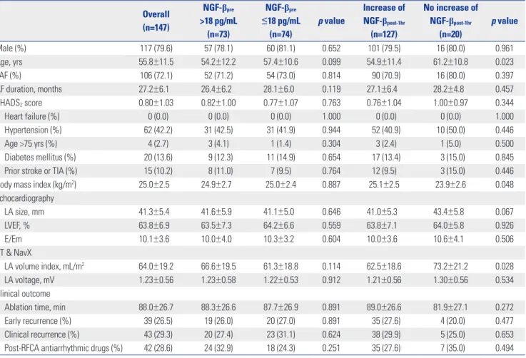
The plasma levels of NGF-β significantly increased 1 hour af- ter RFCA (20.1±11.1 pg/mL vs. 29.6±19.4 pg/mL, p<0.001) (Fig. 1A). There was a significant correlation between the plasma levels of pre-procedural NGF-β and post-procedural NGF-β (R=0.773, p<0.001) (Fig. 1B). Table 1 summarized the baseline characteristics of included patients, and there was no significant difference between the patients with NGF-βpre >18 pg/mL (n=73) and those with ≤18 pg/mL (n=74) based on the median plasma level of NGF-β. When we compared the pa- tients who had an increased plasma NGF-βpost-1hr (n=127) and those did not (n=20), the group with no increase of NGF-βpost-1hr showed older age (p=0.023), lower body mass index (p=0.048), and greater LA volume index (p=0.028) (Table 1). However, higher plasma levels of NGF-β were not related to longer du- ration of total ablation time. In Post AF ablation, mean heart rate and LF was higher than before ablation. In contrast, rMS- SD was higher in pre AF albation (Fig. 1C, D, and E).
High NGF-β
preand NGF-β
post-1hrare associated with high LF/HF and APC frequency in HRV
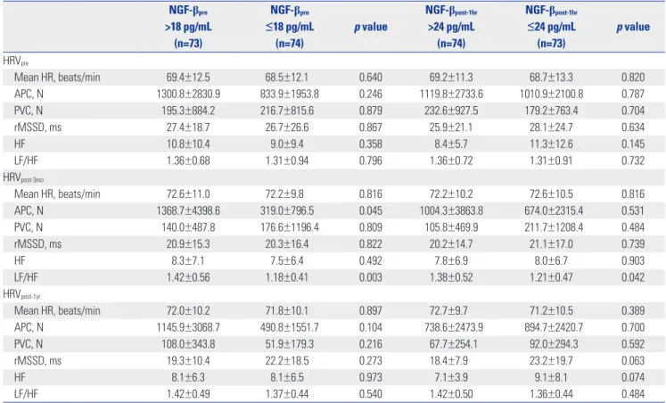
post-3moFig. 2 summarized the changes of mean HR and HRV at pre- RFCA (HRVpre), post-RFCA 3 months (HRVpost-3mo), and post- RFCA 1 year (HRVpost-1yr), depending on median plasma levels of NGF-βpre and NGF-βpost-1hr. After catheter ablation of AF, mean HRs were increased at Holterpost-3mo (68.9±12.3 bpm to 72.4±10.3 bpm, p=0.010) and at Holterpost-1yr (71.9±10.1 bpm, p=0.023). The rMSSD (27.1±22.9 ms to 20.6±15.8 ms, p=0.009 and 20.7±14.8 ms, p=0.010), LF (16.4±19.3 Hz to 10.6±11.4 Hz, p=0.003 and 11.3±10.9 Hz, p=0.010) were reduced at HRVpost- 3mo and HRVpost-1yr, respectively. We compared HRV parame- ters depending on the plasma level of NGF-βpre and NGF-βpost-
1hr using the median values as a cut-off (Table 2). Compared to the patients with NGF-βpre ≤18 pg/mL, NGF-βpre >18 pg/mL group showed higher LF/HF ratio (p=0.003) and higher num- ber of APCs (p=0.045) in HRVpost-3mo (Fig. 2E and F). LF/HF ra- tio was also higher in high NGF-βpost-1hr group than in low NGF-βpost-1hr group (p=0.042). Because both sympathetic (rep- resented by LF) and parasympathetic (represented by rMSSD) activities were reduced after AF catheter ablation, we might not have found significant change of LF/HF ratio in the whole population (Fig. 2).
Fig. 1. (A) Plasma levels of NGF-β before and after AF ablation. (B) Correlation between pre- and post-ablation NGF-βs. (C) Mean heart rate in pre AF abla- tion, post 3 month AF ablation, and post 1 year AF ablation. (D) rMSSD in pre AF ablation, post 3 month AF ablation, and post 1 year AF ablation. (E) LF in pre AF ablation, post 3 month AF ablation, and post 1 year AF ablation. NGF-β, nerve growth factor-β; RFCA, radiofrequency catheter ablation; LF, low fre- quency components.
90
80
70
60 60
40
20
0
100 80 60 40 20 0
Pre-RFCA
NGF-βpre 0 10 20 30 40 50 60 70 Pre-ABL NGF-β (pg/mL)
*
*p<0.001 vs. pre-RFCA
p<0.001 R=0.773
p<0.001
Mean heart rate
Plasma levels of NGF-β after RFCA Correlation between pre- and post-ABL NGF-β
rMSSD LF
*
Post-3 mo NGF-βpost-1hr
Post-1 yr
Mean heart rate (bpm)NGF-β (pg/mL) Post-ABL NGF-β (pg/mL)
60
40
20
0 Pre-RFCA
*
*p<0.001 vs. pre-RFCA
*
Post-3 mo Post-1 yr
rMSSD (ms)
60
40
20
0 Pre-RFCA
*
*p<0.001 vs. pre-RFCA
*
Post-3 mo Post-1 yr LF (ms2)
C
A B
D E
High NGF-β
preand NGF-β
post-1hrare independently associated with high LF/HF in HRV
post-3moIn the uni- and multi-variate linear regression analyses, both NGF-βpre (B=4.240, 95% CI 1.114–7.336, p=0.008) and NGF- βpost-1hr (B=7.617, 95% CI 2.106–13.127, p=0.007) were linearly associated with LF/HF ratio at HRVpost-3mo (Table 3). Both the plasma levels of NGF-βpre (OR=1.159, 95% CI 1.045–1.286, p=0.005) and NGF-βpost-1hr (OR=1.098, 95% CI 1.030–1.170, p=0.004) were independently associated with the increase of LF/HF at HRVpost-3mo (Table 4). However, AF recurrence or ven- tricular arrhythmic events did not vary depending on the plasma levels of NGF-β.
DISCUSSION
The present study demonstrated that catheter ablation of AF increases the plasma level of NGF-β significantly, and NGF-β was closely associated with high sympathetic nerve activity estimated by HRV and high number of APCs in Holter per- formed 3 months after the procedure. However, plasma level of NGF-β was not increased after RFCA in patients with old
age and advanced LA remodeling. We also documented the feasibility of double sandwich ELISA technique for detection of the minimal change of NGF-β in the peripheral blood of patients with AF.
The role of plasma NGF-β in cardiac nerve sprouting after cardiac injury
Following a peripheral nerve injury, a complex and finely reg- ulated sequence of events lead to neurilemma cell prolifera- tion and axonal regeneration.15 Nerve regeneration is trig- gered by re-expression of NGF or other neurotrophic factors in the non-neuronal cells around the site of injury.8 NGF and other neurotrophic factors that are derived from myocardial injuries are transported retrograde to the stellate ganglion, which triggers cardiac nerve sprouting in canine models.16 Acute myocardial infarction causes an immediate (within 30 minutes) increase of transcardiac NGF concentration, where- as mRNA of NGF in the damaged myocardium begins to in- crease 3 days after myocardial infarction and peaks 1 week later.16 NGF over-expression is associated with myocardial hy- per innervation and atrial fibrosis,17 and heterogeneous in- creases in atrial sympathetic innervation contribute to the Fig. 2. Changes of mean heart rate and HRV after catheter ablation of AF. We compared the change of Mean heart rate (A), rMSSD (B), LF (C), HF (D), LF/
HF ratio (E), and number of APCs (F) defending on the NGF-βpre plasma level. HRV, heart rate variability; AF, atrial fibrillation; HF, high-frequency compo- nents; LF, low frequency components; APCs, atrial premature contractions; NGF-β, nerve growth factor-β.
120
80
40
0
120
80
40
0
60
40
20
0
3.0
2.0
1.0
0.0
60
40
20
0
9000
6000
3000
0 Pre-RFCA
Pre-RFCA
Pre-RFCA
Pre
Pre-RFCA
Pre NGF-βpre >18 pg/mL
NGF-βpre≤18 pg/mL
NGF-βpre >18 pg/mL NGF-βpre≤18 pg/mL
NGF-βpre >18 pg/mL NGF-βpre≤18 pg/mL
NGF-βpre >18 pg/mL NGF-βpre≤18 pg/mL
NGF-βpre >18 pg/mL NGF-βpre≤18 pg/mL
NGF-βpre >18 pg/mL NGF-βpre≤18 pg/mL Mean heart rate
HF
rMSSD
LF/HF
LF
Number of APCs
p=0.045 p=0.003
Post-3 mo
Post-3 mo
Post-3 mo
Post-3 mo
Post-3 mo
Post-3 mo Post-1 yr
Post-1 yr
Post-1 yr
Post-1 yr
Post-1 yr
Post-1 yr
Mean heart rate (bpm)HF (ms2) rMSSD (ms)LF/HF LF (ms2)APCs (number)
A
D
B
E
C
F
generation and maintenance of AF by exerting significant ef- fects on automaticity, refractoriness, and conduction veloci- ty.18-20 In this study, elevated NGF level was found to be associ- ated with high sympathetic tone and high frequency of APCs after RFCA.
Catheter ablation and NGF-β
RFCA is a kind of necrotic cardiac injury and induces cardiac
nerve sprouting, which has been known to be related to the rises of the level of NGF.4,16 Kangavari, et al.10 demonstrated that RFCA induces over-expression of NGF mRNA, resulting in increased plasma level of NGF. Although study population and detection methods were different, the current study found consistent elevation of plasma level of NGF-β after RFCA, but, it was not the case in patients with old age, low body mass index, and high LA volume index. We applied the Table 2. Comparisons of HRVpre, HRVpost-3mo, and HRVpost-1yr Depending on the Median Value of NGF-βpre and NGF-βpost-1hr Median Value
NGF-βpre
>18 pg/mL (n=73)
NGF-βpre
≤18 pg/mL (n=74)
p value
NGF-βpost-1hr
>24 pg/mL (n=74)
NGF-βpost-1hr
≤24 pg/mL (n=73)
p value
HRVpre
Mean HR, beats/min 69.4±12.5 68.5±12.1 0.640 69.2±11.3 68.7±13.3 0.820
APC, N 1300.8±2830.9 833.9±1953.8 0.246 1119.8±2733.6 1010.9±2100.8 0.787
PVC, N 195.3±884.2 216.7±815.6 0.879 232.6±927.5 179.2±763.4 0.704
rMSSD, ms 27.4±18.7 26.7±26.6 0.867 25.9±21.1 28.1±24.7 0.634
HF 10.8±10.4 9.0±9.4 0.358 8.4±5.7 11.3±12.6 0.145
LF/HF 1.36±0.68 1.31±0.94 0.796 1.36±0.72 1.31±0.91 0.732
HRVpost-3mo
Mean HR, beats/min 72.6±11.0 72.2±9.8 0.816 72.2±10.2 72.6±10.5 0.816
APC, N 1368.7±4398.6 319.0±796.5 0.045 1004.3±3863.8 674.0±2315.4 0.531
PVC, N 140.0±487.8 176.6±1196.4 0.809 105.8±469.9 211.7±1208.4 0.484
rMSSD, ms 20.9±15.3 20.3±16.4 0.822 20.2±14.7 21.1±17.0 0.739
HF 8.3±7.1 7.5±6.4 0.492 7.8±6.9 8.0±6.7 0.903
LF/HF 1.42±0.56 1.18±0.41 0.003 1.38±0.52 1.21±0.47 0.042
HRVpost-1yr
Mean HR, beats/min 72.0±10.2 71.8±10.1 0.897 72.7±9.7 71.2±10.5 0.389
APC, N 1145.9±3068.7 490.8±1551.7 0.104 738.6±2473.9 894.7±2420.7 0.700
PVC, N 108.0±343.8 51.9±179.3 0.216 67.7±254.1 92.0±294.3 0.592
rMSSD, ms 19.3±10.4 22.2±18.5 0.273 18.4±7.9 23.2±19.7 0.063
HF 8.1±6.3 8.1±6.5 0.973 7.1±3.9 9.1±8.1 0.074
LF/HF 1.42±0.49 1.37±0.44 0.540 1.42±0.50 1.36±0.44 0.484
HRV, heart rate variability; NGF-β, nerve growth factor-β; APC, atrial premature contraction; PVC, premature ventricular contraction; HR, heart rate; HF, high-fre- quency components; LF, low frequency components.
Table 3. Uni- and Multivariate Linear Regression Analyses for Pre-NGF-β, Post-NGF-β1hr, and the Increase of NGF-β1hr
Univariate analysis Multivariate analysis
B 95% CI p value B 95% CI p value
NGF-βpre
HRVpre
PVC, N 0.003 0.001–0.005 0.008 0.000 -0.003–0.002 0.871
HRVpost-3mo
LF/HF 4.234 1.150–7.319 0.007 4.240 1.114–7.336 0.008
NGF-βpost-1hr
HRVpre
PVC, N 0.005 0.001–0.008 0.011 -0.002 -0.007–0.002 0.304
HRVpost-3mo
LF/HF 9.141 3.516–14.765 0.002 7.617 2.106–13.127 0.007
Increase of NGF-βpost-1hr
HRVpost-3mo
LF/HF 4.906 0.235–9.577 0.040
NGF-β, nerve growth factor-β; HRV, heart rate variability; PVC, premature ventricular contraction; HF, high-frequency components; LF, low frequency components.
double sandwich ELISA method to enhance detection sensi- tivity to pg/mL level, and our results of plasma NGF-β level were consistent with other previous reports.21-23 Because AF catheter ablation itself has the effects of cardiac autonomic denervation,24 the change of post-ablation HRV cannot be the result of increased plasma level of NGF-β alone. However, the current study found that plasma level of NGF-β was clearly as- sociated with the change of autonomic nerve activity at HRV-
post-3mo and HRVpost-1yr.
AF and HRV
HRV relies on the principle that the pattern of beat-to-beat control of the sinoatrial node provides a reflection of cardiac autonomic nerve activity.14 Among multiple parameters in HRV, HF components are thought to primarily reflect vagal tone, whereas high LF/HF ratio has been assumed as the in- dex of high sympathetic activity that is known to be pro-ar- rhythmic and higher frequency of PVCs or APCs.14,25 The pres- ent study showed that patients with a higher plasma level of NGF-β had a higher LF/HF ratio and a trend of more frequent APCs in Holter monitoring at 3 months after ablation. Al- though NGF provokes both sympathetic and parasympathetic nerve regeneration, it was associated only with an increased sympathetic nerve activity, but not with an increased para- sympathetic nerve activity in this study. This finding may be due to the fact that RFCA destroyed parasympathetic post- ganglionic cells that are located at or very close to the ablation sites. On the other hand, since sympathetic postganglionic cells are located in the stellate ganglia far from the ablation site, ablation-induced sympathetic nerve axonal damage might recover three months after RFCA. It has been reported that a rapid HR after AF catheter ablation predicts a low recur- rence rate of AF.26 Although it might be associated with an ap- propriate vagal denervation, a high plasma level of NGF-β and an increased post-procedure LF/HF ratio in patients who have less remodeled atrium might contribute to a rapid HR and low recurrence after RFCA.
Study limitations
The present study has several limitations. The patients includ- ed in this study were a highly selected group referred for RFCA, and the number of patients was also limited. We ex- cluded patients with LA >55 mm. HRV analysis requires nor- mal sinus rhythm with normal cardiac function, therefore, we
excluded patients whose Holter could not be analyzed for HRV. We did not evaluate the long-term change in plasma level of NGF-β after AF ablation. Although we could not find statistical difference of clinical recurrence rate depending on the plasma levels of NGF-β in this small sample size and short follow-up period study, we found the differences in the LF/
HF at HRVpost-3mo. Recently, we reported that LF/HF at HRVpost- 3mo is an independent factor predicting clinical recurrence of AF after RFCA.27 We also found higher LF/HF ratio in HRVpost- 3mo and higher number of APCs in patients with NGF-βpre>18 pg/mL than those with ≤18 pg/mL, but we do not have direct evidence for the mechanism or causal-results relationship.
Further study with a large population may be warranted. We chose post-procedure 1 hour to evaluate the plasma level of NGF-βpost-1hr, but it might not be enough time to show in- creased plasma level of NGF-β after RFCA. Because we used irrigated tip ablation catheter, irrigated fluid volume might af- fect the plasma concentration of NGF-β.
In conclusion, AF catheter ablation increases plasma level of NGF-β, and high plasma levels of pre- or post-RFCA NGF-β were associated with high sympathetic nerve activity and the presence of high number of APCs in post-RFCA 3 month Holter. Double sandwich ELISA technique was feasible and acceptable for the detection of the minimal change of NGF-β in the peripheral blood of the patients with AF.
ACKNOWLEDGEMENTS
This work was supported by a grant (A085136 and A120478) from the Korea Health 21 R&D Project, Ministry of Health and Welfare and a grant (7-2013-0362 and 2012027176) from the Basic Science Research Program run by the National Research Foundation of Korea (NRF) which is funded by the Ministry of Science, ICT & Future Planning (MSIP).
REFERENCES
1. Vracko R, Thorning D, Frederickson RG. Fate of nerve fibers in necrotic, healing, and healed rat myocardium. Lab Invest 1990;
63:490-501.
2. Nori SL, Gaudino M, Alessandrini F, Bronzetti E, Santarelli P. Im- munohistochemical evidence for sympathetic denervation and re- innervation after necrotic injury in rat myocardium. Cell Mol Biol (Noisy-le-grand) 1995;41:799-807.
3. Cao JM, Fishbein MC, Han JB, Lai WW, Lai AC, Wu TJ, et al. Rela- Table 4. Uni- and Multivariate Logistic Regression Analyses for Increase of LF/HF Ratio in HRVpost-3mo Compared with HRVpre
Unadjusted Adjusted*
OR 95% CI p value OR 95% CI p value
NGF-βpre 1.019 0.980–1.059 0.343 1.159 1.045–1.286 0.005
NGF-βpost-1hr 1.026 0.998–1.054 0.070 1.098 1.030–1.170 0.004
Increase of NGF-βpost-1hr 1.029 0.993–1.066 0.110 1.065 0.998–1.136 0.057
HF, high-frequency components; LF, low frequency components; HRV, heart rate variability; NGF-β, nerve growth factor-β; AF, atrial fibrillation.
*Adjusted for sex, age, AF subtype (PAF vs. PeAF), hypertension, and estimated glomerular filtration rate.
tionship between regional cardiac hyperinnervation and ventric- ular arrhythmia. Circulation 2000;101:1960-9.
4. Okuyama Y, Pak HN, Miyauchi Y, Liu YB, Chou CC, Hayashi H, et al. Nerve sprouting induced by radiofrequency catheter ablation in dogs. Heart Rhythm 2004;1:712-7.
5. Cao JM, Chen LS, KenKnight BH, Ohara T, Lee MH, Tsai J, et al.
Nerve sprouting and sudden cardiac death. Circ Res 2000;86:816- 21.
6. Chen PS, Chen LS, Cao JM, Sharifi B, Karagueuzian HS, Fishbein MC. Sympathetic nerve sprouting, electrical remodeling and the mechanisms of sudden cardiac death. Cardiovasc Res 2001;50:
409-16.
7. Chen PS, Tan AY. Autonomic nerve activity and atrial fibrillation.
Heart Rhythm 2007;4(3 Suppl):S61-4.
8. Levi-Montalcini R. Growth cntrol of nerve cells by a protein factor and its antiserum: discovery of this factor may provide new leads to understanding of some neurogenetic processes. Science 1964;
143:105-10.
9. Cygankiewicz I, Zareba W, de Luna AB. Prognostic value of Holter monitoring in congestive heart failure. Cardiol J 2008;15:313-23.
10. Kangavari S, Oh YS, Zhou S, Youn HJ, Lee MY, Jung WS, et al. Ra- diofrequency catheter ablation and nerve growth factor concen- tration in humans. Heart Rhythm 2006;3:1150-5.
11. Park JH, Pak HN, Choi EJ, Jang JK, Kim SK, Choi DH, et al. The re- lationship between endocardial voltage and regional volume in electroanatomical remodeled left atria in patients with atrial fi- brillation: comparison of three-dimensional computed tomo- graphic images and voltage mapping. J Cardiovasc Electrophysiol 2009;20:1349-56.
12. Pak HN. Large circular ring catheter ablation versus anatomically guided ablation of atrial fibrillation: back to the future for success- ful catheter ablation of atrial fibrillation? Korean Circ J 2011;41:431-3.
13. Cappato R, Calkins H, Chen SA, Davies W, Iesaka Y, Kalman J, et al. Updated worldwide survey on the methods, efficacy, and safety of catheter ablation for human atrial fibrillation. Circ Arrhythm Electrophysiol 2010;3:32-8.
14. Heart rate variability: standards of measurement, physiological interpretation and clinical use. Task Force of the European Society of Cardiology and the North American Society of Pacing and Elec- trophysiology. Circulation 1996;93:1043-65.
15. Guth L. Regeneration in the mammalian peripheral nervous sys- tem. Physiol Rev 1956;36:441-78.
16. Zhou S, Chen LS, Miyauchi Y, Miyauchi M, Kar S, Kangavari S, et al.
Mechanisms of cardiac nerve sprouting after myocardial infarction
in dogs. Circ Res 2004;95:76-83.
17. Kiriazis H, Du XJ, Feng X, Hotchkin E, Marshall T, Finch S, et al.
Preserved left ventricular structure and function in mice with car- diac sympathetic hyperinnervation. Am J Physiol Heart Circ Physi- ol 2005;289:H1359-65.
18. Chang CM, Wu TJ, Zhou S, Doshi RN, Lee MH, Ohara T, et al.
Nerve sprouting and sympathetic hyperinnervation in a canine model of atrial fibrillation produced by prolonged right atrial pac- ing. Circulation 2001;103:22-5.
19. Jayachandran JV, Sih HJ, Winkle W, Zipes DP, Hutchins GD, Olgin JE. Atrial fibrillation produced by prolonged rapid atrial pacing is associated with heterogeneous changes in atrial sympathetic in- nervation. Circulation 2000;101:1185-91.
20. Olgin JE, Sih HJ, Hanish S, Jayachandran JV, Wu J, Zheng QH, et al.
Heterogeneous atrial denervation creates substrate for sustained atrial fibrillation. Circulation 1998;98:2608-14.
21. Lee BC, Choi IG, Kim YK, Ham BJ, Yang BH, Roh S, et al. Relation between plasma brain-derived neurotrophic factor and nerve growth factor in the male patients with alcohol dependence. Alco- hol 2009;43:265-9.
22. Titanji K, Nilsson A, Mörch C, Samuelsson A, Sönnerborg A, Grutz- meier S, et al. Low frequency of plasma nerve-growth factor detec- tion is associated with death of memory B lymphocytes in HIV-1 infection. Clin Exp Immunol 2003;132:297-303.
23. Aloe L, Bracci-Laudiero L, Alleva E, Lambiase A, Micera A, Tirassa P. Emotional stress induced by parachute jumping enhances blood nerve growth factor levels and the distribution of nerve growth factor receptors in lymphocytes. Proc Natl Acad Sci U S A 1994;91:
10440-4.
24. Mesas CE, Pappone C, Lang CC, Gugliotta F, Tomita T, Vicedomini G, et al. Left atrial tachycardia after circumferential pulmonary vein ablation for atrial fibrillation: electroanatomic characterization and treatment. J Am Coll Cardiol 2004;44:1071-9.
25. Podrid PJ, Fuchs T, Candinas R. Role of the sympathetic nervous system in the genesis of ventricular arrhythmia. Circulation 1990;
82(2 Suppl):I103-13.
26. Pappone C, Santinelli V, Manguso F, Vicedomini G, Gugliotta F, Au- gello G, et al. Pulmonary vein denervation enhances long-term benefit after circumferential ablation for paroxysmal atrial fibril- lation. Circulation 2004;109:327-34.
27. Kang KW, Kim TH, Park J, Uhm JS, Joung B, Hwang C, et al. Long- term changes in heart rate variability after radiofrequency cathe- ter ablation for atrial fibrillation: 1-year follow-up study with irri- gation tip catheter. J Cardiovasc Electrophysiol 2014;25:693-700.