Tuberc Respir Dis 2009;66:136-140
CopyrightⒸ2009. The Korean Academy of Tuberculosis and Respiratory Diseases. All rights reserved.
빠르게 진행하는 종격동의 기관지기원 물혹
순천향대학교 의과대학
1내과학교실,
2흉부외과학교실,
3영상의학교실,
4병리학교실
김 철1, 김양기1, 이영목1, 김기업1, 김현조2, 황정화3, 김동원4, 어수택1Mediastinal Bronchogenic Cyst, which was Grown Rapidly
Chul Kim, M.D.
1, Yang Ki Kim, M.D.
1, Young Mok Lee, M.D.
1, Ki-Up Kim, M.D.
1, Hyun Zo Kim, M.D.
2, Jung Hwa Hwang, M.D.
3, Dong Won Kim, M.D.
4, Soo-Taek Uh, M.D.
1Departments of
1Internal medicine,
2Chest Surgery,
3Radiology,
4Pathology, Soonchunhyang University College of Medicine, Seoul, Korea
Bronchogenic cyst arises from anomalous budding of the primitive foregut during embryonic development and it represents a part of the spectrum of bronchopulmonary foregut malformations. Approximately two-thirds of the malformations are found within the mediastinum, and one-third are found in the lung parenchyma. The prevalence of bronchogenic cyst is unknown, presumably because most patients are asymptomatic. Incidentally detected bronchogenic cysts are usually removed at the time of diagnosis. We do not know how and why bronchogenic cysts grow. We recently experienced a case of rapidly growing mediastinal mass in a young adult, and this presented as a huge mass that had newly developed within one year. This mass was pathologically confirmed to be a bronchogenic cyst. We report on this case of a rapidly growing bronchogenic cyst, which is a rare characteristic of this type of cyst.
Key Words: Bronchogenic cyst, Mediastinum
Address for correspondence: Ki-Up Kim, M.D.
Division of Pulmonary and Allergy, Department of Internal Medicine, Soonchunhyang University College of Medicine, 657, Hannam-dong, Yongsan-gu, Seoul 140-743, Korea Phone: 82-2-709-9027, Fax: 82-2-709-9554
E-mail: [email protected] Received: Oct. 4, 2008
Accepted: Feb. 4, 2009
증 례
환 자: 남자, 20세
주 소: 신체검사에서 우연히 발견된 종격동의 종괴 현병력: 1주 전 개인병원에서 촬영한 단순 흉부방사선 검사에서 종격동에 덩어리가 의심되어 전원 되었다. 환자 는 3개월 전 농구를 하던 중 가슴을 부딪친 적이 있었으나 별다른 치료 없이 회복되었고, 1년 전 촬영하였던 단순 흉부 방사선 촬영에서는 이상소견이 없었다.
과거력: 특이 사항 없었다.
가족력: 특이 사항 없었다.
진찰소견: 활력증후는 혈압 130/80 mmHg, 맥박수 분 당 78회, 호흡수 분당 18회, 체온 36.5oC였다. 건강하게 보였으며 문진 및 전신 진찰에서 이상소견은 없었다.
검사실 소견: 일반혈액검사에서 백혈구 8,900/mm3, 혈 색소 15.3 g/dl, 적혈구 용적 42.5%, 혈소판 374,000/mm3 이었고, 생화학 검사 소견은 총단백 6.5 g/dl, 알부민 4.5 g/dl, AST/ALT 15/24 U/L, 총 빌리루빈은 0.4 mg/dl이었 으며, 소변검사 및 심전도 검사는 정상이었다. 폐기능 검 사는 최대 호기량, 1초간 노력성호기량 그리고 기류-용적 곡선 모두 정상 소견이었다.
영상학적 소견: 단순 흉부 방사선 검사에서 용골(carina) 아래에 약 7.5 cm의 경계가 명확한 덩어리 모양의 음영이 관찰되었으나 폐 실질의 이상소견은 없었다(Figure 1A).
1년 전 개인병원에서 시행한 단순흉부촬영에는 병변이 없 었다(Figure 1B). 흉부 전산화 단층 촬영에서 약 5.6×
4.8×7 cm의 경계가 명확한 저음영의 덩어리가 용골 아래 에 있었다(Figure 2). 덩어리는 식도와 경계를 접하고 있 었으나 주변 혈관이나 심장과는 경계가 명확하였다. 기관
Figure 1. Chest PA finding of 20 year old male at first visit and 1 year ago, well defined mass like opacity was showed at subcarinal area which was almost fist size of child (A). However 1 year ago, the mass was not detected (B).
Figure 2. Chest computed tomography of the presented male. There was about 5.6×4.8×7 cm sized, well de- fined, cystic mass at right subcarinal area. Bronchus in- termedius of right lung was compressed by the cystic
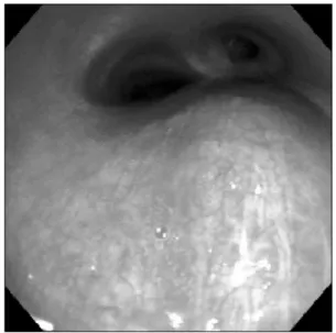
mass. Figure 3. Bronchoscopic finding of right bronchus inter- medius, posterior wall of the bronchus was bulged by extrinsic compression, but mucosal wall looked intact.
과 기관지의 이상소견은 없었으며 폐 실질에 두 개의 작은 석회화병변이 우상엽에 있었다. 그 외 종격동 및 폐문부 의 림프절 비대는 없었다.
기관지내시경 및 식도 초음파내시경 소견: 기관지내시경 (Olympus BF-260, Tokyo, Japan)을 인두 및 후두에 위치 하고 도관을 이용한 국소마취 후 검사를 시작하였다. 오 른쪽 주기관지, 중간기관지 그리고 왼쪽 하기관지 등에서 외부압박에 의한 함입이 관찰되었다(Figure 3).
구강 내 국소마취를 시행한 후 식도 내시경 초음파(GIF- UM2000, Olympus, Tokyo, Japan)검사를 하였다. 내시경
검사 결과 앞니에서 25∼32 cm부위의 식도가 외부 덩어 리에 의하여 눌려 있었다. 식도 초음파 검사에서 하행대 동맥의 반대편, 용골 아래 오른쪽에 6.6 cm의 경계가 분 명하고 대부분에서 낮은 반향(unechoic)의 덩어리가 관찰 되었으나 일부에서는 높은 반향(hyperechoic)을 보이는 부분이 있었으며, 높은 반향 부위는 체위변화에 모양이 변했다(Figure 4).
수술 소견: 급격하게 진행하는 기관지기원 물혹이 의심되
Figure 4. Gastroscopy (A) and endoscopic ultrasound (B) findings of the presented male, there was an in- dentation of esophagus at 25∼32 cm from incisor (arrow). Endoscopic ultra- sound showed about 6.6 cm sized hypoechoic cyst.
Echogenic material of the in- side of cyst was changed their location by position (arrow head).
Figure 5. Gross and microscopic findings after surgical excision from the presented male. A brown red colored unilocular cyst was measured 6×4×1 cm. Wall of the cyst looked grossly smooth and spherical fibrotic tissue which was accom- panied multiple and focal hemorrhage (A). Microscopic finding demonstrated a cyst wall which had contained airway components such as cartilage plate and smooth muscle (B, x40). Magnified photograph showed single layer of ciliated columnar epithelium as like bronchial epithelium with goblet cells (C, H&E stain, x200).
어 개흉을 통한 절제술을 하였다. 덩어리는 용골 바로 아래 왼쪽 기관지에 접하여 있었으며 기관지와 소통은 없었다.
병리학적 소견: 수술로 제거된 덩어리는 6×4 cm의 크 기로 출혈 반을 동반한 주머니 모양의 물혹으로 현미경소 견에서 물혹의 안쪽 벽은 원주상피세포로 둘러 쌓여 있었 으며 편평화생이 동반되어 있고, 물혹의 벽은 섬유조직으 로 이루어져 있었으며 비특이적인 염증소견 및 부분적인 석회화가 관찰되었다(Figure 5).
임상경과: 수술 후 외래 추적검사 중이다.
고 찰
기관지기원 물혹(bronchogenic cyst)은 상대적으로 드 문 질환이나 종격동에 발생하는 가장 흔한 선천성 낭종이 다. 폐 실질에 발생하는 경우는 전체의 15∼20% 정도로
보고 되고 있으며 두경부, 심장 그리고 횡격막 아래까지 골고루 분포한다. 발생학적으로 원시 배쪽 앞장관(primi- tive ventral foregut)에서 기원하는 기관싹(tracheal bud) 의 분화 이상에 의한 것으로 기관지-폐 앞장관 기형 (bronchopulmonary foregut malformation)의 범주에 해 당된다1,2. Maier3에 의하면 1859년에 종격동에 발생한 기 관지기원 물혹의 첫 보고가 있었다고 한다. 국내에서는 You 등4에 의하여 첫 발표 후 지속적인 관심으로 다수의 보고가 있었다. 최근에는 합병증이 동반되거나 전형적이 지 않은 증례들이 보고되었다5-8. 본 증례는 이상이 발견되 기 1년 전에 실시한 흉부방사선 검사에서 정상소견을 보 였던 젊은 성인에서 비교적 짧은 시간에 급격한 물혹의 증가를 확인하였던 흔치 않은 증례로 판단되어 보고한다.
임상증상은 소아에서는 생명을 위협할 정도의 물리적 인 압박이 발생할 수 있다고 알려져 있으나 성인에서는
우연히 발견되는 경우가 많다. 주로 덩어리에 의한 압박 으로 기침, 호흡곤란, 흉통, 객혈 등의 호흡기적 증상과 연하곤란이 있다. 이차 감염이나, 샛길(fistula)이 동반되 었을 경우 호흡기적 증상이 더욱 빈번히 발생할 수 있다.
하지만 성인에서는 무증상인 경우도 많다9. 본 증례에서 도 덩어리가 커서 주변장기를 압박하여 호흡곤란 또는 고 정된 천명이 동반되었을 것으로 추정하였으나 증상이 없 었고 연하곤란 등의 증상도 없었다.
진단은 단순 흉부 촬영과 흉부 전산화 단층촬영을 통하 여 할 수 있다. 단순 흉부 촬영에서 경계가 명확한 균일한 음영의 난원형 또는 원형의 덩어리로 관찰되며, 과거 또는 현재의 감염으로 공기-액체 음영을 보이는 경우도 있다.
전산화 단층 촬영에서는 경계가 분명한 연부조직 또는 액 체의 밀도를 보이는 물혹 덩어리 소견을 보인다. 자기공 명영상을 시행하면 T1 강조영상에서 뇌척수액보다 낮은 신호강도를 보이다가 T2 강조영상에서 신호강도가 증가 하여 뇌 척수액과 동일한 신호강도를 보인다10. 초음파내 시경(endoscopic ultrasonography)도 진단에 도움을 줄 수 있으며 조직검사를 통한 물혹의 악성종양 동반 여부의 확인, 주변조직과의 유착 여부의 확인 또는 치료적 천자 등에 이용될 수 있다11. 본 환자에서 시행한 초음파내시경 에서는 덩어리 내에 낮은 반향 부위와 높은 반향 부위가 혼재된 물혹이 의심되었으며 터질 가능성이 있어 조직검 사는 하지 않았다.
병리조직학적 소견으로 육안적 소견은 대개 반투명한 (translucent) 물혹으로 보이며 내부는 맑은 삼출액부터 젤리모양, 출혈성 액체 등 다양한 성상의 액체로 차 있다10. 현미경적 소견은 호흡상피와 유사한 중층 또는 원주 섬모 상피로 둘러 쌓여 있는 주머니 구조로 연골조직이나 점액 분선(mucous gland) 등이 존재하는 경우도 있으며, 석회 화가 동반되는 경우는 없고 간혹 편평상피화생(squamous metaplasia)을 동반하는 경우도 있다12. 본 증례는 수술 후 병리적 소견에서 주변 기관지와 소통이 없고 악성변이도 없었으나 빠르게 종괴 크기의 증가를 설명할 수 없어 환자 가 3개월 전 가슴을 부딪친 것이 기존의 기관지기원 물혹 에 출혈을 유발하여 낭종의 급격한 비대를 가져왔을 것으 로 추정되었다.
감별진단을 해야 할 질환으로는 폐농양, 감염된 수포 (bullae), 진균감염, 혈관기형, 포충증(hydatidosis), 폐결 핵 등이 있다9.
기관지기원 물혹은 대개는 양성종양으로 수술적 치료 에 대하여는 여러 가지 논란이 있는 것이 사실이나, 덩어
리의 제거를 통한 병리적 진단만이 유일한 확진 방법이고 다른 질환과 감별 진단이 필요하며 악성종양과의 감별이 필요할 수 있어, 수술적 치료가 권고되고 있다12,13. 최근에 는 종격동에 생긴 기관지기원 물혹에 대하여 흉강경을 이 용하여 덩어리를 제거 하는 방법을 많이 사용한다14. 본 증례는 빠른 성장속도를 보이는 덩어리로 판단되어 악성 을 배제하기 어려워 개흉을 통한 제거를 시행하였다.
요 약
기관지기원 물혹은 드물게 발생하는 선천성 질환으로 주로 종격동에 분포하며 기관지와의 소통이 발생하는 경 우, 악성으로 전환된 경우를 제외하고는 크기가 갑자기 커지는 경우는 드문 일이다. 1년 전 검진에서 시행한 단순 흉부 촬영에서 이상이 없었던 젊은 남자가 흉부검진에서 종격동 덩어리가 발견되어 급격한 크기의 증가가 확인되 었던 증례를 경험하였기에 문헌고찰과 함께 보고하는 바 이다.
참 고 문 헌
1. Kirwan WO, Walbaum PR, McCormack RJ. Cystic intra- thoracic derivatives of the foregut and their compli cations. Thorax 1973;28:424-8.
2. Haller JA Jr, Golladay ES, Pickard LR, Tepas JJ 3rd, Shorter NA, Shermeta DW. Surgical management of lung bud anomalies: lobar emphysema, bronchogenic cyst, cystic adenomatoid malformation, and intralobar pulmonary sequestration. Ann Thorac Surg 1979;28:
33-43.
3. Maier HC. Bronchiogenic cysts of the mediastinum.
Ann Surg 1948;127:476-502.
4. You M, Park S. Congenital bronchogenic cyst: report of a case. J Korean Surg Soc 1967;9:537-40.
5. Kim KH, Kwon SS, Yoon SJ, Kim YK, Han KD, Moon HS, et al. A case of congenital bronchogenic cyst with infection. Tuberc Respir Dis 1990;37:323-8.
6. Kim YW, Lee SH, Hong SC, Lee HH, Park SJ, Lee GJ, et al. A case report of a bronchogenic cyst mis- conceived to lung cancer. Tuberc Respir Dis 2003;
55:526-30.
7. Kim GH, Kim KH, Kim MS, Park JE, Kim DJ, Son HS, et al. A case of pulmonary aspergilloma in broncho- genic cyst associated with an actinomycosis. Tuberc Respir Dis 2004;57:584-8.
8. Choi KA, Koh WJ, Lee KS, Han J, Kim K. Multicystic
pulmonary parenchymal lesions in a young adult with hemoptysis. Tuberc Respir Dis 2007;62:71-3.
9. Sarper A, Ayten A, Golbasi I, Demircan A, Isin E.
Bronchogenic cyst. Tex Heart Inst J 2003;30:105-8.
10. McAdams HP, Kirejczyk WM, Rosado-de-Christenson ML, Matsumoto S. Bronchogenic cyst: imaging features with clinical and histopathologic correlation. Radiology 2000;217:441-6.
11. Kramer H, van Putten JW, Douma WR, Smidt AA, van Dullemen HM, Groen HJ. Technical description of en- doscopic ultrasonography with fine-needle aspiration for the staging of lung cancer. Respir Med 2005;
99:179-85.
12. Kawase Y, Takahashi M, Takemura H, Tomita S, Watanabe G. Surgical treatment of a bronchogenic cyst in the interatrial septum. Ann Thorac Surg 2002;
74:1695-7.
13. Ashizawa K, Okimoto T, Shirafuji T, Kusano H, Ayabe H, Hayashi K. Anterior mediastinal bronchogenic cyst:
demonstration of complicating malignancy by CT and MRI. Br J Radiol 2001;74:959-61.
14. Kumar A, Aggarwal S, Halder S, Kumar S, Khilnani GC.
Thoracoscopic excision of mediastinal bronchogenic cyst: a case report and review of literature. Indian J Chest Dis Allied Sci 2003;45:199-201.