Original Article
The Effect of Epiblepharon Surgery on Visual Acuity and With-the-Rule Astigmatism in Children
Na Mi Kim
1, Jae Ho Jung
2, Hee Young Choi
1,31
Department of Ophthalmology, Pusan National University Hospital, Busan, Korea
2
Department of Ophthalmology, Pusan National University Yangsan Hospital, Yangsan, Korea
3
Medical Research Institute, Pusan National University, Busan, Korea
Purpose: To evaluate the effect of epiblepharon surgery on visual acuity and with-the-rule astigmatism in children compared to patients without surgical treatment.
Methods: We undertook a retrospective case control study and reviewed the charts of 202 eyes treated with epi- blepharon surgery and of 142 eyes without surgery. The surgical procedure for epiblepharon correction used ro- tating suture techniques. Data regarding age, best corrected visual acuity, and degree of astigmatism were recorded. Baseline and 1-, 3-, 6-, and 12-month postoperative data were collected. The chi-square test, Student’s t-test and general linear model analysis for repeated measures were applied.
Results: The mean astigmatism in the surgical group decreased from 1.10 ± 1.02 diopter (D) preoperatively to 0.84 ± 1.05 D at 3 months after surgery (p < 0.05). However, there was no statistically significant difference com- pared to the non-surgical group during the first year. The general linear model analysis comparing the mean as- tigmatism between the two groups over time showed a significant group-time interaction (p < 0.05). Within the surgical group, the higher baseline astigmatic subgroup and the 5- to 8-year-old group demonstrated greater cyl- inder reduction over time. The change in mean visual acuity was not significant in either group.
Conclusions: Significant astigmatic reduction was found after surgical correction in epiblepharon patients. Patients with higher baseline astigmatism exhibited greater astigmatic reduction after epiblepharon surgery. These results suggest that, in order to reduce astigmatism, an epiblepharon operation should be considered in patients with a high level of astigmatism.
Key Words: Astigmatism, Epiblepharon, Visual acuity
ⓒ2010 The Korean Ophthalmological Society
This is an Open Access article distributed under the terms of the Creative Commons Attribution Non-Commercial License (http://creativecommons.org/licenses /by-nc/3.0/) which permits unrestricted non-commercial use, distribution, and reproduction in any medium, provided the original work is properly cited.
Received: April 9, 2010 Accepted: August 11, 2010
Reprint requests to Hee Young Choi. Department of Ophthalmology, Pusan National University Hospital, #1-10 Ami-dong, Seo-gu, Busan 602-739, Korea. Tel: 82-51-240-7324, Fax: 82-51-242-7341, E-mail:
[email protected]
Epiblepharon is a congenital lid anomaly in which a fold of skin and the underlying orbicularis muscle tilt the lashes, of- ten pushing them against the globe [1,2]. This condition is frequently observed in Asian infants and children. Patients with epiblepharon suffer not only from tearing, photophobia, ocular irritation, frequent blinking of the eye, and foreign body sensation, but also from visual disturbances caused by corneal injury [3]. In addition to these effects, astigmatism is more frequently observed in patients with epiblepharon com- pared to the normal population [4,5].
Many reports have addressed changes in visual acuity (VA) and astigmatism seen after epiblepharon surgery in children [4,6,7]. However, no case-control study comparing outcomes in epiblepharon patients who underwent surgery compared to those who did not has been conducted.
Moreover, almost all previous studies have been directed at short-term astigmatic changes resulting from epiblepharon correction [4,6,7].
The purpose of this study was to evaluate the efficacy of epiblepharon correction for astigmatic changes in patients with epiblepharon compared to a non-surgical control group.
We also investigated various factors for their correlations with functional outcomes.
Materials and Methods
We performed a retrospective case-control study of con-
Table 1. The distribution of the surgical and non-surgical groups according to age and initial astigmatism at the first visit Surgical group (n=166) Non-surgical group (n=94) Total (n=260) According to age (yr)
Subgroup 1 (< 5) 30 (18.1) 30 (31.9) 60 (23.1)
Subgroup 2 (5 to < 8) 64 (38.6) 54 (57.4) 118 (45.4)
Subgroup 3 (≥ 8) 72 (43.4) 10 (10.6) 82 (31.5)
According to initial astigmatism (diopter)
Subgroup A (< 2) 136 (81.9) 81 (86.2) 217 (83.5)
Subgroup B (2 to < 4) 24 (14.5) 8 (8.5) 32 (12.3)
Subgroup C (≥ 4) 6 (3.6) 5 (5.3) 11 (4.2)
Values are presented as number (%).
secutive eyes that underwent epiblepharon correction to re- duce subjective symptoms and severe punctuate keratopathy at Pusan National University Hospital between July 2002 and June 2007. Data from the epiblepharon population that did not have epiblepharon operations was included as a control.
In the non-surgical control group, a doctor recommended epiblepharon surgery but the parents of the patients refused the operation for reasons such as cost, age, and anticipated complications. Institutional review board policy did not re- quire permission for this study due to the use of medical re- cords that were part of a routine clinical practice.
A total of 344 eyes that met the eligibility criteria were in- cluded in this study. The eligibility criteria for all subjects were 1) subjects younger than 12 years, with more than 12 months’ follow-up and 2) subjects demonstrating prominent corneal touch by cilia and related subjective symptoms. The exclusion criteria were 1) subject had an eyelid problem such as congenital entropion, ptosis, or ectropion; 2) subject had a history of ocular surgery; and 3) subject had some other ocu- lar condition related to VA such as amblyopia, keratoconus, congenital cataract, or glaucoma. Among the 344 eyes diag- nosed with epiblepharon, we excluded 24 eyes with oblique astigmatism and 60 eyes with against-the-rule astigmatism in order to simplify the effect of the operation on astigmatic changes.
Baseline examinations consisted of slit lamp examination, screening for strabismus, VA testing, cycloplegic refractive examination, and fundus examination. VA was converted to a logarithm of the minimum angle of resolution units from decimal notation using the VA conversion chart for statistical analysis. For the cycloplegic refractive examination, 1.0%
cyclopentolate hydrochloride was dropped into the eye 3 times at 10-minute intervals to confirm the loss of pupillary light reflex. A refraction test was then performed. Astigmatism measurement was performed through cycloplegic refraction with an autorefractometer (Auto Ref‐Keratometer RK‐F1;
Canon, Tokyo, Japan) We used the average values of cylinder and axis based on 3 readings. Follow-up visits were sched- uled at 1 month, 3 months, 6 months, and 12 months post- operatively, or after the first visit.
We compared the surgical and the non-surgical groups.
Then, to evaluate factors related to the significant change in
astigmatism in the surgical group, we divided the groups into subgroups based on the subject’s age at the time of surgery (subgroup 1, younger than 5 years; subgroup 2, 5 to 8 years;
subgroup 3, 8 years and older) and initial astigmatism (subgroup A, < 2 diopter [D]; subgroup B, 2-4 D; subgroup C, ≥ 4 D) (Table 1).
All surgeries were performed by a single surgeon (HYC).
The patients who suffered from prominent corneal touch by cilia and related subjective symptoms underwent epiblephar- on surgery regardless of baseline astigmatism. The surgical procedure for epiblepharon correction used rotating suture techniques [8]. After marking the excess skin of the lower eyelid in an elliptical shape, hemostasis of the eyelid was ob- tained by injecting a mixture of 1% lidocaine with 1:100,000 epinephrine. Infralash skin incision and excision of pretarsal orbicularis muscle were performed. The subcutaneous tissue of the skin-muscle flap was then sutured to the exposed tar- asal plate with interrupted and buried 8-0 nylon stitches in or- der to rotate the direction of the lashes. After meticulous he- mostasis, the skin was closed with continuous fast absorbing 6-0 plain gut sutures.
The SPSS ver. 12.0 (SPSS Inc., Chicago, IL, USA) was used for statistical analysis. The chi-square test and Student’s t-test were applied for comparisons of initial demographic features such as age, sex, astigmatism, VA, and follow-up pe- riod between the surgical and the non-surgical groups. A general linear model analysis for repeated measures and the Student’s t-test were used to compare changes in astigmatism over time between the two groups. The general linear model - repeated measures ANOVA analysis was used to evaluate the significance of changes in astigmatism and VA over time among the subgroups. A p-value of less than 0.05 was con- sidered statistically significant.
Results
One hundred and thirty patients were included in the final
study. Of the patients who underwent epiblepharon oper-
ations, 39 were boys and 44 were girls. The control group
(epiblepharon patients without surgery, or the non-surgical
group) included 24 boys and 23 girls. Baseline character-
istics and comparisons between the two groups are shown in
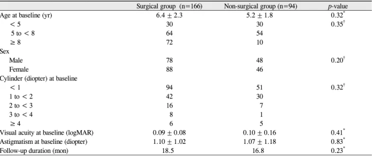
Table 2. Demographic features of the surgical and the non-surgical groups
Surgical group (n=166) Non-surgical group (n=94) p-value
Age at baseline (yr) 6.4 ± 2.3 5.2 ± 1.8 0.32
*< 5 30 30 0.35
†5 to < 8 64 54
≥ 8 72 10
Sex
Male 78 48 0.20
†Female 88 46
Cylinder (diopter) at baseline
< 1 94 51 0.32
†1 to < 2 42 30
2 to < 3 16 7
3 to < 4 8 1
≥ 4 6 5
Visual acuity at baseline (logMAR) 0.09 ± 0.08 0.10 ± 0.16 0.41
*Astigmatism at baseline (diopter) 1.10 ± 1.02 1.07 ± 1.18 0.83
*Follow-up duration (mon) 18.5 16.8 0.23
*logMAR=logarithm of the minimum angle of resolution.
*
t-test;
†Chi-square test.
Table 3. Changes in mean astigmatism and best corrected visual acuity (BCVA) over time in the surgical and non-surgical groups
Baseline 1 mon 3 mon 6 mon 1 yr
Astigmatism (diopter) Surgical group 1.10 0.97 0.84 0.85 0.83
p-value
†- (0.290) (0.046)
*(0.041)
*(0.01)
*Non-surgical group 1.07 1.21 1.09 1.08 1.11
p-value
†- (0.457) (0.871) (0.920) (0.801)
BCVA (logMAR) Surgical group 0.09 0.10 0.07 0.09 0.08
p-value
†- (0.159) (0.458) (0.888) (0.402)
Non-surgical group 0.10 0.11 0.09 0.06 0.07
p-value
†- (0.819) (0.779) (0.083) (0.176)
logMAR = logarithm of the minimum angle of resolution.
*
p<0.05;
†Compared to baseline, t-test.
Table 2. There were no significant differences between the two groups with regard to age, gender, follow-up period, best corrected visual acuity (BCVA), and degree of astigmatism.
The mean astigmatic readings for each group at each fol- low-up visit are shown in Table 3. The mean astigmatic re- fractive error in the surgical group decreased from 1.10 ± 1.02 D preoperatively to 0.84 ± 1.05 D at 3 months post- operatively to 0.83 ± 0.72 D at 1 year postoperatively (p = 0.046, p = 0.01). The average astigmatic refractive error in the non-surgical group was 1.07 ± 1.18 D at baseline and 1.11 ± 1.13 D at 1 year (p = 0.80). There was no statistically significant difference in this group (Table 3). The general lin- ear model analysis, which compared the mean astigmatism between the surgical and the non-surgical groups over time, showed a significant group-time interaction (p = 0.04), in- dicating that the difference in astigmatism between the two groups varied significantly over time. The mean BCVA change was not significant in both groups during follow-up visits.
In the surgical group, after surgery the subgroups (defined by age; subgroup 1, 2, and 3) showed decreasing cylinders over time. The cylinder was significantly decreased in sub- group 2 at 3 months, 6 months, and 1 year after surgery (p = 0.05, 0.04, 0.02). No significant age-related differences were noted in the non-surgical group (Fig. 1). There was also no difference in VA changes according to age in either the surgical group or the non-surgical group.
We also examined the association between astigmatic
changes and initial cylinder values in both the surgical and
non-surgical groups (subgroup A, B, and C) (Fig. 2). In the
surgical group, there was a statistically significant decrease
in astigmatism in subgroups B and C after 3 months postoper
ative. In the non-surgical group, there were no significant as-
tigmatic decreases for any initial astigmatism level. There
was no association between baseline astigmatism and VA
changes in either group.
Fig. 2. Mean cylinder changes in subgroup A, B, and C over 1 year.
Within the surgical group, the higher baseline astigmatic sub- group demonstrated greater cylinder reduction over time com- pared to the lower astigmatic subgroup at 1 year postoperative.
*