• 검색 결과가 없습니다.

RE: Anomalous Cardiac Venous Connection to the Left Atrium Associated with Coronary Sinus Atresia

N/A
N/A
Protected

Academic year: 2022

Share "RE: Anomalous Cardiac Venous Connection to the Left Atrium Associated with Coronary Sinus Atresia"

Copied!
3
0
0

로드 중.... (전체 텍스트 보기)

전체 글

(1)

879

Korean J Radiol 15(6), Nov/Dec 2014

kjronline.org

RE: Anomalous Cardiac Venous Connection to the Left Atrium Associated with Coronary Sinus Atresia

Ersin Ozturk, MD 1 , Cahit Kafadar, MD 1 , Mehmet Dogan, MD 2 , Omer Uz, MD 2

Departments of

1

Radiology and

2

Cardiology, GATA Haydarpasa Teaching Hospital, Istanbul 34668, Turkey

Index terms: Coronary sinus; Coronary venous anomaly;

Coronary CT angiography

Dear Editor,

We read with interest the article of Chou et al. (1). The authors reviewed the multidetector computed tomography (CT) findings of congenital coronary sinus anomalies and reported two cases of coronary sinus anomaly. We would like to share our observations, with respect to coronary sinus anomalies, with our colleagues.

For better clinical outcomes, coronary venous variations should be recognized in the pre-operative planning of numerous cardiac surgical interventions (2). Thus, attention should be directed on the coronary venous anatomy as well as the coronary arterial system. We encountered coronary sinus anomaly in two patients who underwent coronary CT angiography. In the first case, the right atrial coronary sinus ostium was atresic (Fig. 1). Hypoplastic persistent left superior vena cava, great cardiac vein, median cardiac vein,

Received July 25, 2014; accepted after revision August 14, 2014.

Corresponding author: Ersin Ozturk, MD, Department of Radiology, GATA Haydarpasa Teaching Hospital, Uskudar, Istanbul 34668, Turkey.

• Tel: (90) 216 5424654 • Fax: (90) 216 3304388

• E-mail: [email protected]

This is an Open Access article distributed under the terms of the Creative Commons Attribution Non-Commercial License (http://creativecommons.org/licenses/by-nc/3.0) which permits unrestricted non-commercial use, distribution, and reproduction in any medium, provided the original work is properly cited.

Letter to the Editor

and small cardiac veins were joined to a connecting channel.

This channel, probably the levoatriocardinal vein, was found to drain into the left atrium. This case was similar with the case 1 in the article of Chou et al. (1). In the second case, the right atrial ostium of the coronary sinus was again atresi and an abnormal tubular communication between the coronary sinus and the left atrium coexisted and drained into the left atrium. However this case was different from the first one in two ways. Firstly, there were two tortuous, connecting channels, instead of one draining into the left atrium. Secondly, there was a coexisting tortuous tubular connection between the small cardiac vein, which was also draining into the coronary sinus, and the right atrium (Fig.

2). We encountered only one report that was similar with our second case in the English literature (3). Coronary CT angiography provides reliable non-invasive information of cardiac venous anatomy and, since these anomalies are important, especially in cardiovascular surgical interventions, they should be recognized and indicated in the radiological reports.

REFERENCES

1. Chou MC, Wu MT, Chen CH, Lee MH, Tzeng WS. Multidetector CT findings of a congenital coronary sinus anomaly: a report of two cases. Korean J Radiol 2008;9 Suppl:S1-S6

2. Sun JP, Yang XS, Lam YY, Garcia MJ, Yu CM. Evaluation of coronary venous anatomy by multislice computed tomography.

World J Cardiovasc Surg 2012;2:91-95

3. Shum JS, Kim SM, Choe YH. Multidetector CT and MRI of ostial atresia of the coronary sinus, associated collateral venous pathways and cardiac anomalies. Clin Radiol 2012;67:e47-e52 Korean J Radiol 2014;15(6):879-881

http://dx.doi.org/10.3348/kjr.2014.15.6.879

pISSN 1229-6929 · eISSN 2005-8330

(2)

880

Ozturk et al.

Korean J Radiol 15(6), Nov/Dec 2014 kjronline.org Fig. 1. Volume-rendering and maximum intensity projection images show anomalous venous channel (white arrow) connecting coronary sinus (CS) and left atrium (LA). Opening site of anomalous channel (black arrow) and atretic ostium of CS (*) could be seen. Ao

= aorta, GCV = great coronary vein, LAD = left anterior descending artery, LCX = left circumflex artery, LV = left ventricle, MCV = middle coronary

vein, PLSVC = persistent left superior vena cava, PV = pulmonary vein, RCA = right coronary artery, RV = right ventricle, SCV = small coronary vein

(3)

881 Letter to the Editor

Korean J Radiol 15(6), Nov/Dec 2014

kjronline.org

Fig. 2. Volume-rendering images show two anomalous venous channels (white arrows) connecting coronary sinus and left atrium

(LA). Coexisting tortuous tubular connection (black arrow) between small cardiac vein and right atrium could be seen. Ao = aorta, GCV = great

coronary vein, LA = left atrium, MCV = middle coronary vein, PV = pulmonary vein, RA = right atrium, SCV = small coronary vein

수치

Fig. 2. Volume-rendering images show two anomalous venous channels (white arrows) connecting coronary sinus and left atrium  (LA)

참조

관련 문서

Coronary CT angiography revealed a single, common ostium of the right and left coronary artery arising from the right anterior sinus of Valsalva with anomalous course of the

We report here on a case of cardiac hemangioma that originated from the left atrial appendage; this was observed on the CT and coronary angiographic findings and the lesion

Unreported Coronary Artery Anomaly: Association of Right Coronary Artery and Circumflex Coronary Artery with Single Ostium Originate from High Left Anterior Aorta.. Ferhat

Anomalous aortic origin of left coronary artery is a rare congenital coronary anomaly that can cause clinical mani- festations such as ischemic chest pain, arrhythmic syncope or

Management was coronary artery bypass grafting to the left anterior descending artery and obtuse marginal arteries, closure of the left main coronary artery

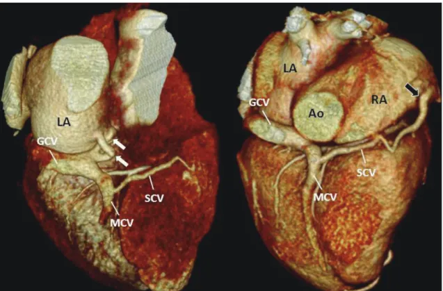
Coronary sinus ostial atresia (CSOA) with persistent left superior vena cava (LSVC) in the absence of an un- roofed coronary sinus is a benign and rare anomaly

Objective: To examine the fractional flow reserve derived from computed tomographic angiography (CT-FFR) in patients with anomalous origin of the right coronary artery from the

mography; LV, left ventricle; LPV, left pulmonary vein; LLPV, left lower pulmonary vein; RLPV, right lower pulmonary vein; RUPV, right upper pulmonary vein; LUPV, left upper