Article Info
Received April 20, 2020 Revised May 25, 2020 Accepted July 15, 2020 Corresponding Author Jong-Duk Choi E-mail: [email protected]
https://orcid.org/0000-0002-9663-4790
Key Words Gait Position sense Postural balance Stroke
Background: Stroke patients have reduced trunk control compared to normal people. The ability to control the trunk of a stroke patient is important for gait and balance. However, there is still a lack of research methods for the characteristics of stroke control in stroke patients.
Objects: The aim of this research was to determine whether trunk position sense has any relation with balance and gait.
Methods: This study assessed trunk performance by measuring position sense. Trunk posi- tion sense was assessed using the David back concept to determine trunk repositioning error in 20 stroke patients and 20 healthy subjects. Four trunk movements (flexion, extension, lat- eral flexion, rotation) were tested for repositioning error and the measurement was carried out 6 times per move; these parameters were used to compare the mean values obtained. Subjects with stroke were also evaluated with clinical measures of balance and gait.
Results: There were significant differences in trunk repositioning error between the stroke group and the control group in flexion, lateral flexion to the affected side, lateral flexion to the unaffected side, rotation to the affected side, and rotation to the unaffected side. Mean flexion error: post-stroke: 7.95 ± 6.76 degrees, control: 3.32 ± 2.27; mean lateral flexion error to the affected side: 6.13 ± 3.79, to the unaffected side: 5.32 ± 3.15, control: 3.57
± 1.92; mean rotation error to the affected side: 8.25 ± 3.09, to the unaffected side: 9.24
± 3.94, control: 5.41 ± 1.82. There was an only significant negative correlation between the repositioning error of lateral flexion and the Berg balance scale score to the affected side (–0.483) and to the unaffected side (–0.497). A strong correlation between balance and gait was found.
Conclusion: The results of this study indicate that stroke patients exhibit greater trunk re- positioning error than age-matched controls on all planes of movement except for extension.
And lateral flexion has correlation with balance and gait.
Copyright ⓒ Korean Research Society of Physical Therapy
This is an Open Access article distributed under the terms of the Creative Commons Attribution Non-Commercial License (http://creativecommons.org/licenses/by-nc/4.0) which permits unrestricted non-commercial use, distribution, and reproduction in any medium, provided the original work is properly cited.
INTRODUCTION
뇌졸중은 환자에게 실제적 혹은 잠재적인 생명의 위협을 경험하게 할 뿐만 아니라 영구적인 기능장애나 합병증을 야기함으로써 신체적
안녕에 위협을 주는 사건이다[1]. 뇌졸중 발생 이후 환자들에게 급성부 터 만성에 이르기까지 근력약화, 근 긴장도의 변화, 선택적이고 분리된 움직임의 소실, 균형유지의 어려움, 머리와 체간의 부정렬, 비대칭적인 체중 지지 등 폭넓게 운동능력과 관련된 증상이 나타난다[2]. 그 중 적
Physical Therapy Korea
PTK https://doi.org/10.12674/ptk.2020.27.3.178 pISSN: 1225-8962 eISSN: 2287-982X Phys Ther Korea. 2020;27(3):178-184
Original Article
정상인과 뇌졸중 환자의 체간 위치감각 비교 및 보행과 균형에 미치는 영향
양해덕1, 김창범2, 최종덕3, 문 영4
1
충남대학교병원 재활센터,
2대한고유수용성신경근촉진법학회 서울 · 경기 남부회,
3대전대학교 보건의료과학대학 물리치료학과,
4더이룸아동발달센터
운동발달실
Comparison Between Stroke Patients and Normal Persons for Trunk Position Sense and It’s Relation to Balance and Gait
Hea-Duck Yang
1, MSc, Chang-beom Kim
2, PhD, Jong-Duk Choi
3, PhD, Young Moon
4, MSc
1
Rehabilitation Center, Chungnam National University Hospital, Daejeon,
2Korea Proprioceptive Neuromuscular Facilitation Association in South Seoul Gyeonggi, Gyeonggi-do,
3Department of Physical Therapy, College of Health & Medical Science, Daejeon University,
4
Department of Movement Development, The ERUM Child Development Center, Iksan, Korea
절한 체간의 움직임은 자세조절, 균형, 보행, 이동, 및 일상생활 활동에 있어서 필수적인 요소이다[2,3].
뇌졸중 환자의 체간 조절은 기능적인 회복을 예측하는 중요한 인자 이기도 하다[4]. 체간 근육들의 상호작용으로 인한 체간의 조절은 균형 능력 향상과 신체의 안정성, 운동성에 중요한 역할을 한다[5]. 체간 조 절의 불완전성은 균형의 기능부전과 관련이 있으며[6], 이는 보행 및 일 상생활에 많은 장애를 갖게 한다[7]. 뇌졸중 환자의 보행 기능을 향상을 위한 중재는 뇌졸중 환자의 전 생애에 걸친 건강과 복지 향상에 매우 중요하기 때문에[8], 일상생활능력과 보행, 삶의 질을 향상시키기 위한 다양한 프로그램이 요구되고 있다.
체간의 안정성은 적절한 신체인식을 필요로 하며, 이는 수동적 요소, 능동적 요소, 신경학적 요소 세 가지의 상호작용으로 이루어진다. 수동 적 요소는 척추뼈, 추간디스크, 인대, 관절낭을 포함하고, 능동적 요소 는 근육과 힘줄이며, 신경학적 요소는 수동적, 능동적 요소에 분포되어 있는 신경섬유와 이를 조절하는 중추신경계이다[9]. 체간의 안정성은 신체에 대한 정확한 인식을 제공하는 고유수용성 감각, 중력과 외력을 버틸 수 있는 충분한 근력, 입력된 정보를 바탕으로 적절한 근육반응을 이끌어내는 신경조절에 있다[10]. 신체에 대한 인식은 시각, 전정감각, 고유수용성 감각에 기초하여 계속 수정되며, 근육활동도 이에 따라 계 속 수정된다[11].
균형이란 안정성 한계 안에서 신체 무게중심을 기저면 위에 유지하 는 능력으로, 다양한 시스템의 통합적인 작용을 필요로 한다[12]. 또한, 보행은 복잡한 균형 작용을 통해 유지되는 것으로, 중력중심이 기저면 밖으로 이탈됨과 동시에 걸음을 디딤으로 새로운 기저면을 만들어 균 형을 회복하면서 진행된다. 보행은 균형을 평가하는 동시에 치료가 될 수 있는 기능적인 수단이며[13], 균형과 보행은 뇌졸중 재활의 주요한 목표 중 하나이다[14,15].
고유수용성 감각, 특히 체간 위치감각은 체간 조절에 있어 필수적인 요소이기 때문에, 손상 시 체간 불안정성이나 근골격계의 손상을 유발 할 수 있으며[10], 뇌졸중 환자들은 정상인에 비해 체간 조절 능력이 감 소되어 있다[7]. 그러나 뇌졸중 환자를 대상으로 한 많은 연구가 상지 기능이나 보행 등의 주제로 이루어져 왔다. 뇌졸중 환자의 재활에 있어 체간 능력의 회복이 중요하나 그 중요성이 자주 간과되어 왔으며, 뇌졸 중 환자의 체간 특성에 대한 연구나 체간 조절을 향상시키는 방법에 대 한 연구는 아직 미비한 실정이다[4,10].
따라서 본 연구의 목적은 뇌졸중 환자의 체간 위치감각을 정상인과 의 비교를 통해 뇌졸중 환자의 체간 움직임 중 어떤 움직임이 가장 많 이 손상 받았는지 알아보고, 균형 및 보행과의 상관성을 알아보아 뇌졸 중 환자의 물리치료적 중재에 있어 중점을 두어야 할 동작이 어떤 것인 지 알아보고자 하였다.
MATERIALS AND METHODS
1. 연구 대상자
본 연구는 대전에 위치한 충남대학교병원에서 입원 또는 외래 로 치료 중인 뇌졸중 환자와 연령대가 유사한 정상 성인을 대상으로 실시하였다. 본 연구의 대상자 수는 G*Power 프로그램(G*Power ver.3.1.9.2; University of Kiel, Kiel, Germany)을 사용하여 산출하 였다. 효과크기를 0.9로 가정하고 유의수준 0.05, 검정력 0.8로 설정한 후 표본크기를 산출한 결과, 대상자의 최소 표본크기는 42명이었다. 이 를 근거로 대상자를 뇌졸중 환자군 21명, 정상인군 21명 총 42명을 모 집하였다. 본 연구의 참가대상자는 (1) 뇌경색 또는 뇌출혈로 편마비 양 상을 보이는 뇌졸중 환자, (2) 한국판 간이 정신상태 검사(mini mental state examinationKorea, MMSEK) 점수가 24점 이상으로 치료사 의 지시를 이해하고 따라 할 수 있는 인지능력을 가진 환자[16], (3) 측 정 장비를 이용하는 데 어려움이 없을 정도의 체간 조절능력을 가진 환 자로 하였으며, 정상인은 최근 1년간 요통을 경험하지 않은 자 중 본 연 구 참여에 동의한 자를 대상으로 하였다. 본 연구의 제외대상은 (1) 척 추 수술을 한 병력이 있는 자, (2) 추간판 탈출증이나 척추관 협착증 같 은 분명한 허리질환이 있는 자, (3) 균형이나 보행에 영향을 주는 다른 신경학적 질환이 있는 자로 하였다. 모든 실험 및 절차는 대전대학교 생명윤리 규정 및 헬싱키 선언에 따라 진행되었으며 모든 대상자들에 게 본 연구에 대해 충분히 설명하였으며, 실험 참여에 동의한 후 연구 를 실시하였다.
2. 측정 방법 및 도구
1) 능동관절범위 및 체간 위치감각 측정
등근육 측정 및 훈련장비인 David back concept (David Health Solutions, Helsinki, Finland)를 이용하여 측정하였다. 이 장비는 흉 요추부의 굴곡을 측정할 수 있는 F130, 신전을 측정하는 F110, 측방 굴곡을 측정하는 F150, 회전을 측정하는 F120의 총 4모델로 구성되어 있다. 각각의 장치는 흉요추부의 움직임만 측정할 수 있도록 하지의 움 직임을 제어하는 고정 장치가 있다. 고관절 90°, 슬관절 90°의 앉은 자 세에서 측정하며, 측정값은 2° 단위로 디지털로 표시된다. 각 대상자들 은 먼저 능동관절범위를 측정하였다. 체간 위치감각은 능동관절범위의 50% 지점을 목표지점으로 하게 되므로 능동관절범위를 먼저 평가하였 다. 각 4개의 장치에 앉아서 측정하였으며, 측정순서는 무작위로 실시 하였다.
본 연구에서는 체간 재위치 오차(trunk repositioning error, TRE) 각도를 재는 방법으로 체간 위치감각을 측정하였다. 먼저 각 환자의 전 체 관절 가동 범위를 측정하고, 전체 관절 가동 범위의 50% 지점을 목 표지점으로 설정하였다[10,17]. 목표지점에 도달한 후에는 3초간 그 지점에 머물게 하였다가 다시 처음 자세로 돌아가게 하였다[10] (Fig
ure 1).
본 측정을 실시하기 전 환자가 측정방법을 제대로 이해하였는지 확
인하기 위해 전체 측정범위의 25% 지점을 목표로 연습을 하였으며, 환 자가 측정방법을 숙지하였음을 확인한 후 바로 본 측정을 실시하였다.
반복 횟수는 오차를 줄이고 정확도를 높이기 위해 6회 반복을 실시하 였다[18]. 장시간 눈을 가리고 있으면 측정값의 편차가 증가하고 오차 가 발생할 수 있어 연습 측정을 할 때는 눈을 뜬 상태에서 실시하였고, 본 측정을 할 때는 눈을 가리고 실시하였다[19]. 또한, 본 실험을 실시 하는 동안에는 어떠한 구어적 피드백도 주지 않았다. 정상인과 뇌졸중 환자의 재위치 오차의 각도는 목표 각도보다 더 지나쳐 가거나 목표 각 도에 도달하지 못한 각도를 절댓값으로 처리하여 분석에 사용하였으 며, 평균과 표준편차를 비교 분석에 사용하였다. 측굴과 회전에서 정상 인은 환측, 건측이 없으므로, 좌우 측정값의 평균값을 분석에 사용하였 다. TRE는 체간 위치감각을 평가하는 신뢰성 있고 타당성 있는 방법이 다[20]. 측정은 장비의 사용이 숙련된 치료사에 의해 수행되었다.
2) 균형능력 측정
균형능력 평가는 버그 균형 척도(Berg balance scale)를 사용하였 다. 0–4점의 5점 척도로 14개 항목으로 되어 있으며, 만점은 56점이 다. 버그 균형 척도는 점수가 높을수록 균형능력이 좋다는 것을 의미한 다. 뇌졸중 환자에게 적용한 버그 균형 척도의 등급 내 상관계수(intra
class correlation coefficients)는 평가자간 신뢰도(interrater reli
ability)가 0.95–0.98이며, 측정자 내 신뢰도(intrarater reliability)는 0.97, 검사재검사 신뢰도(testretest reliability)는 0.98로 뛰어난 신 뢰도를 가지고 있다[21].
3) 보행능력 측정
보행능력 평가는 티네티 수행목적 이동평가(Tinetti performance oriented mobility assessment)의 보행부분으로 평가하였다. 티네티
수행목적 이동평가는 노인들의 균형과 보행능력을 평가하기 위해 만들 어진 평가도구로 균형부분과 보행부분으로 나누어지며, 여기에서는 보 행부분만 평가에 사용하였다. 보행부분은 총 8개 항목의 12점 만점으 로 되어있으며, 점수가 높을수록 기능이 좋음을 의미한다. 뇌졸중 환자 에게 적용한 티네티 수행목적 이동평가의 등급 내 상관계수는 평가자 간 신뢰도가 0.94이다[22].
3. 실험 절차
모든 실험 참가자들의 실험 참가 동의 후 자료 수집이 이루어졌다.
정상인은 면담을 통해 나이, 요통 유무, 신경학적 질환 유무 등 일반적 인 특성을 조사하였고, 뇌졸중군은 본인 또는 대리인의 동의 후 의무 기록지를 통해 뇌졸중 유형 및 부위, 발병날짜, MMSEK 점수 등의 정 보를 얻었다. 두 군 모두 능동관절범위를 먼저 측정한 후 관절 재위치 오차를 측정하였으며, 뇌졸중군은 버그 균형 척도와 티네티 수행목적 이동평가 보행부분을 추가로 실시하였다. 두 군에서 모두 1명씩 중간 탈락자가 발생했으며, 이외의 수집된 모든 자료는 분석에 사용되었다.
4. 분석방법
실험 참가자의 나이, 성별, 마비측, 유병기간 등의 일반적 특성은 기 술통계로 분석하였다. 뇌졸중 환자와 정상인의 능동관절범위와 관절 재위치 오차의 비교, 뇌졸중 환자의 건측과 환측의 비교는 독립 t 검정 을 이용하여 분석하였다. 관절 재위치 오차와 균형 및 보행과의 상관성 은 버그 균형 척도와 티네티 수행목적이동평가의 보행부분 점수를 피 어슨 상관분석을 통해 분석하였다. PASW Statistics 18.0 (IBM Co., Armonk, NY, USA)을 이용하여 분석하였으며, 모든 통계적 유의수준 은 0.05로 하였다.
Flexion (F130) Extension (F110)
Lateral flexion (F150) Rotation (F120)
Figure 1.
Figure 1. Measurment of trunk repositioning error.
RESULTS
1. 연구 대상자의 일반적인 특성
각 군의 연령은 뇌졸중 환자군은 최저 21세부터 최고 74세로 평균 51세이며, 정상인군은 최저 26세부터 최고 60세로 평균 44세였다. 뇌 졸중 환자군의 유병 기간은 최저 1개월부터 최고 120개월로 평균 45.9 개월의 유병 기간을 나타내었다. 성별은 뇌졸중 환자군은 남자가 11명, 여자가 9명이었으며, 정상인군은 남자가 9명, 여자가 11명이었다. 뇌 졸중 유형은 경색이 9명, 출혈이 11명이었고, 마비측은 좌측편마비가 11명, 우측편마비가 9명이었다(Table 1).
2. 뇌졸중군과 정상인군의 능동 관절가동범위 비교
뇌졸중군과 정상인군 간의 능동 관절가동범위 비교에서는 정상인 그 룹이 뇌졸중 그룹보다 굴곡은 4.4도, 신전은 2.6도, 환측 측굴은 11.2도, 건측 측굴은 9.7도, 환측 회전은 23.8도, 건측 회전은 19도 더 많은 가동 범위를 나타내었다. 통계적으로는 뇌졸중군이 신전을 제외한 모든 움직 임에서 정상인보다 더 적은 가동범위를 보였다(p < 0.05) (Table 2).
3. 뇌졸중군과 정상인군의 체간 위치감각 비교
뇌졸중군과 정상인군 간의 체간 재위치 오차 비교에서는 뇌졸중 그룹 이 정상인 그룹보다 굴곡은 4.63도, 신전은 1.88도, 환측 측굴은 2.53 도, 건측 측굴은 1.75도, 환측 회전은 2.84도, 건측 회전은 3.83도 더
큰 오차를 나타내었다. 통계적으로는 뇌졸중군이 신전을 제외한 모든 움직임에서 정상인보다 더 큰 오차범위를 보였다(p < 0.05) (Table 3).
4. 뇌졸중군의 체간 위치감각 환측, 건측 비교
뇌졸중 환자의 건측과 환측의 체간 재위치 오차 비교에서는 좌우 차 이가 측굴은 0.45도, 회전은 0.99도로 측굴과 회전 모두에서 통계적으 로 유의한 차이를 보이지 않았다(p < 0.05) (Table 4).
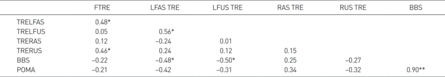
5. 뇌졸중군의 체간 위치감각과 균형, 보행과의 상관성
뇌졸중 환자의 관절 재위치 오차와 균형, 보행과의 상관성에서는 환 측과 건측으로의 측굴이 상관성이 있는 것으로 나타났다(p < 0.05). 또 한, 균형능력과 보행 사이에는 강한 상관관계가 있는 것으로 나타났다 (p < 0.01) (Table 5).
DISCUSSION
체간 위치감각은 주로 정상인이나 만성 요통환자를 대상으로 연구되 어왔다. 기존의 체간 위치감각 연구들에서는 서 있는 자세[17,18,23], 앉은 자세[10,2426], 네발기기 자세[12] 등 다양한 측정 자세가 사용 되었다. 또한, 목표 각도도 전체 관절가동 범위의 50% 지점[10,17], 1/3지점[24], 2/3지점[23,26], 가동범위 끝까지 간 후 중립위치 찾 기[17,25], 전체 가동범위의 20%, 50%, 80% [18], 특정 각도 지정 [27,28] 등 다양하게 시행되었다. 반복 횟수도 3회에서 20회까지 다양 하게 실시되었다[26]. 본 연구에서는 앉은 자세에서 전체 가동범위의 50% 지점, 6회를 반복하여 실시하였다. 앉는 자세는 편안하고, 전정감 각과 다리와 발에서부터 오는 감각정보를 최소화 할 수 있으며, 보상적 Table 1.
Table 1. General characteristics of study subjects
Variable Stroke (n = 20) Normal (n = 20)
Age (y) 50.86 ± 15.93 44.14 ± 12.71
Sex (male/female) 11/9 9/11
Type of stroke (infarction/hemorrhage) 9/11 Side of stroke (right/left) 11/9 Time after stroke (mo) 45.88 ± 43.92
MMSEK 27.34 ± 1.75
BBS 48.01 ± 7.87
POMA 10.49 ± 1.86
Values are presented as mean ± standard deviation or number only.
MMSEK, minimental state Korea examination; BBS, Berg balance scale; POMA, Tinetti performance oriented mobility assessment.
Table 2.
Table 2. Comparison of the AROM between stroke patients and normal
persons
AROM Stroke Normal t
Flexion (degree) 48.13 ± 8.45 52.48 ± 4.15 2.09*
Extension (degree) 31.65 ± 5.96 34.29 ± 2.36 1.81 LFAS (degree) 43.69 ± 12.71 54.91 ± 5.97 3.57*
LFUS (degree) 45.22 ± 13.46 54.91 ± 5.97 2.95*
RAS (degree) 47.49 ± 20.17 71.34 ± 10.51 4.67*
RUS (degree) 52.26 ± 21.75 71.28 ± 10.51 3.51*
Values are presented as mean ± standard deviation. AROM, active range of motion; LFAS, lateral flexion to the affected side; LFUS, lateral flexion to the unaffected side; RAS, rotation to the affected side; RUS, rotation to the unaffected side. *p < 0.05.
Table 3.
Table 3. Comparison of the TPS between stroke patients and normal per
sons
TPS Stroke Normal t
Flexion (degree) 7.95 ± 6.76 3.32 ± 2.27 –2.89*
Extension (degree) 6.05 ± 5.66 4.17 ± 2.02 –1.41 LFAS (degree) 6.14 ± 3.83 3.57 ± 1.92 –2.66*
LFUS (degree) 5.32 ± 3.15 3.57 ± 1.92 –2.13*
RAS (degree) 8.25 ± 3.09 5.41 ± 1.82 –3.54*
RUS (degree) 9.24 ± 3.94 5.41 ± 1.82 –3.95*
Values are presented as mean ± standard deviation. TPS, trunk position sense; LFAS, lateral flexion to the affected side; LFUS, lateral flexion to the unaffected side; RAS, rotation to the affected side; RUS, rotation to the unaffected side. *p < 0.05.
Table 4.
Table 4. Comparison of the TRE between affected side and unaffected
side in stroke patients
TRE Affected side Unaffected side t Lateral flexion (degree) 6.13 ± 3.79 5.32 ± 3.15 0.42 Rotation (degree) 8.25 ± 3.09 9.24 ± 3.94 –0.88 Values are presented as mean ± standard deviation. TRE, trunk reposi
tioning error.
인 움직임을 제한해 척추의 움직임만 측정할 수 있도록 해준다[17,24].
또한, 앉은 자세는 기능적이고, 서 있는 자세보다 더 신뢰할만한 위치 감각 측정을 할 수 있었다[29]. 본 연구에 사용된 DAVID 장비는 하지 고정 장치가 있어, 보다 더 척추의 움직임만 측정하게 되어 있다. 정상 인을 대상으로 한 연구에서 전체 척추 움직임의 50% 지점이 가장 안정 된 TRE 값을 가져오기 때문에 본 연구에서는 체간 재위치 오차의 목표 지점을 전체 가동범위의 50%로 설정하였다[18,23].
연구의 결과 중 뇌졸중 환자와 정상인의 능동관절범위 비교에서는 정상인 그룹이 뇌졸중 환자 그룹보다 굴곡은 4.4도, 신전은 2.6도, 측 굴 환측은 11.2도, 측굴 건측은 9.7도, 회전 환측은 23.8도, 회전 건측 은 19도 더 많은 가동범위를 나타내었다. 통계적으로는 뇌졸중 환자 군이 신전을 제외한 모든 움직임에서 유의하게 더 적은 가동범위를 보 였다(p < 0.05). 이러한 결과는 뇌졸중 환자들의 근력이 정상인에 비 해 저하되어 있기 때문일 것으로 생각된다. 본 연구에서는 근력을 측정 하여 비교하지는 않았지만, 근력의 약화는 뇌졸중 환자들에게 흔히 나 타나는 증상 중 하나이며[30,31], Tanaka 등[31]도 뇌졸중 환자들 체 간의 굴곡신전 근력과 회전근력이 정상인보다 더 낮다고 보고하였다.
Bohannon 등[32]도 뇌졸중 환자의 체간 굴곡과 환측 및 건측으로의 측굴 근력이 정상인보다 더 낮다고 보고하였다. 특히 Ada 등[29]은 뇌 졸중 환자는 근육의 길이가 짧아진 범위에서 근력의 약화가 더 심하게 나타난다고 하였다. 따라서 뇌졸중 환자군은 정상인군에 비해 가동범 위의 제한이 있었을 것이라고 사료된다.
척주의 위치 감각은 나이, 성별, 진동감각, 보조기 착용, 근피로, 체 간의 위치나 움직임 등에 의해 영향을 받는다고 알려져 있다[28]. 본 연 구에서는 뇌졸중이라는 질환이 환자의 체간 위치감각에 어떤 영향을 미치는지 알아보고자 실시하였다. 체간 재위치 오차를 구하는 방법으 로 뇌졸중 환자와 정상인을 비교한 결과 뇌졸중 환자군이 정상인군보 다 굴곡은 4.63도, 신전은 1.88도, 측굴 환측은 2.53도, 측굴 건측은 1.75도, 회전 환측은 2.84도, 회전 건측은 3.83도의 더 큰 오차를 나타 내었다. 통계적으로는 뇌졸중 환자군이 신전을 제외한 모든 움직임에 서 정상인보다 더 큰 오차 값을 나타내었다(p < 0.05). Ryerson 등[9]
의 연구에서는 시상면과 가로면에서의 움직임에서 뇌졸중 환자의 체
간 재위치 오차 값이 정상인보다 2배 정도 큰 오차 값을 가지며, 이마면 에서도 2배 정도의 오차 값을 가지나 통계적으로 유의하지는 않았다고 보고하였다. 본 연구에서는 시상면의 움직임인 신전이 통계적으로 유 의하지 않은 반면 선행연구[10]에서는 이마면의 움직임이 유의하지 않 다고 보고하였다. 측정 장비 및 측정 방법의 차이로 조금 다른 결과를 가져왔으나 Ryerson 등[9]의 연구에서도 이마면의 오차 값이 정상인의 2배에 가깝다고 한 것으로 볼 때 전반적인 뇌졸중 환자의 체간 위치감 각이 정상인과 비교하여 감소되어 있는 것으로 사료된다.
뇌졸중 환자군의 측굴과 회전의 건측과 환측 체간 재위치 오차 비교 에서는 좌우 차이가 측굴은 0.45도, 회전은 0.99도로 건측과 환측이 통 계적으로 유의한 차이가 없었다. 이는 체간 근육은 뇌의 양쪽에서 신경 지배를 받기 때문으로 생각된다[33]. Tanaka 등[31]의 연구에서도 뇌 졸중 환자의 환측과 건측으로의 체간 회전 근력이 유의한 차이가 없는 결론 역시 본 연구의 결과를 설명하는 데 도움이 될 것이다.
체간 위치감각과 균형과의 상관성에서는 측굴이 상관관계가 있는 것 으로 나타났다. 체간 위치감각과 버그 균형 척도와의 상관성에서 측굴 환측과 건측 모두 통계학적으로 유의한 작은 상관성을 나타내었다. 티 네티 수행목적 이동평가 보행부분과 측굴 위치 감각과는 통계적인 유 의성이 나타나지 않았으나 환측으로의 측굴은 p값이 0.06으로 0.05 에 근접한 값을 나타내었다. 그리고 균형과 보행간의 상관성은 통계학 적으로 유의한 강한 상관관계를 보였다. Verheyden 등[3]은 추가적인 체간 운동이 환자의 체간능력을 높이는 데 유익한 영향을 미친다고 하 였다. 따라서 체간의 근육을 강화하는 운동이나 측굴을 위주로 한 체간 운동을 추가로 실시하는 것이, 뇌졸중 환자의 체간 재활에 도움이 될 것으로 생각된다. 본 연구는 대상의 수가 적어 결과를 일반화시키는 것 에 어려움이 있을 것이며, 앞으로 체간 능력 향상에 관한 확대된 연구 가 필요할 것으로 사료된다.
CONCLUSIONS
본 연구는 뇌졸중 환자의 체간 위치감각을 조사하여 정상인과의 비 교를 통해 뇌졸중 환자의 체간 움직임 중 어떤 움직임이 가장 많이 손 Table 5.
Table 5. Relation between TRE+ and balance and gait in stroke patients