Annals of Rehabilitation Medicine
Ann Rehabil Med 2016;40(3):420-431 pISSN: 2234-0645 • eISSN: 2234-0653 http://dx.doi.org/10.5535/arm.2016.40.3.420
Virtual Reality-Guided Motor Imagery Increases Corticomotor Excitability in Healthy Volunteers and Stroke Patients
Hyungjun Im, MD
1, Jeunghun Ku, PhD
2, Hyun Jung Kim, MD
1, Youn Joo Kang, MD
11
Department of Rehabilitation Medicine, Eulji Hospital, Eulji University School of Medicine, Seoul;
2
Department of Biomedical Engineering, Keimyung University, Daegu, Korea
Objective To investigate the effects of using motor imagery (MI) in combination with a virtual reality (VR) program on healthy volunteers and stroke patients. In addition, this study investigated whether task variability within the VR-guided MI programs would influence corticomotor excitability.
Methods The present study included 15 stroke patients and 15 healthy right-handed volunteers who were presented with four different conditions in a random order: rest, MI alone, VR-guided MI, and VR-guided MI with task variability. The corticomotor excitability of each participant was assessed before, during, and after each condition by measuring changes in the various parameters of motor-evoked potentials (MEPs) of the extensor carpi radials (ECR). Changes in intracortical inhibition (ICI) and intracortical facilitation (ICF) were calculated after each condition as percentages of inhibition (%INH) and facilitation (%FAC) at rest.
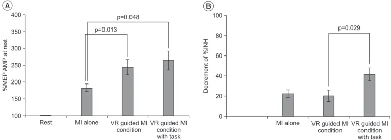
Results In both groups, the increases in MEP amplitudes were greater during the two VR-guided MI conditions than during MI alone. Additionally, the reductions in ECR %INH in both groups were greater under the condition involving VR-guided MI with task variability than under that involving VR-guided MI with regular interval.
Conclusion The corticomotor excitability elicited by MI using a VR avatar representation was greater than that elicited by MI with real body observations. Furthermore, the use of task variability in a VR program may enhance neural regeneration after stroke by reducing ICI. The present findings support the use of various VR programs as well as the concept of combining MI with VR programs for neurorehabilitation.
Keywords Imagery (psychotherapy), Stroke, Transcranial magnetic stimulation, Upper extremity, Virtual reality exposure therapy
Received August 4, 2015; Accepted September 30, 2015 Corresponding author: Youn Joo Kang
Department of Rehabilitation Medicine, Eulji Hospital, Eulji University School of Medicine, 68 Hangeulbiseok-ro, Nowon-gu, Seoul 01830, Korea. Tel:
+82-2-970-8315, Fax: +82-2-979-8268, E-mail: [email protected]
ORCID: Hyungjun Im (http://orcid.org/0000-0002-2360-7775); Jeunghun Ku (http://orcid.org/0000-0002-9610-0078); Hyun Jung Kim (http://orcid.
org/0000-0002-2198-5668); Youn Joo Kang (http://orcid.org/0000-0002-9938-5435).
This is an open-access article distributed under the terms of the Creative Commons Attribution Non-Commercial License (http://creativecommons.org/
licenses/by-nc/4.0) which permits unrestricted noncommercial use, distribution, and reproduction in any medium, provided the original work is properly cited.
Copyright © 2016 by Korean Academy of Rehabilitation Medicine
INTRODUCTION
Because long-term impairments of the upper extremi- ties influence the ability of approximately 65% of patients to perform the activities of daily living following a stroke, exercise programs aimed at restoring function in the up- per extremities are an important part of stroke rehabilita- tion [1]. However, a recent Cochrane review reported that despite the existence of a number of moderate-quality interventions, including virtual reality (VR), mental prac- tice/motor imagery (MI), constraint-induced movement therapy, high doses of repetitive task practice, mirror therapy, and interventions for sensory impairment, there are currently no high-quality evidences for interventions in the field of upper extremity rehabilitation for stroke patients [2].
The observation of other individuals performing skilled movements as well as MI are effective for motor training [3]. Neuroimaging studies have shown that the primary motor cortex (M1) and secondary motor areas, includ- ing the premotor cortex, supplementary motor area, and the parietal cortices, are activated during MI tasks and motor execution [4]. Furthermore, recent random- ized clinical trials evaluating the effectiveness of MI for the improvement of upper extremity motor dysfunction have returned promising results [5]. VR training is also an emerging technology in the field of rehabilitation. Recent randomized controlled trials with large sample sizes that utilized VR training for the upper extremities after stroke have tended to focus on various sensorimotor feedback and augmented reality technologies and have provided encouraging findings [6,7]. Furthermore, functional magnetic resonance imaging (fMRI) studies have demon- strated the occurrence of visuomotor cortical facilitation during VR training [8,9]. Relative to conventional thera- pies for stroke patients, VR training has many advantages.
For example, because it provides a goal-directed task for the patient, VR training is more effective and intensive than self-training, and may also boost the motivation of patients and serve as a pleasurable experience during treatment by controlling the level of difficulty and the variability of the task [10,11].
Because MI and VR applications are increasingly emerging as potentially useful techniques for rehabili- tation after stroke, the present study was designed to determine whether MI combined with a VR program
would have synergistic effects on the patient and provide superior corticomotor facilitation, as compared with MI alone. Thus, we first assessed stroke patients and healthy volunteers to investigate the combined effects of MI and VR training. Second, we investigated whether task vari- ability within the VR-guided MI programs would influ- ence corticomotor excitability or intracortical inhibition (ICI).
It is possible to evaluate corticomotor excitability by applying single- and paired-pulse transcranial magnetic stimulation (TMS). A decreased ICI in the early stages of stroke enhances neural plasticity [12,13]. The assessment of the parameters of motor-evoked potentials (MEPs), such as resting motor threshold (RMT), amplitude, area, ICI, and intracortical facilitation (ICF), allows for the identification of differences in corticomotor facilitation under various experimental conditions.
MATERIALS AND METHODS Participants
The present study included 15 right-handed healthy volunteers (12 males and 3 females; mean age, 31.73±6.22 years) and 15 stroke patients (9 males and 6 females;
mean age, 58.87±10.07 years). Detailed data regarding age, gender, and handedness were provided in Table 1.
Although the mean age of the two groups differed signifi- cantly in the present study, a previous study found that the ability to exhibit MI-induced corticomotor facilitation appears to be largely preserved throughout aging [14].
The healthy volunteers had no history of neurological
or psychological disorders and no abnormalities were
observed during their physical, neurological, and mus-
culoskeletal examinations. According to the Edinburgh
Handedness Inventory [15], all healthy volunteers were
right-handed with a mean laterality quotient (LQ) of
84.77±17.25. Additionally, none of the volunteers had any
contraindications for TMS, such as intracranial metal-
lic pieces or cardiac pacemakers [16]. The stroke group
included patients with a first-ever stroke who were diag-
nosed by MRI or computed tomography scans, had mild
to moderate hemiparesis of an upper extremity with a
Medical Research Council (MRC) grade ≥3 during the
motor examination of the contralesional wrist extension,
and had no upper extremity injuries or deformities. All
subjects were able to sit upright throughout the experi-
ments and did not have any severe cognitive deficits.
The exclusion criteria for the present study were as follows: (1) severe motor weakness (MRC grade of wrist extension ≤2); (2) the absence of MEPs in the affected extensor carpi radialis (ECR) muscle; (3) severe depres- sion, apraxia, and/or cognitive deficits with a score <24 on the Mini-Mental State Examination [17]; (4) a history of seizures; (5) the inability to perform MI using the Viv- idness of Movement Imagery Questionnaire-2 (VMIQ-2) based on an average score ≥4 (vague and dim) [18]; and/
or (6) any contraindications for TMS, such as intracranial metallic implants or cardiac pacemakers [16].
The Institutional Review Board of Eulji Hospital ap- proved the protocol, and all subjects provided written informed consent. Following the experiment, all subjects were assessed to determine if there were any adverse ef- fects from the TMS including dizziness, headaches, or neck pains.
Experimental conditions
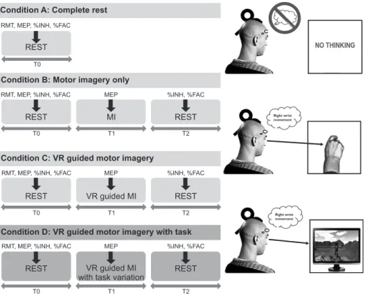
The present study included four different conditions
that were administered in a random order on the same day: rest (Condition A), MI alone (Condition B), VR- guided MI (Condition C), and VR-guided MI with task variability (Condition D). The subjects were asked to sit in a relaxed position during the performance of each condition, and more than 30 minutes of rest was allowed between the performances. Wrist exercise imagery was chosen because this type of movement plays an impor- tant functional role in the recovery of the upper extremity after stroke.
Under Condition A, the subjects were asked to watch a black screen without having other thoughts. Under Condition B, the subjects were asked to imagine wrist extension while observing their own wrist; a metronome (20 beats per minute) was used to cue the MI. Under Condition C, the extension of the wrist was imagined at a constant pace while observing an avatar on the moni- tor screen jump over obstacles at regular time intervals (5 seconds). Under Condition D, the subjects were asked to imagine wrist extension while an avatar on the screen jumped over obstacles at irregular time intervals (ran- Table 1. Demographic and baseline characteristics of subjects
Patient no. Sex Age (yr) Weeks since onset Etiology Site of lesion VMIQ-2
1 M 63 6 Infarction Rt. med. medullary 1.75
2 F 62 3 Infarction Rt. MCA
(cortical and subcortical)
2.25
3 F 67 7 Infarction Rt. MCA
(cortical and subcortical)
2.00
4 F 68 7 Hemorrhage Lt. thalamus (subcortical),
IVH
2.00
5 M 71 4 Infarction Rt. lat. medullary 2.25
6 M 54 4 Infarction Lt. pontine 1.83
7 F 53 5 Infarction Rt. MCA (subcortical) 1.67
8 M 74 3 Infarction Lt. PCA MCA ACA
(cortical and subcortical)
1.83
9 M 52 1 Infarction Lt. MCA (subcortical) 2.00
10 F 59 4 Infarction Rt. pontine 2.25
11 M 35 3 Infarction Lt. MCA
(cortical and subcortical)
1.67
12 F 48 6 Hemorrhage Lt. BG 2.25
13 M 52 33 Hemorrhage Rt. post. medullary 2.25
14 M 62 1 Infarction Rt. MCA
(cortical and subcortical)
1.89
15 F 63 1 Infarction Lt. MCA (subcortical) 2.17
VMIQ-2, Vividness of Movement Imagery Questionnaire-2; MCA, middle cerebral artery; PCA, posterior cerebral ar-
tery; ACA, anterior cerebral artery; IVH, intraventricular hematoma; BG, basal ganglia.
domly between 3 and 7 seconds). During the perfor- mance of each condition, the experimenter instructed the subject not to make any voluntary movements, and muscle activation was monitored by electromyography (EMG) while performing MI.
VR wrist program for MI Set-up
The VR wrist program for MI was developed by biomed- ical engineers, software engineers, and clinicians. The software was operated using a personal computer (PC) with a 19-inch monitor and a resolution of 1280×1024 in Windows 7 (Microsoft, Redmond, WA, USA). All subjects were seated in a comfortable chair with a headrest to se- cure the head position. The right forearm of the healthy volunteers and the affected forearm of the stroke patients were placed in a pronated position with their elbows flexed and secured at 90° over an elbow rest on a desk (Fig.
1). The experimenter inspected and confirmed that there was no voluntary upper extremity movement during the experiments.
Description of the VR program
To elicit the effective MI, we provide specific experience within 3D VR simulation which attract user’s attention using character, goal directed tasks (avatar running along a track and jumping over obstacles) and vivid acoustic
sound. The VR program for MI was composed of an ava- tar running along a track and jumping over obstacles; the intervals at which the obstacles would appear could be selected by the experimenter and could appear at either regular or irregular intervals in a variable manner. With this program, the subjects could recognize the moment that the character would have to jump or keep running.
They were asked to imagine the performance of a wrist extension as the avatar jumped and maintained MI while the avatar was staying in the air (Fig. 1). The subjects lis- tened to sound of foot step and jerk associated with run- ning or jumping action.
Transcranial magnetic stimulation
The TMS was applied with a butterfly figure-of-eight coil (MC-B70; diameter, 97 mm) attached to a MagPro R30 stimulator (MagVenture, Farum, Denmark). The MEPs were recorded from the contralateral ECR using the Dantec Keypoint EMG/NCS/EP Workstation (Alpine Biomed ApS, Skovlunde, Denmark). TMS was applied at the hot spot, which was the dominant M1 for the healthy volunteers and the ipsilesional M1 for the stroke patients, and the coil position was adjusted before starting the next condition. The coil was placed with the handle at a 45
oposterolaterally with respect to the mid sagittal axis of head by the same experimenter, and the hot spot was marked on a close-fitting cap with a 1-cm grid to ensure that the coil could be maintained at a constant position and checked by the experimenter throughout the pro- cedure. The MEPs were recorded by a surface disc using Ag/AgCl electrodes (diameter, 20 mm) with the active electrode placed on the motor point of the ECR and the reference electrode attached to the tendon of the ECR muscle. The EMG signals were amplified and filtered (10 Hz to 1 kHz) and then sampled at 5 kHz.
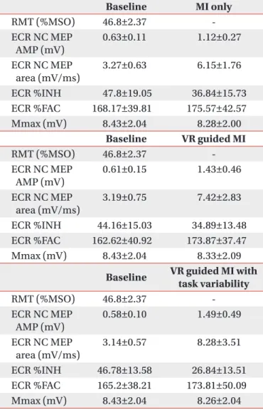
The RMT was defined as the lowest current that evoked MEPs with a peak-to-peak amplitude greater than 50 μV for at least four of eight consecutive stimuli. Subsequent- ly, the test stimulus intensity was set to 120% of the RMT for each subject. The corticomotor excitability of each subject was compared before, during, and after each of the different conditions by assessing changes in the MEP amplitude (mV), MEP negative area (mV/ms), ICI, and ICF (Fig. 2). When testing the paired-pulse paradigm, the conditioning stimulus intensity was set to 80% of the RMT and the inter-simulation interval (ISI) was set at 2
Imagine of moving your wrist when the avatar jumps
in the screen
Fig. 1. The subjects were required to imagine wrist exten-
sion when the avatar jumped over obstacles in a virtual
reality-guided motor imagery program. During the motor
imagery, motor-evoked potentials were recorded.
ms for ICI and 15 ms for ICF to allow for maximal inhibi- tion and facilitation. ICI and ICF were determined based on the relationship between the sizes of the conditioned MEPs. TMS was delivered at intervals of at least 5 seconds and 10 MEPs were recorded per parameter under each of the four experimental conditions.
Measurements were taken at baseline (T0), during MI (T1), and post-MI (T2) during Conditions B, C, and D.
The RMT, peak-to-peak MEP amplitude, negative MEP area, ICI, and ICF were recorded at baseline (T0), and the MEP amplitude and MEP area were measured during MI in wrist extension phase (T1). Finally, to assess changes in ICI and ICF, these variables were recorded immedi- ately after the MI was finished (T2) and calculated as percentages of inhibition (%INH) and facilitation (%FAC) from baseline (T0). The order of stimulation for each MEP parameter was randomized. The time points for the TMS applications were shown in Fig. 2.
To assess peripheral nerve excitability, the supramaxi- mal M waves (Max) of the median nerve of the wrist were evaluated after the performance of each condition. A 1-ms circular electrical stimulus was applied to the medi- an nerve between the palmaris longus and the flexor car- pi radialis tendons, and the resulting Max were recorded
from the abductor pollicis brevis (APB) using paired Ag/
AgCl disc electrodes (diameter, 20 mm) that were placed on the center of the APB muscle belly and tendon after preparing the skin.
Assessment of vividness of movement imagery
The MI ability of the subjects was measured using the VMIQ-2 [18], a 12-item questionnaire that brings certain images to mind and requires the subject to rate them us- ing a five-point scale according to the degree of clearness and vividness. A score of 1 represents a perfectly vivid image, as in normal vision, whereas a score of 5 repre- sents no image at all. During the assessment, three types of MI are performed per item while the eyes are closed:
(1) external visual imagery, or watching oneself perform the movement; (2) internal visual imagery, or looking through one’s own eyes while performing the movement;
and (3) kinesthetic imagery, or feeling oneself do the movement.
Data analysis
Ten MEPs were recorded per parameter (peak-to-peak amplitude, negative area the MEPs, ICI, and ICF) under each condition and were then averaged to mean values.
Condition A: Complete rest
RMT, MEP, %INH, %FACREST
T0
Condition B: Motor imagery only
RMT, MEP, %INH, %FAC MEP %INH, %FAC
REST MI REST
T0 T1 T2
Condition C: VR guided motor imagery
RMT, MEP, %INH, %FAC MEP %INH, %FAC
T0 T1 T2
REST VR guided MI REST
Condition D: VR guided motor imagery with task
RMT, MEP, %INH, %FAC MEP %INH, %FAC
T0 T1 T2