• 검색 결과가 없습니다.

Extramedullary plasmacytoma in the maxillary sinus: a case report

N/A
N/A
Protected

Academic year: 2021

Share "Extramedullary plasmacytoma in the maxillary sinus: a case report"

Copied!
5
0
0

로드 중.... (전체 텍스트 보기)

전체 글

(1)

Ⅰ.

서 론

머리와 목 영역에서의 형질세포종은 전체 형질세포종양 의 3%를 차지하는 드문 병소이다

1

. 드물게 연조직에서 발 생하는 경우 골수외 형질세포종이라는 용어를 사용한다.

골수외 형질세포종의 80-90%는 상기도의 점막관련 림프 양조직에 호발하고, 75%는 코과 상악동에 호발한다

1

. 형질 세포종은 보통 평균나이 55세 이상에서 발견되며 남녀 3:1 의 비율로 남성에서 호발한다

2

. 방사선학적으로 경계가 명 확한, 단방성의 방사선투과성을 보이며

3

, 경화성 경계를 가 지지 않는다. 조직학적으로 두경부 점막하조직 내에 많은 단세포성의 형질세포가 국소화되어 있다

2,4,5

. 몇몇 연구자 들은 이 병소의 약 15%에서 다발성 골수종으로 이환될 가 능성이 있다고 보고 하였는데

4

, 이 경우 예후가 매우 불량

하며 이는 임상적으로 중요하다

6-8

.

이에 저자 등은 59세 남자환자에서 우측 상악동 부위에 발 생한 골수외 형질세포종을 경험하였기에 보고하는 바이다.

Ⅱ.

증례보고

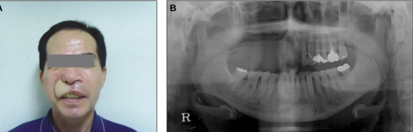
남자환자(59세)가 우측 상악의 잇몸 및 중안면부의 갑작 스런 부종을 주소로 본과에 내원하였다. 내원하기 전까지 환자의 특이할 만한 전신적 병력은 없었으며, 본과 내원 2 일 전 개인병원에서 우측 상악 치은의 조직검사를 위한 절 개 후 출혈경향이 있어 조직검사는 하지 못하였으며, 병리 과와의 긴밀한 협진의 필요성과 출혈조절, 병소에 대한 평 가 및 치료를 위해 본과로 의뢰되었다. 환자는 내원 5일 전 부터 통증 없이 우측 상악 치은과 중안면부의 부종을 호소 하였다. 임상검진결과 상악 우측 협부에 경결감이 있는 연 조직 종괴가 촉진되었다.(Figs. 1, 2) 촉진 시 동통을 호소하 였고, 출혈이 쉽게 되는 양상을 보였다. 상악 우측 견치, 제 1소구치와 제2 구치에는 1도의 동요도를 보였다.(Fig. 3)

임상병리적 검사결과 일반 혈액검사(complete bood cell count, CBC)의 혈소판 수치가 40,000/μL로 정상치인 150,000-400,000/μL보다 매우 낮았다. 자세한 평가 및 치료 김 형 준

120-752 서울시 서대문구 성산로 250 연세대학교 치과대학 구강악안면외과학교실 Hyung Jun Kim

Department of Oral and Maxillofacial Surgery, College of Dentistry, Yonsei University

250 Seongsanno, Seodaemoon Gu, Seoul, 120-752, Korea TEL: +82-2-2228-3132 FAX: +82-2-2227-8022 E-mail: [email protected]

상악동에 발생한 골수외 형질세포종: 증례보고

김형근∙함태훈∙남 웅∙차인호∙김형준 연세대학교 치과대학 구강악안면외과학교실

Extramedullary plasmacytoma in the maxillary sinus: a case report

Hyoung Keun Kim, Tae-hoon Hahm, Woong Nam, In-ho Cha, Hyung Jun Kim Department of Oral and Maxillofacial Surgery, College of Dentistry, Yonsei University, Seoul, Korea

Plasmacytoma is a rare malignant neoplasm in the head and neck region and comprises approximately 3% of all plasma cell tumors. This lesion is a unifocal, monoclonal, neoplastic proliferation of plasma cells that usually arises within the bone. Infrequently, it is observed in soft tissue, in which case, the term extramedullary plasmacytoma is used. Approximately 80-90% of extramedullary plasmacytomas involve the mucos-Associated- Lymphoid Tissue of the upper airways with 75% of these involving the nasal and paranasal regions. The plasmacytoma is usually detected in adult males, with an average age at diagnosis of 55 years. The male-to-female ratio is 3:1.Radiographically, the lesion may be seen as a well-defined, unilocularradioluceny with no evidence of a sclerotic border. Some investigators believe that this lesion represents the least aggressive part of the spectrum of plasma cell neoplasms that extend to multiple myeloma.Therefore, plasma cytoma is believed to have clinical importance.

We report a case of extramedullary plasmacytoma in the right maxillary sinus of a 59-year-old male with review of the relevant literature.

Key words: Extramedullary plasmacytoma, Maxillary sinus

[paper submitted 2010. 7. 29 / revised 2010. 11. 26 / accepted 2010. 12. 14]

Abstract (J Korean Assoc Oral Maxillofac Surg 2010;36:543-7)

(2)

를 위해 혈액내과에서 복부초음파검사 후 골수생검을 시 행하였고 말초혈소판감소증 가진하에 전신적 스테로이드 투여 및 주기적인 경과관찰결과 2주 후 혈소판 수치가 190,000/μL로 회복되었다. 내과와의 협의진료결과 수술이 가능하며 술후 혈소판감소증이 발생 가능하여 지속적인 추적관찰이 필요하다는 답변을 받았다.

진단을 위해 시행한 computed tomography (CT)(Fig. 4) 우 측 상악동 협측 피질골흡수 소견 및 파괴적 골성 변화를 관 찰할 수 있었고, 자기공명영상촬영검사결과(Fig. 5) 우측 상악동의 내부 및 전방 경계와 경구개의 일부분을 침범한 직경 3.6 cm 가량의 악성 종양이 의심되는 연조직 종물이 관찰되었다. 11일 후 재촬영한 자기공명영상검사결과 종

Fig. 1. Extraoral view showing facial swelling on right buccal cheek. A: Frontal view, B: Lateral view.

Fig. 2. Intraoral view showing soft tissue mass on right buccal cheek mucosa.

Fig. 3. Preoperative panoramic view showing radiolucent lesion on right maxillary sinus.

Fig. 4. Axial and coronal computed tomography images showing resorpted buccal plate of right maxillary sinus and destructive bony change. A: Axial view, B: Coronal view.

Fig. 5. Axial and coronal MRI showing tissue mass on right buccal cheek and maxillary sinus. A: Axial view, B:

Coronal view. (MRI: magnetic resonance imaging)

A B

A B

A B

(3)

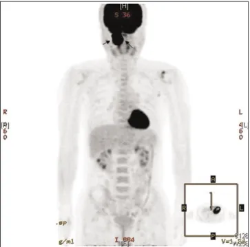
물의 크기가 커짐을 확인할 수 있었다.(Fig. 6) 확진을 위해 F

18

-fluorodeoxyglucose를 정주한 positron emission tomogra- phy (PET) CT를 시행하였으며(Fig. 7) 전이는 관찰되지 않 는 우측 상악동의 원발성 악성 종양으로 판독되었다. 따라 서 임상적, 방사선학적, 핵의학적으로 혈관내피종, 상악동 내의 악성 종양 또는 비부비동의 비분화성 악성 종양으로 가진하였다.

혈관종 가능성 및 병소의 출혈성향 때문에 술전 조직검 사는 시행하지 않았으며 전신마취하 수술 도중 시행한 동 결절편 조직검사결과 비분화성 편평상피세포암종으로 진 단되어 우측 상악골 부분절제술 후 요골전완 유리피판을

이용하여 재건하였다. 재건 시 하나의 피판을 사용하여 구 강 내외의 연조직 결손부를 동시에 재건하였다.(Figs. 8, 9) 술후 최종 조직검사결과 다양한 정도의 이형성을 보이는 치밀하고 균질한 형질세포의 침투가 관찰되었으며, 풍부 한 핵소체, 분산된 염색질, 불규칙한 핵 및 다핵성을 포함 하는 소견도 관찰되었다. 면역세포화학검사결과 Kappa chain이 제한된 IgG의 발현을 나타내었다.(Figs. 10. A, B) 이상의 조직검사결과 및 면역세포화학검사를 토대로 골수 를 침범하지 않고 분화도가 낮은 골수외 형질세포종으로 확진하였다. 절개한 조직의 전후방 경계 및 구개골도 종양 에 포함되어 술후 우측 상악골에 대한 방사선치료를 시행

Fig. 7. F

18

-FDG PET showing uptake of FDG on right buccal cheek and maxillary sinus.(F

18

-FDG PET: F

18

-fluorode- oxyglucose positron emission tomography)

Fig. 6. Enlarged mass (arrow) compared with previous MRI.

A: Axial MRI on first visit, B: Coronal view on second visit.

(11 days after)

(MRI: magnetic resonance imaging) Fig. 8. Perioperative images.

A: Preoperative margin design, B: After mass removal, C:

Before reconstruction, D: After reconstruction on extraoral soft tissue defect.

Fig. 9. Mass removal and reconstruction using RFFF.

A: Removed mass by partial maxillectomy, B: Flap design on right forearm, C: RFFF, D: After reconstruction on intra- oral and extraoral soft tissue defect using one RFFF.

(RFFF: radial free forearm flap)

A B

A B

C D

A B

C D

(4)

하였다. 방사선치료는 술후 1개월째부터 4개월째까지 총 3 개월에 걸쳐 시행하였으며, 회당 선량 (cGy) 180으로 하루 4회, 주당 5회, 처방선량 (cGy) 4,500으로 계획하였다.

술후 1개월에 내원하여 임상검사 및 방사선검사를 시행 하였으며 특이할 만한 소견은 관찰되지 않았고, 재건된 조 직피판의 양호한 치유가 관찰되었다.(Fig. 11)

Ⅲ.

고 찰

두경부에 발생하는 골수외 형질세포종은 형질세포와 관 련된 전체 종양 중 약 3% 정도 되는 매우 드문 악성 종양으 로 편평세포암종보다 낮은 유병률을 나타내지만 상악동에 발생한 골수외 형질세포종은 임상적, 방사선학적으로 편 평세포암종과 거의 유사한 특징을 보인다

7

. 이처럼 임상양 상이 골수외 형질세포종과 유사한 병소로 감별진단을 요 하는 질환은 혈관종, 혈관기형, 혈관내피종, 평활근육종,

비분화성 비부비동 악성 종양, 상악동의 편평세포암종 등 이다

9,10

.

조직학적으로 골수외 형질세포종의 명확한 특징은 치밀 하고 균질한 단세포성 형질세포가 골수외의 영역에 국소 화된 침투양상을 보이며 면역학적으로 Kappa chain이 제한 된 IgG의 발현을 보인다

7

. 이것은 상악동 내에 발생하는 편 평세포암종과 감별할 수 있는 가장 큰 특징이다. 혈액검사, CT와 자기공명영상검사 등의 방사선검사, 핵의학검사 및 골수검사를 바탕으로 골수외 형질세포종은 질환의 확장 정도에 따라 3단계로 분류될 수 있다. 편측에 한정된 경우 는 1기이고 국소적 확장을 보이거나 임파선을 침범한 경우 는 2기이며 전이성 확장을 보이는 경우가 3기이다

7

. 본 증 례의 환자는 상악동의 전벽 및 구개골을 침범하였으나 골 수를 침범하거나 전이성 확장을 보이지 않았으므로 2기에 해당한다.

골수외 세포종의 치료에 대한 다양한 논의가 있는데 국

Fig. 10. Histologic findins.

A: Dense, homogenous infiltration of plasma cells showing varying degrees of dyspla- sia, including prominent nucleoli, dispersed chromatin, irregular nuclei and multinu- cleation.(H&E staining, original magnification ×200)

B: IgG(+), Kappa(-), Lambda(-).(Immunohistochemical staining ×100)

Fig. 11. Postoperative 1 month clinical and panoramic view showing well reconstructed soft tissue defect and partial maxillectomy site on right maxilla area. A: Post operative photo, B: Post operative panoramic view.

A B

A B

(5)

소화된 골수외 형질세포종은 방사선에 대한 민감도가 매 우 높으므로

3

수술적 제거, 방사선치료 또는 이 2가지 방법 의 조합이 추천된다

11

. Gross 등

5

은 상악 전방부에 발생한 골 수외 형질세포종을 수술 및 술후 방사선치료로 성공적으 로 치료한 증례를 보고한 바 있으며, Lomeo 등

12

은 병소의 크기가 작은 경우 수술적 제거만으로도 효과적이지만 크 기가 큰 경우 수술적 제거와 방사선치료를 함께 시행할 경 우 생존율이 높아진다고 주장했다. Soutar 등

13

이 British Journal of Haematology에서 제시한 지침에 따르면 국소화 된 골수외 형질세포종의 1차 치료는 방사선요법이지만

Katodritou 등

14

의 연구에 의하면 골수외 형질세포종의 치료

시 방사선치료 없이 thalidomide와 dexamethasone의 병용투 여 후 자가줄기세포이식술을 이용하여 성공적인 결과를 얻었다고 하였다. 일반적으로 다수의 전이소견을 보이는 골수외 형질세포종의 경우에는 전신적 화학치료를 추천하 지만 그 예후가 국소화된 단일 병소의 골수외 형질세포종 보다 나쁘다고 알려져 있다

7

. 국소화된 골수외 형질세포종 의 5년 생존율은 약 50%로 보고된 바 있다

3

.

상악 우측 치은과 중안면부의 갑작스런 부종을 주소로 본 과에 내원한 59세 남자환자의 방사선학적, 핵의학적 검 사결과와 임상검사결과가 편평세포암종과 유사하였으며 전신마취하에 수술 도중 시행한 동결절편 조직검사결과도 편평세포암종으로 진단되어 우측 상악동 부분 절제술 후 요골전완 유리피판을 이용하여 연조직을 재건하였다. 그 러나 술후 최종 조직검사결과 분화도가 낮은 골수외 형질 세포종으로 최종 확진되어 술후 방사선치료를 시행하였으 며 1개월 후 시행한 임상검사 및 방사선검사결과 재발이나 특이한 소견은 없었으며 중안면부의 연조직 및 경조직의 재건은 성공적으로 이루어졌다. 상악 결손부의 재건 시 1 개의 요골전완 유리피판을 사용하여 구강 내외의 연조직 결손부를 모두 재건하고 성공적인 결과를 얻었으며 이는 수술적 기법에서 주목할 만하다.

상악동에 발생한 골수외 형질세포종은 극히 드문 질환이 지만

11

상악의 종양을 감별진단할 때 간과해서는 안된다.

임상 및 방사선학적 양상은 편평세포암종과 거의 유사하 지만 치료를 위한 접근법은 매우 다르므로 주의 깊은 감별 진단이 필요하다

7

. 이 2가지 질환은 조직학적 검사로 감별 하여야 하며 치료접근법 또한 달라야 한다

3

.

골수외 형질세포종은 방사선치료를 우선적으로 고려하 고

15

병소의 크기가 작은 경우 수술적 제거도 함께 고려할

수 있다

8,11,12

. 편평세포암종은 수술적 제거 및 재건으로 치

료한다. 골수외 형질세포종과 편평세포암종을 확실하게 구분하기 위해서는 조직검사를 할 때 Kappa-lambda 항체 로 염색하면 종양세포 내에 Kappa chain이 제한된 IgG의 발 현으로 구분할 수 있다

7

. 이와 같이 면역조직학적 검사가 골수외 형질세포종과 편평세포암종의 감별 시 중요한 역 할을 하므로 술전 조직검사를 추천한다

7

.

본 증례의 경우 임상검사 시 출혈성향을 보여 술전 조직

검사를 시행할 수 없었고 수술 도중 시행한 동결절편 조직 검사결과에서 편평세포암종으로 진단되어 편평세포암종 의 치료법에 준하여 수술적 제거 및 요골전완 유리피판을 이용한 재건술을 시행하였으나 술후 조직검사결과에서 골 수외 형질세포종으로 확진되어 술후 방사선치료를 시행하 였다. 술후 주기적인 경과관찰 시 재건된 부위에 양호한 소 견을 보이며 재발소견은 관찰되지 않았으며 환자는 방사 선치료 및 경과관찰 도중 술후 5개월째에 자택에서 자연사 하였으나, 사망원인과 본 증례와의 연관성은 없는 것으로 사료되었다.

References

1. Attanasio G, Viccaro M, Barbaro M, de Seta E, Filipo R.

Extramedullary plasmacytoma of paranasal sinuses. A combined therapeutic strategy. Acta Otorhinolaryngol Ital 2006;26:118-20;

discussion 120.

2. Cumbo V, Gallina G, Messina P. Extramedullary plasmacytoma of the maxillary sinus: report of a case. J Oral Maxillofac Surg 1988;46:406-9.

3. Majumdar S, Raghavan U, Jones NS. Solitary plasmacytoma and extramedullary plasmacytoma of the paranasal sinuses and soft palate. J Laryngol Otol 2002;116:962-5.

4. Bruns F, Janssen S, Laenger F, Dobbelstein C, Meyer A.

Extramedullary plasmocytoma: a rare case with bifocal manifes- tation at uncommon sites. Anticancer Res 2010;30:1779-81.

5. Gross M, Eliashar R, Maly B, Sichel JY. Maxillary sinus plasma- cytoma. Isr Med Assoc J 2004;6: 119-20.

6. Tamada A, Araki T, Makimoto K, Takatsuki K. Plasmacytoma in nasal cavity and maxillary sinus. Arch Otorhinolaryngol 1982;

237:83-91.

7. Megat Shiraz MA, Jong YH, Primuharsa Putra SH. Extramedu- llary plasmacytoma in the maxillary sinus. Singapore Med J 2008;49:e310-1.

8. Greenberg P, Parker RG, Fu YS, Abemayor E. The treatment of solitary plasmacytoma of bone and extramedullary plasmacy- toma. Am J Clin Oncol 1987;10:199-204.

9. Jaffe ES, Harris NL, Stein H, Vardiman JW, eds. World Health Organization Classification of tumours: pathology and genetics of tumours of haematopoietic and lymphoid tissues. Lyon:

International Agency for Research on Cancer (IARC) Press;

2001.

10. Barnes L, ed. Surgical pathology of the head and neck. 2nd ed.

New York: Marcel Dekker; 2000.

11. Michalaki VJ, Hall J, Henk JM, Nutting CM, Harrington KJ.

Definitive radiotherapy for extramedullary plasmacytomas of the head and neck. Br J Radiol 2003;76:738-41.

12. Lomeo PE, McDonald JE, Finneman J, Shoreline. Extramedu- llary plasmacytoma of the nasal sinus cavities. Am J Otolaryngol 2007;28:50-1.

13. Soutar R, Lucraft H, Jackson G, Reece A, Bird J, Low E, Samson D. Guidelines on the diagnosis and management of solitary plas- macytoma of bone and solitary extramedullary plasmacytoma. Br J Haematol 2004;124:717-726.

14. Katodritou E, Speletas M, Pouli A, Tsitouridis J, Zervas K, Terpos E. Successful treatment of extramedullary plasmacytoma of the cavernous sinus using a combination of intermediate dose of thalidomide and dexamethasone. Acta Haematol 2007;117:20-3.

15. Tsang RW, Gospodarowicz MK, Pintilie M, Bezjak A, Wells W,

Hodgson DC, et al. Solitary plasmacytoma treated with radio-

therapy: impact of tumor size on outcome. Int J Radiat Oncol

Biol Phys 2001;50:113-20.

수치

Fig. 1. Extraoral view showing facial swelling on right buccal cheek. A: Frontal view, B: Lateral view.
Fig. 9. Mass removal and reconstruction using RFFF.
Fig. 11. Postoperative 1 month clinical and panoramic view showing well reconstructed soft tissue defect and partial maxillectomy site on right maxilla area

참조

관련 문서

Measurements items between the maxillary second premolar and the first molar: A, The occlusal plane and reference lines which were above 4, 6, 8 mm from cementoenamel junction

The thickness of the maxillary sinus lateral wall according to tooth site and measurement level was measured by using image-processing software and its histologic

With the increase of the dental implant procedure, there are many cases of Maxillary Sinus Floor Elevation Surgery(MSFES). Additionally, its side effect has

Methods to overcome insufficient bone due to poor bone quality, the pneumatization of a maxillary sinus and other anatomical limitations of implant placement

Time series of vertical cross section of potential vorticity and wind vector calculated by Case 1 along the A-A' line indicated at Fig.. Same

Long-term outcomes of short dental implants supporting single crowns in posterior region: a clinical retrospective study of 5-10 years.. Osteotome sinus

The OSFE (osteotome sinus floor elevation) technique has been used for maxillary sinus augmentation.. The implants were clinically and radiographically followed

The average position of the posterior superior alveolar artery, the wall thickness of the lateral wall, and the average volume of the maxillary sinus will