하천은 어류의 성장과 번식에 필수적으로 요구되는 여러 가지 조건을 가지고 있기 때문에 수질이 오염될 경우 어류의 생활에 미치는 영향은 아주 크다. 특히 하 천의 수질오염은 공업단지로부터 중금속, 농약, 포르말린 같은 자극성 오염물질이 직접 방출되고 인간의 생활 하 수가 직접∙간접적으로 유입되기 때문에 수질 오염은 가속화되고 있는 실정이다. 최근 하천오염과 개발로 인 하여 하천의 수질환경이 극히 악화되는 과정에서 그곳 에 서식하는 어류와 많은 수중생물의 생존은 크게 위협 받고 있으며 심한 경우에는 폐사하거나 생존이 불가능
한 상태로 변화된다. 따라서 수질오염에 따른 어류의 병 리조직학적 연구는 수질오염의 평가 뿐 아니라 치료제 개발에 중요한 수단으로 이용되어 왔다. 특히 아가미 및 신장의 조직학적 변화가 주로 알려져 왔으나(Davis, 1946; Hickman and Trump, 1968; Sarig, 1971; Skidmore and Tovell, 1972; Cawley, 1975; Landolt, 1975; Smith and Piper, 1975 ; Molnar, 1979; Morrison, 1981), 국내에 서는 오염수역에 서식하는 어류의 조직학 또는 병리학 적 연구는 매우 미흡한 실정이다.
따라서 본 연구는 오염수에서 어류에 미치는 조직학 Korean J. Ichthyol. 14(1), 70~75, 2002
─
─ 70 ──
만경강 오염수에서 서식하는 붕어의 아가미와 신장의 비정상적인 조직
김 익 수∙김 선 영∙박 종 영 전북대학교 자연과학대학 생물과학부
The Abnormal Tissues of Gill and Kidney of
Crucian Carp Carassius auratus in the Polluted Waters of Mangyong River, Korea
Ik-Soo Kim, Seon-Young Kim and Jong-Young Park
Faculty of Biological Sciences, Chonbuk National University, Jeonju, 561-756, Korea
Gill and kidney tissue of Carassius auratus are used to investigate the effects of the water pollution on aquatic organisms inhabiting the Jeonju stream near a sewage disposal plant on the Mangyong River, Korea. Around the sewage disposal plant the water temperature is high and pollutants flow into the streams frequently. In the Jeonju stream, Carassius auratus shows abnormal structures in the gills: edema of primary gill lamella, epithelial lifting or separation, complete necrosis, increase of secondary lamella thickness, lamellar hypertrophy, hyperplasia, and secondary gill lamellar fusion. The kidneys of the fishes also have abnormal structures: acute tubular necrosis, shrunken renal tubules and glomerulus, thickened capillary basement membrane of the renal glomerulus. In addition, the kidney shows pathological structures such as enlarged melanomacrophage centres with golden and dark brown pigments, vacuoles, and giant cells. It was considered that these abnormal tissues of the gills and kidneys in the crucian carps were affected by the parasites or bacteria in the polluted waters.
Key words : Gill, kidney, abnormal structures, water pollution, Carassius auratus
적 영향을 파악하기 위하여 비교적 심각한 수질오염 상 태를 보이는 만경강 지류인 전주천 하수종말처리장(전 주시 팔복동)에서 비교적 우세하게 출현하고 있는 붕어 의 아가미 및 신장을 조사하였다. 본 실험에 사용된 붕 어는2001년 3월에서6월까지 만경강 중류인 전주천 하 수종말처리장에서 채집된 개체 가운데 20개체를 조사하 였으며, 이들의 체장은 65.1~118.9 mm이었다. 일반적인 paraff in method를 통해서 조직학적 조사를 수행하였다.
전주천 하수종말처리장에서 채집한 붕어의 외부형태 는 일반적으로 지느러미는 약간 손상되어 있었고, 일부 피부의 탈락과 궤양을 보였으며, 일부 개체에서는 눈과 지느러미의 기부 등에서 출혈을 보였으며, 그리고 새개 (operculum) 및 세사(gill f ilament)의 파손 등이 발견되 었다(Fig. 1). 2001년 3월에 채집된 붕어 가운데 복강에 하얀 쌀 모양의 Myxobolus sp.의 포낭(cyst), 복부의 피 부에 함몰된 가느다란 실 모양의 기생충 등이 발견되었 다.
신장은 척추 밑 복막 후강에 위치해 있었고, 신장의 색깔은 엷거나 짙은 갈색을 띠고 있었다. 중간과 뒷부분 만이 유합되었으며, 유합된 부분은 둥근 모양이었다. 신
장은 크게 전신(head kidney)과 중신(trunk kidney)으 로 구분되는데, 본 실험에서는 중신을 사용하였다. 신장 에서는 외부 형태적인 손상은 발견되지 않았다.
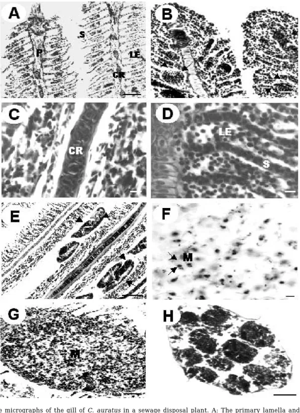
이러한 개체들의 아가미를 조직학적으로 조사한 결과 여러가지 병리학적 특징이 나타났다. 조직학적 특징으로 는 일차새변은 상대적으로 두꺼워져 있었으며(Fig. 2, A), 특히, 기저부와 말단부분이 불룩한 팽대부를 이루고 있었다. 일차 및 이차새변의 호흡성 상피(epithelium)에 서는 부종(edema), 증식(hyperplasia)이 나타났다(Fig.
2, A, D). 이차새변 사이에는 호염기성인 점액세포(mu- cous cell)와 드물게 연두색과 검은색의 색소세포(pig- ment cell)가 나타났다. 이차새변은 양쪽이 유합(fusion) 된 것, 한 쪽만이 분화가 이루어진 것, 또한 완전히 유합 되어 곤봉과 같은 모양을 띠는 곤봉화(clubbing)가 나타 났다(Fig. 2, C). 곤봉화가 나타난 새변은 두꺼운 상피층 을 이루고, 표면에 다수의 점액세포를 가졌다. 이러한 현 상 등은 이차새변의 유합, 새변간 공간의 소실, CO2교환 율 저하로 인해 호흡성 산증(respiratory acidosis)과 새 변의 유합으로 인해 호흡면적의 손실과 호흡기능의 악 화가 나타날 수 있다(Ferguson, 1989). 이차새변은 말단 Fig. 1. External apprearance of Carassius auratus in a sewage disposal plant. A: The fin was damaged, and the lateral skin near dorsal fin was fell off; B: The skin near ventral f in fell off; C: The anal and pectoral fins were congested, and no operculum (or gill cover) existed due to damage; D: The scale fell severely off and the pectoral fin was severely congested; E: Blood was trasparented in proximal segment of fins and ventral region; F: Some of skin got removed, identified parasite was invaded.
72 김익수∙김선영∙박종영
Fig. 2. The micrographs of the gill of C. auratus in a sewage disposal plant. A: The primary lamella and lamellar epithelium were thicken and swollen. Bar = 4µm; B: Telangiectatic secondary lamella (arrowed) was appeared in the gill and the secondary lamella was fused. Bar = 3µm; C: Clubbing of the gill lamella, and cartilaginous rod was thicked. CR, cartilaginous rod. Bar = 2.5µm; D: Primary lamella, terminal segments of the second lamella and lamellar epithelium were thicked. Bar = 3µm; E: Identified parasites were appeared between the clubbing of the gill lamella. Primary lamella was largely swollen, one or two parasites (arrowed) nearly adhered among lamella.
Bar = 5µm; F: Cyst of Myxobolus sp. in the abdominal cavity and its shape looks like a grain. spores of Myxobolus sp.; M: Bar = 2µm; G: Cyst of Myxobolus sp.; M: was situated at the distal portion of the gill lamella. Bar = 4µm; H:
Identified parasites were invaded to the skin, and looks like a slender duct with brown color. Unknown parasite has thin membrane, and the empty space of volvox shaped mass. Bar = 5µm.
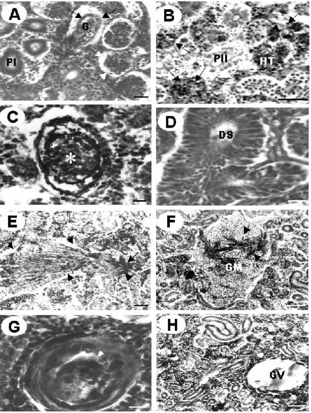
Fig. 3. The micrographs of the kidney of Carassius auratus in a sewage disposal plant. A: Blood capillaries and basement membrane of glomerulus became thick (arrowed); G, glomerulus; PI, first proximal segment. Bar = 3µm; B: Renal tubule showed tubular necrosis, showing deep basophilic; HT, haemopoietic tissue; P II, second proximal segment.
Bar = 6µm; C: Stroma of haemopoietic tissue was necrotic, basophilic cell was oval (*). Bar = 3µm; D: Necrosis of the distal segment. DS, distal segment. Bar = 4µm; E: Flexibacter sp. (arrowed) is widely distributed in the haemopoietic tissue. Bar = 3µm; F: Flexibacter sp. (arrowed) is located in granulromata; arrowed, Flexibacter sp.;
GM, granulromata. Bar = 4µm; G: Ureters were increased in diameter several fold and filled with soft caseous material (arrowed). Bar = 4µm; H: GV, eosinophilic globular vacuole. Bar = 4µm.
부위에 lamellar telangiectasis이 나타났는데 이 부위에 서는 이차새변의 분화가 거의 일어나지 않았고, 이는 호 산성으로 솔방울 모양을 하고 있었다(Fig. 2, B). 이들은 외상, 파열(rupture), 출혈(haemorrhage) 등을 가져와 결 국에는 호흡장애를 일으키는 것으로 알려져 있다 (Skidmore and Tovel, 1972; Cawley, 1975; Smith and Piper, 1975, Roberts, 1989). 주로 일차새변의 말단부분 과 기저부에서 기생충이 발견되었고, 3월에 채집된 한 개체에서 복강에 쌀알모양의 포낭(cyst)이 나타났다. 발 달된 기생충의 대부분은 수많은 상피세포에 의해 둘러 싸여 있었다. 원생동물의 일종인, Myxobolus sp.의 특징 적인 포자(spore)는 특히 filament 꼭대기로부터 포낭 내부에 풍부했다(Fig. 2, F-G). 포자는 레몬 모양으로, 연 두색의 형광을 띠고 있었고, polar capsule 안에는 호산 성인 polar filament가 있었다. spore valve를 가지고 있 어, 포자는 이중막으로 보였다. 또한, 곤봉화가 나타나는 새변 간에서 짚신모양 혹은 구형의 기생충이 발견되었 다(Fig. 2, E). 3월에 채집된 한 개체에서 피부에 연한 갈색의 긴 실 모양의 기생충이 관찰되었고(Fig. 1, F), 조 직학적 관찰에서는 타원형인 바깥쪽 막을 가지고 있었 으며, 내부에는 호산성으로 volvox 형태를 띠고 있었다 (Fig. 2, H). 원생동물인Myxobolus sp.는 조직의 파괴, 괴 사를 유발하며, 출혈, 빈혈증, 2차 감염을 일으키거나, 심 할 경우 숙주의 죽음까지 이른다고 보고된 바 있으며 (Pacha and Ordal, 1970; Molnar, 1979; Roberts, 1989;
Landberg, 1993), 일부 담수 어류의 아가미에서도 보고 되었다(Sanaullah and Ahmed, 1980).
또한 신장에서는 사구체(glomerulus)의 기저막이 두 꺼워졌고(Fig. 3, A), 신사구체와 신세관(renal tubule)은 축소되었다(Fig. 3, F, H). 또한 haemopoietic tissue에서 괴사(necrosis) 또는 섬유증(fibrosis)이 발생하였으며 (Fig. 3. B), 과립모양의 호염기성 세포(Fig. 3, C), 공포 (vacuole)나 호산구(eosinophilie)의 형태도 나타났다 (Fig. 3, H). 특히 신장에서 나타나는 관상의 괴사는 수은 혹은 카드뮴 같은 중금속과 함께 신장세포에 치명적인 병리적인 현상을 일으키고 있다(Hickman and Trump, 1968). 조혈조직(haemopoietic tissue)의 간질(stroma)에 있는 신사구체와 신세관에서는 황갈색의 melanomacro- phage centres가 나타났다(Fig. 3, B). 신장 표면 위의 뇨 관은 몇 개의 주름을 이룬 직경이 증가하고 있었고, 부 드러운 치즈모양의 물질로 채워져 있었다(Fig. 3, G). 이 러한 병리현상은 포유류의 만성 사구체 신염(chronic glomerulonephritis)과 유사하다고 보고된 바 있다 (Fennel and Pardo, 1967). 또한 세균의 일종인 Flexi-
bacter sp.가 신장조직에서 발견되었는데 이는 호산성으
로서 eosin이 진하게 염색되며, 가늘고 긴 사상형태를
갖는다(Fig. 3, E-F). 또한 Flexibacter sp.는 보통 군체를 이루면서 신사구체나 신세관 혹은 조혈조직에 넓게 걸 쳐서 분포하거나(Fig. 3, E), 육아종(graluromata) 내부에 분포했다(Fig. 3, F). Flexibacter sp.는 Davis (1946)에 의 해 처음으로 무지개 송어에서 발견된 그람음성의 간균 으로 알려져 있다(Wolke, 1975; Roberts, 1989). 이러한 세균성 질병은 야생과 양식 어류에서 높은 사망률을 초 래한다. Fennel and Pardo (1967)는 신장의 두꺼워진 기 저막 그리고 사구체의 수축은 포유류의 만성 사구체 신 염(chronic glomerulonephritis)과 유사하다고 보고한 바 있다. 경골어류의 신장은 독성의 암모니아 같은 성분의 배설에는 관여하지 않고 있어서 물고기가 죽을 때까지 병변들이 발달할 가능성이 있다. 특히 신장에서 나타나 는 관상의 괴사는 수은 혹은 카드뮴 같은 중금속과 함 께 신장세포에 치명적인 병리적인 현상을 일으키고 있 다(Hickman and Trump, 1968). 붕어는 다른 어종에 비 하여 오염에 관한 저항력이 강하여 서식은 가능하지만 오염수역에 서식하는 붕어의 아가미와 신장조직은 원생 생물과 세균감염에 의하여 비정상적인 구조를 이루고 있는 것으로 추측되었다. 추후 정밀한 조사를 통하여 아 가미와 신장의 비정상적인 조직형성의 직접적인 원인 구명이 요구된다.
적 요
수질오염이 수중 생물에 미치는 영향을 조사하기 위 하여 전주천의 하수종말처리장에 서식하는 붕어의 아가 미, 신장을 사용하였다. 하수종말처리장 주변의 온도는 높고 오염물질은 빈번하게 유입되었다. 이와 같은 환경 조건에서, 붕어는 아가미에서 일차 새변(primary la- mella)의 부종(edema), 상피 탈락 혹은 분리(epithelial lifting or separation), 완전한 괴사(complete necrosis), 이차 새변(secondary lamella) 두께의 증가, 새변의 비대 (lamellar hypertrophy), 증식(hyperplasia) 그리고 이차 새변의 유합(fusion)되는 등의 비정상적인 구조를 보여 주었다. 신장에서는 급성의 관상의 괴사, 신세관과 사구 체가 위축되었고, 사구체의 모세관의 기저막이 두꺼워졌 다. 또한, 노랗고 어두운 갈색의 색소를 가지는 확장된 melanomacrophage centres와 공포, 거대세포(giant cells)같은 병변이 나타났다. 붕어의 아가미와 신장조직 의 비정상적인 구조는 오염수역의 원생생물 및 세균 감 염에 의한 영향이라고 생각된다.
74 김익수∙김선영∙박종영
인 용 문 헌
Cawley, G.D. 1975. Some aspects of gill disease in framed rainbow trout (Salmo gairdneri). M. Sc. Thesis, Univer- sity of Stirling. 90 pp.
Davis, H.S. 1946. Care and disease of trout. Res. Rep. U. S.
Fish Wildl. Serv., no. 12.
Fennel, R.H. and V.M. Pardo. 1967. Experimental glo- merulonephritis in rats. Laboratory Investigation, 17 : 483~488.
Ferguson, H.W. 1989. Gill and pseudobranchs In: Systemic pathology of fish. Iowa State University Press/Ames, pp. 11~40.
Hickman, C.P. and B.F. Trump. 1968. The kidney. In:
Hoar, W.S. and D.J. Randall (ed.), Fish Physiology, Vol.
1. Academic Press, London. pp. 91~239.
Landsberg, J.H. 1993. Myxosporean Parasites of Common Snook in Florida. Journal of Aquatic Animal Health, 5 : 102~109.
Landolt, M.L. 1975. Visceral granuloma and rephrocalcina- sis of trout. In: W.R. Ritelin and G. Migaki (eds.), The pathology of fishes, Madison, Wis: University of Wis- consin Press. pp. 793-801.
Molnar, K. 1979. Gill sphaerosporosis in the common carp and grasscarp. Acta Veterinaria Academiae Scientia- rum Hungaricae 27 : 99~113.
Morrison, C.M. 1981. Microsporidian parasites in the gills
of salmonid fishes. Journal of Fish Deseases, 4 : 371~ 386.
Pacha, R.E. and E.J. Ordal. 1970. Myxobacterial diseases of salmonids. In: S. F. Snieszko (ed.), A Symposium on Diseases of Fishes and Shellfishes. Special publication no. 5. Washington, D.C. : American Fisheries Society, pp. 243~257.
Roberts, R.J. 1989. Fish Pathology. 2nd ed., Bailliere Tin- dall. London. 318 pp.
Sanaullah, M. and A.T.A. Ahmed. 1980. Gill myxoboliasis of major carps in Bangladesh. Journal of Fish Diseases, 3 : 349~354.
Sarig, S. 1971. The Prevention and Treatment of Diseases of Warmwater Fishes under Subtropical Conditions, with Special Emphasis on Intensive Fish Farming. T.
F. H. Publications, Neptune City, N. J.
Skidmore, J.F. and P.W.A. Tovell. 1972. Toxic effects of zinc sulphate on the gills of rainbow trout. Water Res., 6 : 217~230.
Smith, C.E. and R.G. Piper. 1975. Lesions associated with chronic exposure to ammonia. In: Ribelin, W.E. and G.
Migaki (ed.), The Pathology of Fishes. Madison, Wis.:
University of Wisconsin Press, pp. 497~514.
Wolke, R.E. 1975. Pathology of bacterial and fungal disea- ses affecting fish. In: Ribelin, W.E. and G. Migaki (ed.), The Pathology of Fishes. Madison, Wis. : University of Wisconsin Press, pp. 33~116.
Received : January 12, 2002 Accetped : February 9, 2002