Apoptotic Effect of Rubia cordifolia Dichloromethane Extracts on Human Acute Jurkat T Cells
Ji-Hye Kim, Jong-Hwan Lee
1, Young Ho Kim
2and Kwang-Hyeon Kim*
Department of Life Science and Biotechnology, College of Natural Science, Dong-eui University, Busan 614-714, Korea
1Department of Biotechnology and Bioengineering, College of Engineering, Dong-eui University, Busan 614-714, Korea
2School of Life Sciences and Biotechnology, College of Natural Sciences, Kyungpook National University, Dae-gu 702-714, Korea Received October 10, 2008 /Accepted February 4, 2009
To understand cytotoxic activity of Rubia cordifolia L. (Rubiaceae), which has been used as a traditional oriental medicine, the mechanism underlying cytotoxic effect of its extract on human acute Jurkat T cells was investigated. The methanol extract of roots (3 kg) of R. codifolia was evaporated, dissolved in water, and then extracted by dichloromethane. The substances in the chloroform extract showing the most cytotoxic activity were further purified by a series of preparative HPLC. The extracted active substance (65 mg) was designated as CCH1. When Jurkat T cells were treated with CCH1 at concen- tration ranging from 0.5 to 2.0 μg/ml, apoptotic phenomena of cells companying several subsequent biochemical reactions such as mitochondria cytochrome c release, activation of casapase-8, -9, and cas- pase-3, degradation of PARP and DNA fragmentation occurred via mitochondria-dependent pathway.
However, abrogation of apoptosis was observed in an ectopic expression of Bcl-xL, which is a sup- pressor for mitochondrial cytochrome c release. These results demonstrate that the cytotoxicity of CCH1 against Jurkat T cells is attributable to apoptosis mediated by mitochodria-dependent death-sig- naling regulated by Bcl-xL. In addition, the CCH1 is more potent to leukemia Jurkat T cell than to human peripheral blood monocyte cells (PBMC).
Key words : Anticancer, apoptosis, cytotoxicity, Jurkat T cells, Rubia cordifolia
*Corresponding author
*Tel:+82-51-890-1533, Fax:+82-51-890-1532
*E-mail : [email protected]
Introduction
Rubia cordifolia L. (Rubiaceae) was one of the best candidates. R. cordifolia have been reported for an important oriental medicine plant which is used for anti-inflammatory [1], immunomodulatory [6], anticonvulsant and anxiolytic [8] and anti-tumor activity [13]. It has reported that cyclic hexapeptide isolated from R. cordifolia (Rubiaceae) shows high anti-tumor activity [4]. However, anti-tumor effect of other chemicals isolated from R. cordifolia has not been elucidated. We have found that a potential cytotoxic sub- stance except hexapeptide also exists in roots of R. cordifolia.
The substance was partial purified, and designated as CCH1.
In this report, we investigated signaling pathways during CCH1-induced apoptosis in Jurkat T cells, with particular focus on mitochondria-dependent cell death and whether it could be protected by the ectopic expression of anti-apop- totic protein, Bcl-xL. These results showed that the CCH1-induced apoptotic cell death was mediated by mi- tochondrial cytochrome c release with resultant activation
of caspase-8, -9 and -3, and cleavage of poly (ADP-ribose) polymerase (PARP), which was suppressed by Bcl-xL.
In the present study, we investigated approximately 80 medicine plants to find potent cytotoxicity material toward human Jurkat T cells.
Material and Methods
Isolation of a cytotoxic component from Rubia cordifolia
Rubia cordifolia Linn. (Rubiaceae) was purchased from Hyun-dae Pharmacological Company (Busan, Korea). A sample has been deposited in the author’s laboratory (Kim).
The dried roots (3 kg) from R. cordifolia were extracted with
100% methanol. The methanol extract was evaporated (295.2
g), dissolved in water, and then extracted with dichloro-
methane (CH
2Cl
2). The CH
2Cl
2fraction (82.5 g) had cytotox-
icity against Jurkat T cells. The fraction was then subjected
to silica gel column chromatography using a solvent system
of CH
2Cl
2:EtOAc (50:1) to afford 60 fractions (100 ml/frac-
tion). Analysis of each fraction was performed by a thin lay-
er chromatography (TLC, 20 cm×20 cm, kiesel 60 F254,
Merck, Germany) with a solvent system (CH
2Cl
2:EtOAc=
assay. The active fraction (4.1 g) was applied on secondary silica gel column chromatography using a solvent system (n-hexane:ethyl acetate=10:1), and subsequently on a prepa- rative HPLC GS320 column (Japanese Engineering Instru- ment Co., Japan) chromatography (eluted with CH
3CN:
H
2O=10:1, 3.0 ml/min, 245 nm). The active fraction by the HPLC GS320 column chromatography afforded 65 mg of cy- totoxic compound, of which the IC50 value was 1.7 μg/ml against Jurkat T cells. The cytotoxic compound was des- ignated as CCH1. The IC50 was expressed as concentration of sample that inhibits 50% of growth compared with a drug-free control at 37
oC for 20 hr.
Reagents, antibodies, and cells
ECL Western-blotting kit was purchased from Amersham Biosciences (Arlington Heights, IL, USA). Antibodies used in this study were anti-cytochrome c (Pharmingen), anti-cas- pase-8, anti-caspase-9 (Cell signaling Technology, N.K, USA), anti-caspase-3, anti-PARP and anti-β-actin (Santa Cruz Biotechnology, Inc., USA). A broad-range caspase inhibitor z-VAD-fnk was obtained from Calbiochem (San Diego, CA, USA). The human T cell lymphoma line, Jurkat T E6.1 cells were obtained from Albert A Nordin (Gerontology Research Center, NIA/NIH, Baltimore, MD, USA). The Jurkat T E6.1 cells were grown in RPMI1640 medium (Sigma, USA) sup- plemented with 10% fetal bovine serum (FBS, Invitrogen, USA), β-mercaptoethanol, and 100 μg/ml G418 (Invitrogen, USA). Expression vectors for J/Neo and J/Bcl-xL were a gift from Dennis Taub (Gerontology Research Center, NIA/NIH, MD, USA).
Cytotoxicity assay with Jurkat E6.1 T cell
Cytotoxicity of CCH1 against Jurkat T cell was determined by using 3-(4,5-dimethylthiazol-2-yl)-2,5-diphenyl- tetrazo- lium bromide (MTT) assay as described previously [2].
Briefly, the cells (4×10
4) were added to a serial dilution of the CCH1 in 96-well plates. After incubation for 20 hr, 50 μl of the MTT solution (1.0 mg/ml) was added to each well and incubated for an additional 4 hr. After centrifugation, the supernatant was removed from each well and then 150 μl of demethyl sulfoxide (DMSO) was added to dissolve the
centration of CCH1 for 20 hr. The cells were washed with phosphate buffered saline (PBS), and trypan blue dye sol- ution was then added to the cell suspension. After that, the viable cells were counted with a haemocytometer.
Immunoblotting
Cells pretreated with or without CCH1 were lysed with lysis buffer [137 mM NaCl, 15 mM EDTA, 1 mM sodium orthovanadate, 15 mM MgCl
2, 0.1% Triton X-100, 25mM 3-N-morpholino-propanesulfonic acid (MOPS, Sigma, USA), and 2.5 μg/ml proteinase inhibitor E-64, pH 7.2]. Cell sus- pension was sonicated for 30 min at 4
oC and centrifuged.
Protein concentration was measured by BCA (Peirce, USA).
After 4-12% gradient SDS-PAGE, the samples were trans- ferred onto immobilon-P membranes. The membranes were soaked in a blocking solution (5% skim milk and 0.2%
Tween 20-PBS) for 1 hr, and then incubated with primary antibodies. After being washed with Tween 20-PBS, mem- branes were incubated with appropriate HRP-conjugated secondary antibodies for 1 hr. Specific bands were visualized by an ECL method (ECL+Amersham Biosciences)
Detection of cytochrome c released from mitochondrial cytosol
To detect cytochrome c, cytosolic proteins of CCH1 treat- ed cells were extracted as described elsewhere [15]. Briefly, cells were harvested, washed with ice-cold PBS, and then incubated with 500 μl of lysis buffer (250 mM sucrose, 10 mM KCl, 1.5 mM MgCl
2, 1 mM DTT, 1 mM PMSF, 2.5 μg/ml proteinase inhibitor E-64, 20 mM HEPES, pH 7.2) on ice for 30 min. Cells were passed through the 26-G needle with 20 strokes. Then, the disrupted cells were centrifuged at 750 g for 10 min. The supernatant was centrifuged at 10,000 g for 25 min. After centrifugation, cytosolic fraction was frozen at -70
oC. Total lysates were used as the sample of the cyto- chrome c release.
Results
CCH1 has the cytotoxicity against Jurkat T cells
We tested CCH1 for cytotoxicity by using Jurkat T cells.
Fig. 1. Effect of CCH1 on viability and apoptotic phenomea in Jurkat T cells. CCH1 shows cytotoxicity dose- dependently in Jurkat T cells. Continuously growing Jurkat T cells (4×10⁴) were incubated with indicated concentrations of CCH1 in a 96-well plate for 24 hr and further incubated with MTT for 4 hr. The cells were sequentially processed to assess the colored formazan crystal produced from MTT as an index of cell viability.
The cells (4×10
4) were incubated with CCH1 solution con- taining 0.5-2.0 μg/ml concentration in 96-well plate for 24hr.
The CCH1 (0.5 μg/ml) treated-cells showed the cell viability about 90% and the CCH1 (2.0 μg/ml) treated-cells showed the cell viability less than 50% (Fig. 1). This means that CCH1 induces the apoptosis of Jurkat T cells.
Apoptosis is abrogated in J/Bcl-xL cells treated with CCH1.
Bcl-xL is well known to block a cell apoptosis phenomenon [10]. We tested protection of cell apoptosis in Bcl-xL over-ex- pressed cell (J/Bcl-xL) stimulated with CCH1. Cells (4×10
4) treated at concentration ranging 0.5 to 2.0 μg/ml CCH1 were incubated for 20 hr and carried out with MTT assay. As shown in fig. 2, 0.5 μg/ml treated-control cells (J/Neo) show cell viability about 90%, but cell viability was decreased dose-dependently with increased CCH1 concentration.
However, J/Bcl-xL cells show no significant cell viability changes at indicated several concentrations (Fig. 2). These results show that over-expressed Bcl-xL proteins abrogate cytochrome c release from mitochondria and subsequent acti- vation of apoptosis signaling. Therefore, these experiments demonstrate that CCH1 facilitates the activation of mitochon- dria-dependent death-signaling pathway in Jurkat T cells.
Apoptosis pathway triggered by CCH1 is through mitochondrial cytochrome c release
We investigated the apoptosis mechanism by utilizing
Fig. 2. CCH1 facilitates the activation of mitochondria-depend- ent death-signaling in Jurkat T cells. (A) Apoptosis of Bcl-xL over-expressed cells have no influence on CCH1.
J/Bcl-xL and J/Neo cells (4×10⁴ cells) were incubated with indicated concentrations of CCH1 in microplate for 20 hr. The cells were further incubated with DMSO for 4 hr to solve the colored formazan crystal produced from MTT as an index of cell viability.
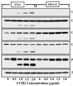
dose-dependent treated J/Bcl-xL cells and J/Neo cells stimu- lated by CCH1 to find molecular mediators. Significant dif- ferences from antibody reaction related to mitochondrial cy- tochrome c release pathway [14] were detected in J/Neo cell compared to J/Bcl-xL cells. Release level of mitochondrial cytochrome c to cytosol was increased in J/Neo cells in- cubated with 0.5-2.0 μg/ml of CCH1 (Fig. 3A). Subsequently, released cytochrome c switched pro-caspase 9, downstream partner of cytochrome c related to apoptotic pathway, to ac- tive-caspase 9, dose-dependently (Fig. 3B) in J/Neo cells. The amount of active-caspase 3 was increased in the range from 1.0 μg/ml to 2.0 μg/ml of CCH1 (Fig. 3D) in J/Neo cells.
Next, we performed Western blotting for PARP, which is the downstream target of active-caspase 3. As pro-caspase 3 ranged from 1.0 μg/ml to 2.0 μg/ml of CCH1 was acti- vated, PARP was degraded (Fig. 3E) in J/Neo cells. In this study, it was found that cleavages of caspase-8 and -9 in Jurkat T cells were also induced by CCH1 treatment (Fig.
3B, 3C). Recent reports [9,17] suggest that pro-caspase-3 can be activated through active caspase-8 in turn can cleave Bid, leading to cytochrome c release from mitochondria [12] and setting up a self-amplification loop to amplify caspase-9.
Together, these findings support the notion that apoptotic signaling of CCH1 in Jurkat T cell is regulated by mitochon- drial cytochrome c release pathway.
Cytotoxic effect of CCH1 on PBMC
Cytotoxic effect of CCH1 was tested with normal PBMC
C Caspase-8 (p57) (p43/p41)
D
β-actin PARP (p116)
(P85) Caspase-3 (p32)
(P19)
E
F
CCH1 Concentration (μg/ml) 0 0.5 1.0 1.5 2.0 0 0.5 1.0 1.5 2.0
Fig. 3. CCH1 induced-cell death is through mitochondrial cyto- chrome c release. The cells (5×10⁶) were incubated with indicated concentrations of CCH1 for 20 hr and prepared for the cell lysates. Equivalent amounts of cell lysates were electrophoresed on 4~12% SDS gradient poly- acrylamide gels and electrotransferred to Immobilon-P membrane. Western blot analysis was performed as de- scribed in materials and methods using the PIERCE Western blot detection system. Cytochrome c (A), cas- pase-9 (B), caspase-8 activation (C), caspase-3 activation (D), cleavage of PARP (E), and β-actin (F).
at concentration ranging from 0 to 130 μg/ml for 48 hr. As shown in fig. 4, cell viability of Jurkat T cells was inhibited by 70% at 8.0 μg/ml of CCH1, however, PBMCs were dead by 34% at the same concentration of CCH1.
Thus, acute Jurkat T lymphoma showed sensitivity to CCH1, but normal PBMCs were resistant against CCH1. It seems that CCH1 is used as evaluation for chemotherapeutic
Fig. 4. Comparative cell viability of PBMCs and Jurkat T cells treated with CCH1. PBMCs and Jurkat T cells were in- troduced at a density of 2.5×10⁵ cells in each well of 96-well plate and cultured for 48 hr. Cell viability was calculated by MTT assay.
purified phytochemical ingredient designated as CCH1 from
R. cordifolia, which has been used as tonic materials or fork
remedies in several countries including Korea, stimulates
human acute Jurkat T cell apoptosis via mitochondria de-
pendent caspase signaling. With dried roots (3 kg) of R. cor-
difolia, the purification procedure employed in this study fi-
nally afforded 65mg of CCH1 as described in material and
methods. Further analysis to examine the purity of CCH1
by analytical HPLC C18 column chromatography revealed
that CCH1 is composed of the mixture of two substances
(data not shown). The CCH1 appear to be a steroid sub-
stance because it positively reacts with antimony tri-
chloride-CHCl
3based on spraying chemical reagent on TLC,
but it fails to react with ninhydrine and FeCl
3. In addition,
CCH1 appeared to contain carbohydrate because of pos-
itively reacting with anisaldehyde, 2,4-dinitrophenyl hydra-
zine, and aniline phthalate reagents on TLC. Taken together
with these chemical properties, it seems that CCH1 is a ste-
roid derivative. For the chemical structure and pure sub-
stance, further instrument analysis is on track. Cytotoxicity
of R. cordifolia against acute Jurkat T cell lymphoma was ex-
pressed as IC
50during purification, and the IC
50was de-
termined at each step fractionated. The results showed that
IC
50at methanol extract, CH
2Cl
2fraction, silica gel chroma-
tography, and preparative HPLC GS320 chromatography
was 65 μg/ml, 32 μg/ml, 6.3 μg/ml, and 1.7 μg/ml,
respectively. Although it has been reported that bicyclic hex-
apeptides (RA-IX and RA-Z) in R.cordifolia have potential an-
ticancer activity [4], but little is known that other bio-
chemical including a steroid derivative in R. cordifolia shows
potential cytotoxicity against cancer cells, and especially sig-
naling pathways during apoptosis is still unknown. In this
report, we have understood the cytotoxic effect, the apop-
totic signaling pathway, and cell cycle distribution of CCH1
by taking advantage of antiapoptotic protein, Bcl-xL whose
major function is known to suppress mitochondrial cyto-
chrome c release [3,14]. In Jurkat T cells exposed to CCH1,
mitochondrial cytochrome c release, activation of caspase-3,
-8 and -9, and cleavage of PARP were detected. These results
indicated that CCH1-induced apoptotic DNA fragmentation
was triggered by mitochondria-dependent death-signaling
pathway including cytochrome c release and caspase activation. Recent reports [9,16] suggest that caspase-8 acti- vation, when triggered downstream of the mitochondrial pathway of apoptosis, may amplify caspase-9 activation through the cleavage of the pro-apoptotic protein Bid, which, in turn, elicits a further efflux of cytochrome c from mitochondria [12]. Although the current results indicate that CCH1-induced apoptosis is accompanied by mitochondrial cytochrome c release and subsequent activation of caspase cascade in Jurkat T cells, we employed two experimental approaches to confirm that these cellular events are pre- requisite for the CCH1-induced apoptosis; one is for taking advantage of antiapoptotic protein Bcl-xL whose major func- tion is known to suppress mitochondrial cytochrom c release [11,18] and the other is to use z-VAD-fmk, a well known broad-range caspase inhibitor [17]. The CCH1-induced apop- tosis was abrogated by an ectopic over-expression of Bcl-xL.
In addition, sensitivity of Jurkat T cells on CCH1 is more than that of normal PBMCs.
Collectively, these results demonstrate that CCH1, a parti- ally purified constituent from medicinal plant R. cordifolia, induces apoptosis of Jurkat T cells via mitochondria depend- ent pathway by release of mitochondria cytochrome c, acti- vation caspase-8, -9, and subsequent cascade events. These things will be helpful and useful for evaluation of its chemo- therapeutic potency and tonic material.
References
1. Antarkar, D. S., T. Chinwala, and N. Bhatt. 1983. Anti-in- flammatory activity of Rubia codifolia Linn. in rats. Indian Journal of Pharmacology 15, 185-188.
2. Ciapetti, G., E. Cenni, L. Pratelli, and A. Pizzoferrato. 1993.
In vitro evaluation of cell/biomaterial interaction by MTT assay. Biomaterials 14, 359-364.
3. Chou, J. J., H. Li, G. S. Salvesen, J. Yuan, and G. Wagner.
1999. Solution structure of BID, an intracellular amplifier of apoptotic signaling. Cell 96, 615-624.
4. Itokawa, H. and Y. Yamomiya. 1992. New antitumor bicy- clic hexapeptides RA-IX and RA-Z from R. cordifoliapart-3, conformation antitumor activity relationship. Journal of the Chemical Society Perkin transactions I 4, 455-459.
5. Jang, M. H., D. Y. Jun, S. W. Rue, K. H. Han, W. Park, and Y. H. Kim. 2002. Arginine antimetabolite L-canavanine induces apoptotic cell death in human Jurkat T cells via caspase-3 activation regulated by Bcl-2 or Bcl-xL. Biochemical Biophysical Research Communications 295, 283-288.
6. Joharapurkar, A. A., N. M. Deode, S. P. Zambad, and S.
N. Umathe. 2003. Immunomodulatory activity of alcoholic extract of Rubia cordifolia Linn. Indian Drugs 40, 179-181.
7. Gale, J. S., A. A. Proulx, J. R. Gonder, A. J. Mao, and C.
M. L. Hutnik. 2004. Comparison of the in vitro toxicity of indocyanine green to that of trypan blue in human retinal pigment epithelium cell cultures. American Journal of Ophthalmology 138, 65-69.
8. Kasture, V. S., V. K. Desmukh, and C. T. Chopde. 2000.
Anticonvulsant and behavioral actions of triterpine isolated from Rubia cordifolia Linn. Indian Journal of Experimental Biology 38, 675-680.
9. Kim, R. S., K. Tanabe, Y. Uchida, M. Emi, H. Inoue, and T. Toge. 2002. Current status of the molecular mechanisms of anticancer drug-induced apoptosis. Cancer Chemotherapy Pharmacology 50, 343-352.
10. Kim, Y. H., J. J. Proust, M. J. Uchholz, F. J. Chrest, and A. A. Nordin. 1992. Expression of the murine homologue of the cell cycle control protein p34 cdc2 in T lymphocytes.
Journal of Immunology 149, 17-23.
11. Kluck, R. M., E. Bossy-Wetzel, D. R. Green, and D. D.
Newmeyer. 1997. The release of cytochrome c from mi- tochondria: a primary site for Bcl-2 regulation of apoptosis.
Science 275, 1132-1136.
12. Luo, X., J. Budihardjo, H. Zou, C. Slaughter, and X. Wang.
1998. BID, a Bcl-2 is an inner mitochondrial membrane pro- tein that blocks programmed cell death. Nature 348, 334-336.
13. Manohar, K., M. K. Adwankar, and M. P. Chitnis. 1982. In vivo anticancer activity of RC-80, a plant isolate from Rubia cordifolia Linn. against a spectrum of experimental tumor models. Chemotherapy 28, 291-293.
14. McDonnell, J. M., D. Fushman, C. L. Milliman, S. J.
Korsmeyer, and D. Cowburn. 1999. Solution structure of the proapoptotic molecule BID: a structural basis for apoptotic agonists and antagonists. Cell 96, 625-634.
15. Park, C. N., H. S. So, C. H. Shin, S. H. Baek, B. S. Moon, S. H. Shin, H. S. Lee, D. W. Lee, and B. K. Park. 2003.
Quercetin protects the hydrogen peroxide-induced apopto- sis via inhibition ofmitochondrial dysfunction in H9c2 car- diomyoblast cells. Biochemical Pharmacology 66, 1287-1295.
16. Perkins, C. L., G. Fang, C. N. Kim, and K. N. Bhalla. 2000.
The role of Apaf-1, caspase-9 and BID proteins in etoposide- or paclitaxel-induced mitochondrial events during apoptosis. Cancer Research 60, 1645-1653.
17. Slee, E. A., H. Zhu, S. C. Chow, M. MacFarlane, D. W.
Nicholson, and G. M. Cohen. 1996. Benzyloxycarbonyl- Val-Ala-Asp (OMe) fluoromethylketone (z-VAD-fmk) in- hibits apoptosis by blocking the processing CPP32.
Biochemical Journal 315, 21-24.
18. Yang, J., X. Liu, K. Bhalla, C. N. Kim, A. M. Ibrado, J. Cai, T. I. Peng, D. P. Jones, and X. Wang. 1997. Prevention of apoptosis by Bcl-2: release of cytochrome c from mitochon- dria blocked. Science 275, 1129-1132.