Inhibitory Effect of Extract from Acanthocoris sordidus on Oxidative Damage
Young Mi Park
1, Jae Hwan Lim
1, Jong Eun Lee
1and Eul Won Seo
1*
1
Department of Biological Science, Andong National University, Andong 760-749, Korea Received July 23, 2014 /Revised September 6, 2014 /Accepted September 12, 2014
Here, we showed that Acanthocoris sordidus extract inhibited both cell and DNA damage caused by oxidative stress. In a radical scavenging assay, the scavenging activity of the A. sordidus extract against 1,1-diphenyl-2-picrylhydrazyl (DPPH) and hydroxyl radicals was 48.9% and 37.8%, respectively, that of ascorbic acid, which was used as a positive control. The ferrous iron chelating activity of the A.
sordidus extract was 80.0% compared to that when ethylenediaminetetraacetic acid (EDTA) was used a control. To verify the inhibitory effect of the extract on oxidative cell damage induced by reactive oxygen species (ROS), a lipid peroxidation assay was performed. The results showed that peroxidation was completely inhibited in an extract-treated group compared to a radical-treated group. The level of p21 protein expression was 68.1% that of a control sample. The DNA cleavage-inhibiting property of the A. sordidus extract-treated group was 53.3% that of a control group. Moreover, the phosphor- ylation of the H2AX protein was reduced to 39.0% of that treated with radical agents, indicating that the extract might inhibit the DNA damage that causes radical oxidation. Taken together, our findings suggest that the A. sordidus extract is effective not only in repressing oxidation by free oxygen radicals and hydroxyl radicals but also in decreasing cell and DNA damage caused by oxidative stress.
Key words : Acanthocoris sordidus, antioxidant activity, hydroxyl radical, oxidative stress
*Corresponding author
*Tel : +82-54-820-5462, Fax : +82-54-820-7705
*E-mail : [email protected]
This is an Open-Access article distributed under the terms of the Creative Commons Attribution Non-Commercial License (http://creativecommons.org/licenses/by-nc/3.0) which permits unrestricted non-commercial use, distribution, and reproduction in any medium, provided the original work is properly cited.
Journal of Life Science 2014 Vol. 24. No. 10. 1078~1084 DOI : http://dx.doi.org/10.5352/JLS.2014.24.10.1078
서 론
모든 호기성 호흡을 하는 생명체는 생명 유지에 필요한 에 너지를 생성하기 위하여 산소를 필요로 한다. 그러나 산소를 이용한 물질 대사 과정 중 미토콘드리아 내의 산화환원 효소 계나 외부 항원에 노출된 면역 세포 및 각종 물리· 화학적 요인 들에 의해 체내 활성산소가 과다하게 생성되어 심각한 산화적 스트레스에 시달리고 있다. Hydroxyl radical, hydrogen per- oxide, superoxide anion 등에 의해 유발되는 산화적 스트레스 는 여러 가지 대사 질병과 세포 사멸의 주요 요인으로 작용한 다. 특히 생체막에 존재하는 지질은 체내에서 발생하는 유리 라디칼의 공격을 받아 여러 종류의 과산화물을 형성하는데, 이 과산화물들과 분해산물들은 높은 반응성으로 주변 생체 분자들의 구조와 기능을 변화시키게 된다[3, 4]. 이와 같은 산 화적 대사 부산물인 활성산소는 세포 내 여러 구성성분인 지 질, 단백질, 핵산 및 DNA를 산화시켜 노화 및 다양한 퇴행성 질환을 유발하기도 하며[18], 세포 내 여러 조직을 손상시켜 암, 동맥경화, 류마티스 관절염 및 알레르기 등을 유발하기도 한다[1]. 그러나 생체에 존재하는 superoxide dismutase, cata-
lase, glutathione peroxidase 등과 같은 항산화효소는 세포 활 동 중 생성되는 활성산소의 유리기를 제거함으로써 산화-항산 화의 균형을 유지시켜 줌으로서 산화적 스트레스로부터 생체 를 보호하고 있다[10]. 이 외에도 활성산소를 소거할 수 있는 화합물 또는 과산화물 생성 억제 물질과 같은 항산화제들은 활성산소로 인한 산화적 손상으로부터 세포를 보호함으로써 산화스트레스에 의해 유발되는 질병의 예방 또는 치료 효과를 나타내고 있다[12]. 그러나 현대인은 흡연, 각종 환경오염물질, 알코올, 약물, 방사선, 격렬한 운동 등 물리, 화학적 요인들에 의해 발생되는 체내 활성 산소종의 생성과다로 산화-항산화의 균형이 깨져 심각한 산화적 스트레스에 시달리고 있다[20]. 이 에 따라 최근 산화적 스트레스와 이로 인해 유발되는 건강 문제를 해결할 수 있는 물질로서 항산화제에 대한 관심이 집 중되고 있다. 일반적인 항산화제는 지질과산화 작용의 억제, DNA 손상 회복, 노화 억제, 식품 품질 유지 등의 기능을 갖고 있어 다양한 용도로 사용되고 있으나, 부작용과 같은 안전성 문제가 제기됨에 따라 산화적 스트레스에 의한 성인병 예방 및 치료에는 매우 제한적이다[16]. 이에 따라 효능이 우수하고 안전성 면에서 입증된 천연 항산화제를 개발하기 위한 연구들 이 수행되고 있으며[14], 항산화 효과가 풍부한 식품을 일상적 으로 섭취함으로써 이들 식품구성 성분의 생체조절기능에 대 한 효과를 병행하는 방법 등이 다양하게 검토되고 있는 실정 이다.
곤충의 경우 매우 높은 종 다양성과 특이성을 가지고 있으
나 재료 확보의 어려움과 상대적으로 알려지지 않아 많은 연
구 분야에 있어 제한을 받기 때문에 국내외적으로 많은 연구
가 이루어지지 못하고 있었으나 최근 들어 곤충을 유용생물자 원으로 인식하면서 이를 활용하고자 하는 연구가 활성화되고 있다. 따라서 종 다양성이 가장 풍부할 뿐만 아니라 개체가 작고 세대가 짧아 시간적, 공간적, 인력적인 면에서 효율적 관리의 탁월한 장점을 지닌 곤충을 생물자원으로 활용해야 할 필요성이 절실히 요청되고 있다.
본 연구의 소재인 꽈리허리노린재(Acanthocoris sordidus)는 허리노린재과의 곤충으로 줄기에 뾰족한 침을 박고 즙액을 빨아먹어 고추나 고구마 등의 생육을 저해하고 탄저병 포자를 타작물에 전염시키는 해충이다. 최근 잡곡재배면적의 증가에 따라 노린재로 인한 건강기능성 물질의 수량과 품질이 크게 떨어져 그 피해는 점점 심각해지고 있어 노린재 방제에 대한 연구는 지속적으로 이루어지고 있으나, 이러한 해충류의 곤충 류로부터 유래되는 생리활성물질에 관한 연구는 전무한 실정 이다. 따라서 본 연구는 꽈리허리노린재 추출물의 항산화능을 조사하고 세포와 DNA의 산화적 손상에 미치는 영향을 분석 하고자 하였다.
재료 및 방법
시료 추출
본 연구에 사용된 꽈리허리노린재(Acanthocoris sordidus)는 경북 안동시 송천동 주변에서 채집하였으며 Won 등[21]과 Yoon [22]의 연구 자료를 참고하여 동정하였다. 꽈리허리노린 재로부터 시료를 추출하기 위하여 공시충을 동결 건조 후 분 쇄하여 분말화하였다. 시료의 추출은 공시충 분말 30 g에 물 100 ml를 가하고 25℃에서 총 3회 환류 추출 후 여과하여 농축 하였으며, -70℃에 보관하면서 시료로 사용하였다.
꽈리허리노린재 추출물의 항산화활성 측정
DPPH (1,1-diphenyl-2-picrylhydrazyl) 라디칼 제거능의 측 정은 Hsu 등[7]의 방법에 따라 DMSO (Dimethyl sulfoxide, Sigma-Aldrich, USA)로 희석한 농도별 꽈리허리노린재 추출 물 40 μl와 300 mM DPPH (1,1-diphenyl-2-picrylhydrazyl, Sigma-Aldrich, St. Louis, MO, USA) 760 μl를 혼합하여 37℃
에서 반응시킨 후 515 nm에서 흡광도를 측정하였다. 또한 수 산화 라디칼 제거 활성은 1.5 mM FeSO
4(Sigma-Aldrich, USA) 250 μl, 6 mM H
2O
2(Sigma-Aldrich, USA) 175 μl와 농도 별 시료 75 μl를 혼합하여 반응시킨 후 560 nm에서 흡광도를 측정하였다[19]. Fe
2+-chelating 활성은 시료에 2 mM FeCl
2(Sigma-Aldrich, USA) 15 μl와 5 mM ferrozine (Sigma- Aldrich, USA) 30 μl를 혼합하여 상온에서 15분 반응시킨 후 562 nm에서 Fe
2+-ferrozine 용액의 흡광도를 측정하였다[7]. 각 실험 항목에서 농도에 따른 추출물의 항산화활성은 다음의 식으로 %를 계산하였다.
소거능(%) = {1- (추출물 처리군의 흡광도 / 추출물 무처리
군의 흡광도)} ×100
세포배양
본 연구에 사용한 NIH-3T3 마우스 섬유아 세포주는 한국세 포주은행(KRIBB, Taejeon, Korea)에서 분양 받았으며 Dulbec- co’s modified eagle medium (DMEM, Gibco Inc., USA)에 10% fetal bovine serum (FBS, Gibco Inc., USA)을 첨가한 배지 를 이용하여 37℃, 5% CO
2배양기에서 배양하였다. 꽈리허리 노린재 추출물이 NIH-3T3 세포 생존에 미치는 영향은 MTT (3-(4,5-dimethylthiazol-2-yl)- 2,5-diphenyl-tetrazolium bro- mide) 분석을 통해 실시하였다. NIH-3T3 세포(5×10
3cells/
well)를 96-well cell plate에 분주한 후 시료, FeSO
4, H
2O
2를 각각 농도별로 처리하여 24시간 배양 후 MTT (Sigma-Aldrich, St. Louis, MO, USA) 용액(1 mg/ml)을 처리하여 4시간 반응 시킨 다음 DMSO로 세포 내 염색액을 용출시킨 후 microplate reader (Infinite
®200, Tecan Trading AG, Switzerland)를 이용 하여 540 nm 파장에서 흡광도를 측정하였다.
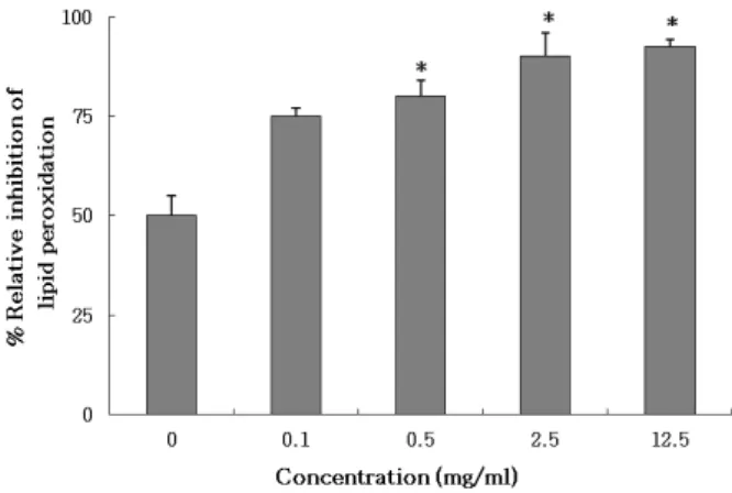
지질과산화 억제능 분석
지질과산화 억제능은 NIH-3T3 세포(2×10
5cells/ml)에 시 료와 FeSO
4, H
2O
2를 처리하여 배양한 후 회수하여 1.15% KCl 로 균질화시켰으며, 0.2 ml sodium dodecyl sulfate (8.1%), 1.5 ml acetic acid (20%) 및 1.5 ml thiobarbituric acid (0.8%)를 첨가하여 95℃에서 2시간 반응시켰다. 반응 후 5 ml n-buta- nol/pyridine mixture (15:1, v/v)로 분획을 통해 얻은 상등액 의 흡광도를 532 nm에서 측정하였다[11].
DNA 손상 억제능 분석
꽈리허리노린재 추출물이 유리 라디칼에 의한 유전정보의 손상을 유발하는 DNA 분절에 미치는 영향을 조사하기 위하 여 먼저 ΨX-174 RF I plasmid DNA (New England BioLabs, County Road Ipswich, MA) 분석을 실시하였다. ΨX-174 RF I plasmid DNA 5 μl, 농도별 시료 10 μl, FeSO
4와 H
2O
25 μl 를 혼합하여 37℃에서 반응시킨 후 50% glycerol (v/v), 40 mM EDTA, 0.05% bromophenol blue을 첨가하여 전기영동 후 분석하였다.
Western blot 분석
NIH-3T3 세포(1×10
6cells/well)를 6-well plate에서 분주하
여 16시간 배양한 후 농도별 시료, FeSO
4, H
2O
2를 처리하여
24시간 반응시켰다. 이 후 NIH-3T3 세포로부터 lysis buffer
(50 mM Tris–HCl, pH 7.4, 150 mM NaCl, 1 mM EDTA,
1mM EGTA, 10 mg/ml aprotinin, 10 mg/ml leupeptin, 5
mM phenylmethylsulfonyl fluoride [PMSF] and 1 mM DTT)
이용하여 추출한 단백질을 15% SDS-PAGE로 전기영동 후
PVDF membrane (Sigma-Aldrich, St. Louis, MO, USA)에
A
B
Fig. 1. (A) DPPH free radical and hydroxyl radical scavenging activities and (B) Fe
2+-chelating activities of extract from A.sordidus. (A) DPPH radical reaction mixture containing 40 μl of A.sordidus extract and 760 μl of 300 mM DPPH ethanol solution were incubated and absorbance was measured at 515 nm. Ascorbic acid was used for the pos- itive control. The DPPH free radical scavenging activity was 48.9% at 12.5 mg/ml. Hydroxyl radical reaction mix- ture contained 250 μl of 1.5 mM FeSO
4, 175 μl of 6 mM H
2O
2, 300 μl of 20 mM sodium salicylate and varying concentrations of the extract. The absorbance was meas- ured at 562 nm. Ascorbic acid was used for the positive control. Hydroxyl-radical scavenging activity was found to be enhanced with concentration-dependent manner, exhibiting 37.8% at 12.5 mg/ml. (B) Fe
2+-chelated re- action mixture contained 15 μl of 2 mM FeCl
2, 30 μl of 5 mM ferrozine, 150 μl of four different concentrations of the extract and 605 μl of distilled water. The absorb- ance of the Fe
2+-ferrozine complex was measured at 562 nm. EDTA was used for the positive control. The Fe
2+- chelating activity was revealed 80.0% at 12.5 mg/ml. In all experiments, the absorbance values were converted to scavenging and chelating effect (%) and data plotted as the means of replicate scavenging and chelating activ- ity (%) values ± S.E.
transblot 하였다. 이 후 5% BSA로 blocking 시키고, p21 rabbit polyclonal IgG (Cell Signaling Technology, Beverly, MA)와 P-histone H2AX rabbit antibody (Cell Signaling Technology, Beverly, MA)로 1차 처리 후 IgG HRP-linked anti-rabbit anti- body (Cell Signaling Technology, Beverly, MA)를 2차 처리하 여 반응시켰다. 이 후 ECL detection polaroid camera (Amersham Biosciences, USA)를 사용하여 촬영하였으며, 발 현 수준은 Un-SCAN-IT gel Version 5.1 (Silk Scientific, Inc.).
프로그램을 통해 분석하였다.
통계처리
모든 실험은 독립적으로 3회 이상 실시하였으며 각 실험에 서 얻어진 결과는 평균±표준오차로 나타내었다. 통계처리는 SPSS version 12.0로 분석한 후 t-검정을 실시하여 분산과 평균 의 동일성 여부를 검정하였으며, 분석결과는 일원분산분석 (one way ANOVA)에 의한 Duncan 검정을 실시하여 p값이 0.05 미만일 때 유의한 것으로 간주하였다.
결 과
꽈리허리노린재 추출물의 항산화능
본 연구에서 꽈리허리노린재 추출물의 항산화활성을 분석 하기 위하여 DPPH 유리 라디칼과 수산화 라디칼의 제거 활성 및 Fe
2+-chelating 효과를 조사하였다. 꽈리허리노린재 추출물 의 DPPH 유리 라디칼 제거능은 양성대조군인 ascorbic acid 의 모든 농도별 활성에 비해서는 다소 낮게 나타났으나 시료 의 농도가 높아짐에 따라 활성이 증가하여 0.1 mg/ml에서 5.3%, 0.5 mg/ml에서 7.0%, 2.5 mg/ml에서 25.2%, 12.5 mg/
ml에서 48.9%의 제거 효과를 나타내었다. 또한 수산화 라디칼 제거능은 대조군에 대해 12.5 mg/ml에서 37.8%의 활성을 보 였다(Fig. 1A). Fe
2+-chelating 분석 결과 양성대조군인 EDTA 에 비해서는 모든 농도에서 활성이 다소 낮았으나 시료의 농 도가 0.1 mg/ml에서 17.2%, 0.5 mg/ml에서 41.4%, 2.5 mg/
ml에서 50.4%, 12.5 mg/ml에서 80.0%로 농도가 높아질수록 활성이 증가하는 것으로 나타났다(Fig. 1B).
꽈리허리노린재 추출물의 산화적 세포 손상 억제능 꽈리허리노린재 추출물이 ROS에 의해 유발되는 산화적 세 포 손상에 미치는 억제 효과를 분석하기 위하여 MTT 분석, 지 질과산화 분석 및 p21 단백질 발현 수준을 분석하였다. MTT 분석에서 세포 생존율은 Fe
2+와 H
2O
2만 처리한 라디칼 처리군 의 세포 생존율은 대조군에 비해 71.2%의 활성을 보여주고 있으나 꽈리허리노린재 추출물 12.5 mg/ml를 처리한 실험군 에서는 96.9%의 생존율을 나타내주고 있어 세포에 대한 시료 의 독성은 거의 없는 것으로 조사되었다(Fig. 2). 이러한 조건 에서 꽈리허리노린재 추출물을 처리하였을 때 지질과산화 억
제능은 0.1 mg/ml에서 75.4%, 0.5 mg/ml에서 80.3%, 2.5
mg/ml에서 89.8%, 12.5 mg/ml에서 92.4%로 농도가 높아짐
에 따라 억제능이 증가하는 경향을 나타내었다(Fig. 3). 또한
꽈리허리노린재 추출물이 산화적 스트레스에 의한 p21 단백
Fig. 2. The effect of A. sordidus extract on NIH-3T3 cell viability.
The NIH-3T3 cells (5×10
3cells/well) were cultured in 96-well plate at 37℃ for 24 hr. The extract from A.sordi- dus were treated by concentration dependent manner to each well with FeSO
4and H
2O
2.. After then 50 μl of MTT solution (1 mg/ml) was treated to each well for 4 hr, and then 100 μl of DMSO was treated to each well. The observance was measured with a microplate reader at 540 nm. The cell viability of radical treated group with Fe
2+and H
2O
2only was 71.2% compare to that of the radical untreated control group. However, the cell via- bility treated with A.sordidus extract was restored to 95.0% at the concentration of 12.5 mg/ml treatment.
*p<
0.05 indicate significant difference between the only radi- cal treated group (treated with FeSO
4, H
2O
2without ex- tract) and extract-treated group (treated with FeSO
4, H
2O
2and varying concentration of extract). Data were shown as mean ± SE.
Fig. 3. The inhibitory effect of A. sordidus extract on the lipid peroxidation induced by hydroxyl radical in NIH-3T3 cellsl. The NIH-3T3 cells were cultured in a 6-well plate at 2×10
6cells/well for 16 hr. After plating, the cells were treated with the varying concentration of A.sordidus ex- tract, FeSO
4and H
2O
2were added to the plate. The cell lysate was mixed with 0.1 ml of 8.1% sodium dode- cylsulfate, 0.75 ml of 20% acetic acid, and 0.75 ml of 0.8%
thiobarbituric acid. The supernatant fractions were iso- lated and the absorbance was measured at 532 nm. The inhibition level of lipid peroxidation was shown to 92.4%
at 12.5 mg/ml, exhibiting an increasing tendency as the concentration rose.
*p<0.05 indicate a significant differ- ence between the only radical treated group (treated with FeSO
4, H
2O
2without extract) and extract-treated group (treated with FeSO
4, H
2O
2and varying concen- tration of extract). Data were shown as mean ± SE.
질의 발현에 미치는 영향을 조사하기 위하여 p21 단백질 발현 율 수준을 Western blot에 의해 분석하였다. Fe
2+와 H
2O
2만 처리한 라디칼 처리군의 p21 단백질의 발현 수준은 대조군에 비해 31.2%의 활성을 보여주고 있으나 꽈리허리노린재 추출 물을 처리한 경우에 p21 단백질의 발현 수준은 0.1 mg/ml에 서 45.3%, 0.5 mg/ml에서 58.1%, 2.5 mg/ml에서 65.0%, 12.5 mg/ml에서 68.1%로 나타나 농도가 높아질수록 대조군 수준 으로 회복하는 경향을 나타내었다(Fig. 4).
꽈리허리노린재 추출물의 산화적 DNA 손상 억제능 꽈리허리노린재 추출물이 산화적 스트레스에 의한 DNA 손상에 미치는 영향은 ΨX-174 RF I plasmid DNA를 사용한 in vitro DNA cleavage 분석을 통해 평가하였다. ΨX-174 RF I plasmid DNA는 대조군에서 supercoiled (SC) DNA로 확인 되고 있으나, Fe
2+와 H
2O
2만 처리한 라디칼 처리군에서는 su- percoiled plasmid DNA가 DNA의 분절로 인하여 open circu- lar (OC) DNA로 전환되고 있다. 이러한 조건에서 시료를 처리 하였을 때 supercoiled plasmid DNA는 0.1 mg/ml에서 6.0%, 0.5 mg/ml에서 17.4%, 2.5 mg/ml에서 25.0%, 12.5 mg/ml에 서 53.3% 수준으로 조사되어 시료의 농도가 높아질수록 su-
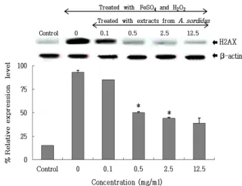
percoiled plasmid DNA로부터 open circular plasmid DNA 로 전환되는 것을 억제시키는 것으로 나타났다(Fig. 5). 꽈리허 리노린재 추출물이 DNA 손상에 미치는 간접적인 항산화 효 과를 조사하기 위하여 NIH 3T3 세포의 히스톤 H2AX의 인산 화 수준을 phospho-H2AX 특이 항체를 사용한 immuno-anal- ysis로 분석하였다. 꽈리허리노린재 추출물의 농도에 따른 H2AX 인산화비는 Fe
2+와 H
2O
2만 처리한 음성 대조군에 대해 0.1 mg/ml에서 85.0%, 0.5 mg/ml에서 50.1%, 2.5 mg/ ml에 서 44.4%, 12.5 mg/ml에서 39.0%로 나타나 시료의 농도가 높 아짐에 따라 인산화비는 감소하는 경향을 보여주고 있다(Fig.
6). 즉, 이러한 결과는 꽈리허리노린재 추출물을 처리하였을 경우 NIH 3T3 세포 염색체에서 DNA 손상 빈도가 라디칼 처리군에 비해 약 60.9%의 억제 효과를 나타낼 것으로 분석되 었다.
고 찰
산소를 소비하여 에너지를 획득하는 호기성 유기체들은 산
소에 의한 산화적 스트레스에 항상 노출되어 있으며 이러한
산화적 스트레스는 생체막의 손상과 고분자 단백질 및 핵산
변성, 세포소기관의 기능 상실 등을 유발한다. 따라서 활성산
Fig. 6. The effect of A.sordidus extract on the phosphorylation level of H2AX in NIH-3T3 cells damaged by hydroxyl radical. Lane 1 is control group and lane 2 is only radical treated group. Lanes 3-6 were treated with varying con- centration of the extract. The phosphorylation ratio of H2AX over different concentration of extract were 85.0%
at 0.1 mg/ml, 50.1% at 0.5 mg/ml, 44.4% at 2.5 mg/ml, and 39.0% at 12.5 mg/ml compared to those ratio in neg- ative control treated with Fe
2+and H
2O
2. The occurrence of DNA damage was inhibited in NIH-3T3 chromosome when extract treated, with approximately 83.3% compar- ing to radical treated group.
*p<0.05 indicates a sig- nificant difference between only radical treated group and extract-treated group. Data were shown as mean ± SE.
Fig. 4. The effect of A. Sordidus extract on the expression of p21 protein in NIH-3T3 cells damaged by hydroxyl radical.
Lane 1 is control and lane 2 is treated with only radical treated group. Lanes 3-6 were treated with varying con- centration of extract (0.1, 0.5, 2.5 and 12.5 mg/ml). The level of p21 expression was measured by western blot analysis. The level of p21 expression showed 12.5% in radical treated group with Fe
2+and H
2O
2only. On the other hand, the amounts of expression of p21 were in- creased from 45.3% to 68.1% at 12.5 mg/ml in A.sordidus extract treated group. *p<0.05 indicates significant differ- ence between the only radical treated group and ex- tract-treated group. Data were shown as mean ± SE.
Fig. 5. The effect of A.sordidus extract on the relaxation of the ψX-174 RF I plasmid DNA by hydroxyl radical. SC, su- percoiled DNA; OC, open circular DNA. Lane 1 is con- trol group and lane 2 is only radical treated group. Lanes 3-6 were treated with varying concentration of the ex- tract (0.1, 0.5, 2.5 and 12.5 mg/ml). The ψX-174 RF I plas- mid DNA was identified as SC form DNA in the control group. In contrast, the SC plasmid DNA had trans- formed to OC form due to DNA segmentation in radical treated group with Fe
2+and H
2O
2. *p<0.05 indicates sig- nificant difference between the radical treated group and extract-treated group. Data were shown as mean ± SE.
소에 의한 산화적 스트레스를 감소시키는 항산화능은 호기성 생물에 있어 질병과 노화에 있어 매우 중요한 의미를 가진다.
본 연구에서는 꽈리허리노린재 추출물의 항산화능 및 산화적 스트레스에 의함 세포와 DNA 손상 억제력을 분석하였다. 최 근 연구에 따르면 청동풍뎅이(Anomala alopilosa) 에탄올 추출 물의 DPPH 유리 라디칼 소거능은 3 μg/ml의 농도에서 약 90%의 활성을 나타내어 우수한 항산화 효과를 가진다고 하였 으며, 풀색꽃무지(Gametis jucunda)와 칠성무당벌레(Coccinella septempunctata)는 추출용매에 상관없이 높은 수산화 라디칼 소거 활성을 가져 혈중 지질과산화 수준을 저하시키는 데 효 과적이라고 보고된 바 있다[23]. 또한 방아깨비(Acrida cinerea cinerea)와 호랑나비(Papilio xuthus)의 경우 물 추출물에서 가장 우수한 chelating 효과를 보여 활성산소에 의한 생체 손상을 감소시킨다고 알려져 있다[18]. 본 연구에서도 꽈리허리노린 재 추출물의 DPPH 유리 라디칼과 수산화 라디칼 제거능은 다소 낮았으나 Fe
2+-chelating 효과는 시료의 농도가 높아짐에 따라 증가되어 12.5 mg/ml에서 80% 이상의 활성을 보여주고 있어 꽈리허리리노린재 추출물이 Fenton 반응에 의한 활성산 소의 생성을 억제하는 데 효과적인 것으로 확인되었다.
산화적 세포 손상에 대한 기존의 연구에서 Heo 등[5]은 칠
성무당벌레(Coccinella septempunctata)와 무당벌레(Harmonia axyridis) 추출물의 경우 시간에 따른 산화 속도의 차이는 있으 나 메탄올 추출물에서 가장 높은 활성을 보여 지질과산화 억 제에 유효하다고 한 바 있다. 본 연구에서 꽈리허리노린재 추 출물이 세포의 산화적 손상에 미치는 영향을 조사한 결과 라 디칼 처리군의 세포는 70.7%의 생존율을 보여주고 있으나, 시 료를 처리한 실험군에서는 96.9%의 세포 생존율을 보였으며, 12.5 mg/ml 농도에서 92.4%의 지질과산화 억제효과를 보였 다. 따라서 꽈리허리노린재 추출물은 세포에 대한 독성을 가 지지 않으면서 활성산소에 의한 체내 과산화물의 생성을 감소 시키는 데에 매우 효과적인 것으로 나타났다.
세포의 비정상적 증식을 저해하는 것으로 알려져 있는 p21 단백질은 세포주기 상 G1 기에서 암세포의 증식을 억제하는 가장 중요한 세포 주기조절 인자 중 하나이다[17]. 최근 Kim 등[13]은 동충하초(Cordyceps militaris) 열수 추출물의 경우 인 체 간암세포주인 HepG2 세포의 성장에 미치는 영향을 조사한 결과 처리 농도가 높아짐에 따라 p21의 발현 증가가 관찰되어 간암세포의 증식 억제에 연관성을 지닐 수 있다고 한 바 있다.
본 연구에서도 산화적 세포 손상에 대한 p21 단백질의 발현 수준을 조사한 결과 시료의 농도가 높아질수록 p21 단백질의 발현이 증가되었으며 이는 NIH-3T3 세포에서 hydroxyl radi- cal 처리시 apoptotic 또는 necrotic cell death를 유발하는 것으 로 생각된다.
유전 정보를 지니고 있는 핵 안의 DNA는 자발적으로 일어 나는 nucleotide의 화학적 변화, 림프세포에서 일어나는 프로 그램화된 이중 나선 손상(programmed double strand breaks, DSB)과 같은 불가피한 내부요인과 자외선, 방사선과 같은 외 부 환경요인에 의해 계속적인 손상을 겪게 된다[2]. 이 때 손상 된 DNA가 복원되거나 사멸하지 못하고 돌연변이 세포로 될 경우 암을 비롯한 여러 질병의 직접적인 요인으로 작용할 수 있는데, 특히 암에 있어 개시단계는 생체 내에서 대사가 활성 화된 발암인자가 유전자의 핵산에 결합함으로써 정상세포의 DNA를 손상시키기 때문에 DNA의 산화적 손상 억제력은 매 우 중요한 의미를 가진다[15]. Jeong 등[8]은 오미자(Schizandra chinensis)의 에탄올 추출물은 농도가 높아질수록 DNA 손상 억제능도 증가하여 약 93%의 억제 활성을 보여 DNA의 분절 을 억제하는 데 유효할 것이라고 하였으며, Hong 등[6]은 밀몽 화(Buddleja officinalis) 추출물이 산화적 DNA 손상 분석에서 supercoiled DNA에서 open circular DNA로 전환되는 것을 80% 억제하여 DNA 분절의 감소에 효과적이라고 보고한 바 있다. 또한 Jeong 등[9]은 DNA 이중나선의 분절에 의한 히스 톤 단백질의 변이 중 하나인 phospho-H2AX의 발현에 대하여 보리(Hordeum vulgare) 씨앗에서 정제된 3,4-dihydroxybenzal- dehyde는 200 μg/ml에서 80%의 H2AX 인산화비 억제율을 보여 DNA의 인산화를 감소시키는 데 매우 효과적이라 시사 한 바 있다. 본 연구의 꽈리허리노린재 추출물도 NIH 3T3 세
포 염색체에서 DNA 손상 빈도가 라디칼 처리군에 비해 약 60.9%의 억제 효과를 나타내고 있어 산화적 스트레스에 의한 DNA의 손상을 억제하는데 효과적일 것으로 생각된다.
본 연구를 요약하자면 꽈리허리노린재 추출물은 유리 라디 칼과 수산화 라디칼 제거능은 다소 낮았으나 Fenton 반응에 의한 라디칼 생성을 억제하는데 매우 효과적인 것으로 나타났 다. 또한 2차 산화물의 생성을 억제하고 세포의 비정상적인 증식을 조절하는 것으로 알려진 p21 단백질의 발현 수준을 증가시켰으며 활성산소에 의한 DNA 분절화를 감소시키고 DNA의 손상을 억제시키는 것으로 조사되었다. 따라서 본 연 구의 꽈리허리노린재 추출물은 산화적 스트레스로 인한 생체 독성으로부터 세포와 DNA를 보호하는 데 매우 효과적일 것 으로 사료된다.
감사의 글
이 논문은 2013학년도 안동대학교 학술연구조성비(연구교 수)에 의하여 연구되었음.
References