위령선 추출물의 항염활성 및 멜라닌 생성 억제효과
김예림·홍윤정·양기숙#
숙명여자대학교 약학대학
(Received November 30, 2013; Revised February 12, 2014; Accepted February 17, 2014)
Anti-inflammatory Effect and Inhibition of Melanin Biosynthesis of Clematis mandshurica
Ye Rim Kim, Yun Jung Hong and Ki Sook Yang#
College of Pharmacy, Sookmyung Women’s University, Seoul 140-742, Korea
Abstract — Clematis mandshurica (Ranunculaceae) has traditionally been used as a remedy for antidiuretic, antifungal, rheumatic conditions and alleviate pain. We carried out to evaluate the anti-oxidative effect, anti-inflammatory effect and anti-melanogenic effect of ethanol extract and solvent fractions of Clematis mandshurica. The ethanol extract and the dichlo- romethane fraction of Clematis mandshurica showed an anti-oxidative effect in DPPH assay, the inhibitory activity of nitric oxide (NO) production in lipopolysaccharide (LPS) activated RAW 264.7 cell, and melanin synthesis and tyrosinase activity of B16F10 melanoma cells. They reduced NO production and melanin content in a dose-dependent manner at con- centrations of 2.5~10µg/ml. They also suppressed iNOS and tyrosinase protein and m-RNA expressions dose dependently, assayed by western blot analysis and RT-PCR experiment.
Keywords □ Clematis mandshurica, NO, iNOS, melanin, tyrosinase
위령선(Clematis mandshurica)은 미나리아재비과 으아리속에 속하는 다년생의 덩굴성 초본으로서 뿌리를 약용하는데, 전통적 으로 신경통, 근육통, 편두통과 부종의 치료제로 사용되어 왔고,1) 항염작용, 항균작용, 이뇨작용 및 진통작용에 대한 연구가 보고 되어 있다.2,3)위령선의 성분으로는 clematoside A, A', B, C 등 의 배당체와4) hederagenin5)이 있고, 그 외에도 anemonol,6) steroids, saponin, amino acid, phenol, flavonoid7)등을 함유하 고 있으며 배당체나 saponin이 주 약효 물질로 볼 수 있다.위령 선의 항염활성에 대한 연구로서 Park 등은 위령선의 에탄올 추 출물이 LPS 및 interferon-γ(IFN-γ)로 유도한 macrophage에서 pro-inflammatory cytokine인 NO와 prostaglandin E2(PGE2)를 억제함을 보고하였고,8) Suh 등은 rat adjuvant arthritis 모델에 서 위령선 물 추출물이 염증을 감소시키고 interleukin-1(IL-1) 및 tumor necrosis factor-α(TNF-α) 생성을 억제함을 보고하였 다.9)본 실험에서는 위령선 추출물과 각 분획에 대한 약리작용
을 평가하고자, 항염 활성을 측정하기 위하여 RAW 264.7 세포에 추출물과 각 분획을 LPS와 처리하여 NO 생성 억제 효과를 측정
하였고,10-15)멜라닌 생성 억제 작용에 대해 B16F10 melanoma
세포를 이용하여 α-MSH와 위령선 에탄올 추출물 및 각 용매분 획을 처리하여 melanin을 정량하였으며,16-19)각각의 Western blot assay와 RT-PCR법을 이용하여 protein과 mRNA 수준의 억 제효과를 측정하였다.
실험방법
실험재료
본 실험에서 사용한 위령선은 경동시장내 동경종합상사에서 구입하여 사용하였으며 확증 표본은 본 대학 생약표본실에 보관 하였다.
추출 및 분획
세말한 위령선(4.2 kg)을 에탄올로 4시간씩 5회 가온 추출 후 온시 여과하고 여액을 감압 농축하여 에탄올 추출물(254 g, 수득 률: 6.04%)을 얻었다. 이 에탄올 추출물을 물 가용부로 나눠 각 각을 헥산 분획, 디클로로메탄 분획, 에틸아세테이트 분획, 부탄 올 분획, 물 분획으로 분획하였다. 감압 농축하여 가용부 헥산 분
#Corresponding Author Ki Sook Yang
College of Pharmacy, Sookmyung Women's University, Seoul 140-742, Korea
Tel.: 02-710-9578 Fax.: 02-715-9498 E-mail: [email protected]
종설
획 22.25 g(수득률: 11.13%), 디클로로메탄 분획 12.46 g(수득률:
6.23%), 에틸아세테이트 분획 28.59 g(수득률: 14.30%), 부탄올 분획 58.88 g(수득률: 29.44%), 물 분획 66.66 g(수득률: 33.33%) 을 얻어 시료로 사용하였다.
DPPH를 이용한 항산화능 측정
시료를 각 농도별로 조제한 용액 0.1 ml(control: 99.5%
ethanol)에 0.1 mM DPPH 용액 1.9 ml을 가하였다. 각 시료를 농도별로 조제하여 시험관 진탕기로 10초간 진탕한 후 37oC에 서 30분 동안 반응시키고 spectrophotometer를 이용하여 515 nm에서 흡광도를 측정하였다. 시료의 항산화 활성을 측정하기 위하여 전자 공여능(electron donating ability, EDA%)에 의한 환 원력으로 표시하였다. 양성 대조약물로는 ascorbic acid를 농도 별로 용시 조제하여 사용하였다. 각 시료의 항산화 활성을 비교 검토하기 위하여 EDA가 50%에 이르도록 하는 데 필요한 시료 의 양(IC50)을 측정하였다.
RAW 264.7 세포에서의 NO생성 저해 작용 측정
세포주의 배양 − Mouse macrophage RAW 264.7 세포는 한국 세포주 은행에서 분양받아 10% FBS(fetal bovine serum), penicillin G(100 IU/ml), streptomycin(100 µg/ml), amphotericin (0.25µg/ml)을 포함한 DMEM 배지를 이용하여 온도 37oC와 5%
의 CO2를 유지하면서 배양기에서 배양하였다.
MTT 시험법을 이용한 세포 독성 측정 − RAW 264.7 세포에 대한 세포독성 및 실험시 처리농도를 결정하기 위해 MTT 시험 법을 실시하였다. 세포를 1.5×10⁵cells/ml로 48 well plate에 분 주하여 24시간 부착시킨 후 LPS를 1 µg/ml이 되도록 가하였다.
시료는 최종농도 0.1% DMSO 용액이 되도록 단독으로 또는 LPS 와 동시에 배양액에 2.5~10 µg/ml의 농도를 가한 다음 37oC, 5%
CO2 incubator에서 22시간 배양시킨 후, MTT 용액(0.5 mg/ml) 을 함유한 새로운 배지로 교환하고 4시간 더 배양하였다. 생성 된 MTT-formazan은 DMSO에 용해시켜 ELISA reader를 이용 하여 570 nm에서 흡광도를 측정하였다.
NO 생성 저해 작용의 측정 − RAW 264.7 세포를 1.5×10⁵ cells/ml로 48 well plate에 분주하여 24시간 부착시킨 후 LPS를 1µg/ml이 되도록 가하였다. 시료는 LPS와 동시에 배양액에 2.5~
10µg/ml의 농도를 가한 다음 37oC, 5% CO2 incubator에서 22 시간 배양시킨 후 배양액에 생성되어 있는 NO의 양을 Griess reagent를 이용하여 570 nm에서 흡광도를 측정하여 정량하였다.
Western blot 법에 의한 iNOS 및 COX-2 단백질의 측정 − RAW 264.7 세포를 5×10⁵cells/ml로 분주한 뒤 24시간 후에 LPS와 시료를 농도별로 처리하고 18~20시간 동안 배양하였다.
배양액을 제거하고 lysis buffer 를 넣어 세포질을 얻고 단백질 정량을 통해 각각 20 µg을 얻어 western blot용 시료로 준비하
였다. 이 시료를 10% SDS-PAGE를 이용하여 전기영동 한 후, PVDF membrane에 transfer하고 i-NOS, COX-2, β-actin 항체를 결합시킨 후, horseradish peroxidase 결합성의 2차 항 체를 결합시킨 후 ECL kit를 이용하여 LAS-3000으로 측정하 였다.
RT-PCR법에 의한 iNOS 및 COX-2의 mRNA 측정 − RAW 264.7 세포를 1×10⁶cells/ml로 분주하고 하루 동안 부착시킨 후 LPS와 시료를 농도별로 처리하고 18~20시간 동안 배양하였다.
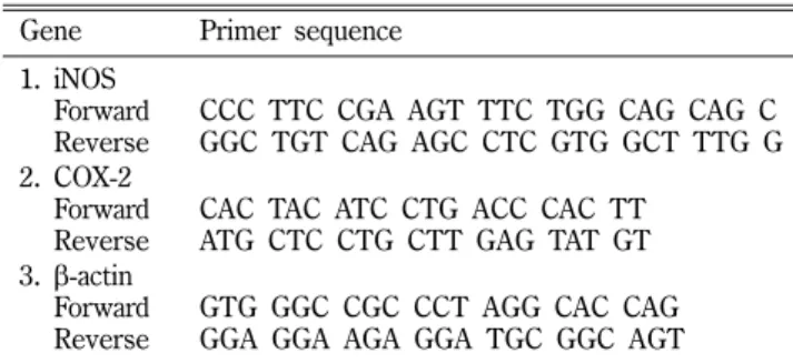
배양액을 제거하고 신속히 1 ml Trizol(Invitron)을 가한 후 -70oC deep freezer에 24시간 보관하였다. 시료를 녹인 후 세포를 lysis 시키고 200 µl의 CHCl3을 가한 뒤 원심분리 후 수층 400 µl를 취하고 동량의 iso-propanol을 가하였다. -20oC에서 1시간 이상 보관하고 원심분리 후 미색침전으로 얻어진 RNA에 superscript transcriptase II와 hexamer를 가한 후 42oC에서 1시간 반응시켜 cDNA로 전환하였다. 얻어진 cDNA 일정량과 primer, polymerase 를 가한 후 PCR thermal cycler에서 DNA를 증폭하였다. 증폭 된 DNA의 특정 band를 1% agarose gel에 전개시켜 DNA image reader로 확인함으로써 mRNA의 발현 정도를 관찰하였다 (Table I).
B16F10 세포에서의 멜라닌 생성 저해 작용 측정
세포주의 배양 − B16F10 멜라닌 세포는 10% FBS, 100 units/
ml penicillin, 100 µg/ml streptomycin, 0.25 µg/ml amphotericin 이 첨가된 DMEM을 사용하여 37oC, 5% CO2 incubator에서 배 양하였다.
MTT 시험법을 이용한 세포 독성 측정 − B16F10 melanoma 세포를 48 well plate에 1.5×10⁴cells/ml 농도로 접종한 후 37oC, 5% CO2 incubator에서 하루 동안 안정화 시킨 후 시료를 농도별로 처리한 후 48시간 동안 배양하였다. 그 후 각 well의 배지들을 제거 하고 MTT solution(0.5 mg/ml in PBS)을 첨가하 고, 37oC에서 4 시간 동안 반응 시킨 후 MTT solution을 제거 하고, 각 well에 400 µl의 dimethylsulfoxide(DMSO)를 첨가하여 1시간 동안 plate shaker로 흔들어 준 다음 microplate reader을 사용하여 대조파장을 650 nm로 하고 측정 파장을 570 nm로 하 여 흡광도를 측정하였다.
Table I− Sequences of primers for iNOS and COX-2 Gene Primer sequence
1. iNOS
1.
Forward1.
ReverseCCC TTC CGA AGT TTC TGG CAG CAG C GGC TGT CAG AGC CTC GTG GCT TTG G 2. COX-2
1.
Forward1.
ReverseCAC TAC ATC CTG ACC CAC TT ATG CTC CTG CTT GAG TAT GT 3. β-actin
1.
Forward1.
ReverseGTG GGC CGC CCT AGG CAC CAG GGA GGA AGA GGA TGC GGC AGT
멜라닌 생성 억제 작용의 측정 − 6 well plate에 5×10⁴cells/ml 로 접종한 후 37oC, 5% CO2 incubator에서 24시간 동안 배양하 였다. 배지를 제거 하고 0.4 µM α-MSH(alpha-melanocyte stimulated hormone)와 시료를 농도별로 처리하여 배지를 교체 한 후 48시간 배양하였다. 그 후 배지를 제거한 세포를 물에 불 용성인 melanin의 액화를 일으키기 위하여 10% DMSO가 함유 된 1 N NaOH를 넣고 70oC에서 1분간 반응 시킨 후 microplate reader 를 이용하여 405 nm에서 흡광도를 측정하였다.
Western blot 법에 의한 Tyrosinase 단백질의 측정 − B16F10 cell을 1×10⁵cells/ml로 분주한 후 24시간 후 α-MSH, 시료처리 군으로 나눠 농도별로 처리하고, 48시간 동안 배양하였다. 배양 이 끝난 후 배양액을 제거하고 lysis buffer를 넣고 4oC에서 1시 간 반응시킨 뒤 12000 rpm에서 20분 동안 원심 분리하였다. 단 백질 정량은 Bradford 법으로 하였으며, 20 µg의 단백질을 이용 하여 10% SDS-PAGE 이용하여 전기영동을 한 후, PVDF membrane에 transfer하고, Tyrosinase(SantaCruz), β-actin (Cell Signaling) 항체를 결합시킨 후, 2차 항체를 2시간 동안 반 응 시키고, ECL kit를 이용하여 LAS-3000으로 측정하였다.
자료의 통계적 처리
실험결과에서 얻은 모든 값은 평균±표준편차로 나타내었다.
통계적 유의적 차이의 정도는 Student's t-test를 사용하여 p<0.01, p<0.001인 값에 대해 유의적인 것으로 처리하였다.
실험결과 및 고찰
DPPH를 이용한 항산화능 측정
위령선 뿌리의 에탄올 추출물과 각 분획에 대한 DPPH를 이 용한 항산화능을 측정한 결과 에탄올 추출물은 697.9 µg/ml, 디
Fig. 1− Effect of ethanol extract and solvent fractions from Clematis mandshurica on antioxidation. EDA(%)=(Control O.D.- Sample O.D.)/Control O.D.×100. IC50: Required sample amount (µg) for 50% reduction of 0.1 mM DPPH solution.
Each value represents the mean±S.D. (A: ascorbic acid, E:
ethanol extract, H: hexane, D: dichloromethane, EA: ethyl acetate, B: butanol, W: H2O).
Fig. 3− Inhibition of NO production in RAW 264.7 cells by ethanol extract and solvent fractions of Clematis mandshurica.
RAW 264.7 cells (1.5×10⁵cells/ml) were incubated for 1~2 hrs in DMEM containing 10% FBS and were treated for 22 hrs with ethanol extract and its fractions of Clematis mandshurica with LPS (1 µg/ml). NO released into the cell culture medium was measured by the Griess reagent in the nitrite form. Each value represents the mean±S.D. (n=3).
Significantly different from LPS control, * p<0.01, ** p<
0.001 (E: ethanol extract, H: hexane, D: dichloromethane, EA: ethylacetate, B: butanol, W: H2O).
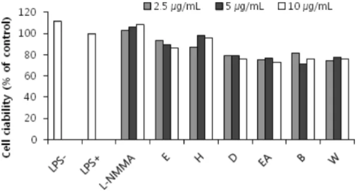
Fig. 2− Effects of ethanol extract and solvent fractions of Clematis mandshurica on the viability of RAW264.7 cells with LPS (1µg/ml). The viability of the cells was measured by MTT assay. Results were expressed as % of control absorbance.
Each value represents the mean±S.D. (n=3). (E: ethanol extract, H: hexane, D: dichloromethane, EA: ethylacetate, B: butanol, W: H2O).
클로로메탄 분획은 206.9 µg/ml 그리고 에틸아세테이트 분획은 253.4µg/ml로 항산화능이 나타났다. 양성 대조약물인 아스코르 빈산의 IC50은 37.0 µg/ml로 측정되었다(Fig. 1).
RAW 264.7 세포에서의 NO생성 저해 작용 측정
MTT 시험법에 의한 세포 독성 − 위령선 뿌리의 에탄올 추출 물과 각 분획의 RAW 264.7 cell에 대한 세포독성을 MTT 분석 법으로 측정한 결과 2.5~10 µg/ml의 농도에서 모두 80% 이상 의 생존율을 나타내어 정상세포에는 독성이 나타나지 않음을 알 수 있었다(Fig. 2).
NO 생성 저해 활성 − LPS로 활성화한 대식세포주인 RAW 264.7 cell에서의 NO 생성 저해 작용을 관찰한 결과 각 분획과 LPS를
동시에 처리했을 때, 에탄올 추출물과 디클로로메탄 분획, 에틸아 세테이트 분획에서 농도 의존적인 저해활성이 나타났다(Fig. 3).
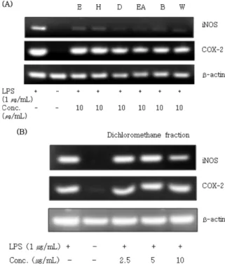
Western blot 법에 의한 iNOS 및 COX-2 단백질의 측정 − 위령선의 단백질 발현 단계에 미치는 영향을 알아보기 위하여 iNOS 항체와 COX-2 항체를 이용하는 western blot 실험 결과, 세포 독성이 없는 농도에서 디클로로메탄 분획과 에틸아세테이 트 분획이 iNOS 발현을 억제하는 것으로 나타났다. 또한, 디클 로로메탄 분획을 농도별로(2.5~10 µg/ml) 처리하여 western blot 실험을 하였을 때, 10 µg/ml에서는 iNOS와 COX-2모두 단백질 발현을 억제하는 것으로 나타났다(Fig. 4).
RT-PCR법에 의한 iNOS 및 COX-2의 mRNA 측정 − 위령선 의 에탄올 추출물과 용매 분획이 mRNA발현 단계에 미치는 영 향을 알아보기 위해 iNOS와 COX-2 primer를 이용하여 RT-PCR 실험을 한 결과 에탄올 추출물과 각 용매분획의 10 µg/ml 농도 에서 iNOS 발현을 현저히 억제하였고, 디클로로메탄과 에틸아세 테이트 분획은 COX-2 발현 또한 억제하였다. 디클로로메탄분획 의 투여 농도에 따른 결과에서는 iNOS의 발현이 농도의존적으 로 억제됨을 확인할 수 있었다(Fig. 5).
B16F10 세포에서의 멜라닌 생성 저해 작용 측정
MTT 시험법에 의한 세포 독성 − 위령선의 에탄올 추출물과 각 분획의 멜라닌 세포(B16F10)의 사멸효과를 측정한 결과 10~30
µg/ml의 시료농도에서 세포독성을 보이지 않았다(Fig. 6).
멜라닌 생성 억제 작용의 측정 − 위령선의 에탄올 추출물과 각 분획의 5~15 µg/ml 시료농도에서의 멜라닌 생성 억제작용을 측 Fig. 4− The protein levels of iNOS and COX-2 were determined
from the 24 hrs culture of cells stimulated with LPS (1µg/
ml) and ethanol extract and solvent fractions of Clematis mandshurica (10 µg/ml) (A) and several concentrations of dichloromethane fraction of Clematis mandshurica (2.5~10µg/ml) (B) (E: ethanol extract, H: hexane, D:
dichloromethane, EA: ethylacetate, B: butanol, W: H2O).
Fig. 5− Effects of ethanol extract and solvent fractions of Clematis mandshurica on iNOS and COX-2 mRNA expression in LPS-activated RAW 264.7 cells (A) and Effects of dichloromethane fractions on iNOS and COX-2 mRNA expression (B). RAW 264. 7 cells (1×10⁶cells/ml) were incubated for 24 hours in DMEM containing 10% FBS and were treated for 6 hours with ethanol extract and fractions (10µg/ml) of Clematis mandshurica with LPS (1 µg/ml). (E:
ethanol extract, H hexane, D: dichloromethane, EA:
ethylacetate, B: butanol, W: H2O).
Fig. 6− Effects of ethanol extract and solvent fractions of Clematis mandshurica on the viability of B16F10 cells with α-MSH (0.4µM). B16F10 cells (1.5×10⁴cells/ml) were incubated for 1~2 hrs in DMEM containing 10% FBS and were treated for 2 days with α-MSH (0.4 µM) and solvent fractions (10~30µg/ml). Results were expressed as % of negative control. Each value represents the mean±S.D (n=3). (K: kojic acid, E: ethanol extract, H: hexane, D:
dichloromethane, EA: ethylacetate, B: butanol, W: H2O).
정하였다. α-MSH를 처리한 군과 비교할 때, 10 µg/ml 농도에서 양성대조물질인 kojic acid를 처리한 경우는 37.3%, dichloro- methane 처리군은 32.6%, ethylacetate 처리군은 7.1%, butanol 처리군은 22.2%, H2O 처리군은 7.6%의 멜라닌 생성을 감소시 켰다(Fig. 7).
Western blot 법에 의한 Tyrosinase 단백질의 측정 − 위령선 의 에탄올 추출물과 각 분획들이 멜라닌 생성과정에서 중요한 tyrosinase의 단백질 발현에 어떠한 영향을 나타내는지 알아보기 위해 α-MSH(0.4 µM)로 B16F10 melanoma 세포를 자극시킨 후 시료를 분획별(10 µg/ml)로 처리한 후 western blot assay를 이 용하여 단백질 발현 양상을 측정하였고, tyrosinase 발현 억제효 과가 우수한 dichloromethane 분획을 농도별로(2.5~10 µg/ml) 처리하였을 때 농도 의존적으로 억제하는 효과를 나타내었다 (Fig. 8).
결 론
위령선 뿌리의 에탄올 추출물과 분획물들의 항산화 활성을 확 인하고자 DPPH를 이용한 유리기소거작용을 측정한 결과 디클 로로메탄과 에틸아세테이트 분획에서 강한 항산화 활성을 나타 내었다. 추출물들의 RAW 264.7 cell에 대한 세포독성을 평가하 기 위한 MTT 분석 결과 2.5~10 µg/ml 처리 농도에서 독성을 나타내지 않는 것을 확인 하였다. LPS에 의해 유도된 iNOS에 의한 NO 생성 억제 활성 측정결과 디클로로메탄 및 에틸아세테 이트 분획에서 농도 의존적으로 유의성 있는 억제효과가 있었으 며, 양성대조군인 L-NMMA보다 우수한 활성을 나타내었다. 또 한 western blot법에 의한 iNOS 단백질 발현을 측정한 결과 디 클로로메탄과 에틸아세테이트 분획의 10 µg/ml 투여군에서 강한 억제활성을 나타내었고, 디클로로메탄 분획의 2.5~10 µg/ml 투 여군에서 농도 의존적인 iNOS 및 COX-2 억제활성을 나타내었 다. RT-PCR법에 의한 iNOS의 mRNA 발현을 측정한 결과 또한 디클로로메탄과 에틸아세테이트 분획에서 강한 억제활성을 나타 내었다.
위령선 뿌리의 에탄올 추출물 및 분획의 B16F10 melanoma cell에 대한 세포독성 실험에서 10~30 µg/ml 투여농도에서 독성 을 나타내지 않았고, α-MSH로 유도한 B16F10 cell에서 디클로 로메탄 분획이 멜라닌 생성과 tyrosinase 단백질 발현을 농도의 존적으로 억제하였다. 따라서 위령선 추출물 및 그 분획에 대한 생리활성을 검색한 결과 디클로로메탄 및 에틸아세테이트 분획 에서 강한 항산화 및 항염활성을 보였으며, 디클로로메탄 분획 에서 우수한 미백 활성이 있음을 확인하였다.
참고문헌
1) 이창복 : 대한식물도감, 향문사, 서울 p. 303 (1980).
2) Park, E. K., Ryu, M. H., Kim, Y. H., Lee, Y. A., Lee, S. H., Woo, D. H., Hong, S. J., Han, J. S., Yoo, M. C., Yang, H. I. and Kim, K. S. : Anti-inflammatory effects of an ethanolic extract from Clematis mandshurica Rupr. J. of Ethnopharmacol. 108, 142 (2006).
Fig. 7− Inhibition of melanin synthesis by ethanol extract and solvent fractions of Clematis mandshurica. B16F10 cells (5×10⁴cells/ml) were incubated 24 hrs after α-MSH (0.4µM) and sample treatment. Melanin was lysed with 1 N NaOH in 10% DMSO solution and the content was measured by ELISA. Each value represents the mean±S.D (n=3). Significantly different from α-MSH control, *p<
0.01, **p<0.001 (K: kojic acid, E: ethanol extract, H:
hexane, D: dichloromethane, EA: ethylacetate, B: butanol, W: H2O).
Fig. 8− Effect of ethanol extract and solvent fractions (A) and effect of various concentrations of dichloromethane fraction (B) on protein expression of tyrosinase in B16F10 melanoma cells. B16F10 cells were treated for 48 hours with α-MSH (0.4µM) and ethanol extract and fractions (10 µg/ml). The tyrosinase protein level in lysates was determined by western blot analysis (E: ethanol extract, H: hexane, D:
dichloromethane, EA: ethyl acetate, B: butanol, W: H2O).
3) Ding, Q., Yang, L., Yang, H., Jiang, C., Wang, Y. and Wang, S. : Cytotoxic and antibacterial triterpenoids derivatives from Clematis ganpiniana. J. of Ethnopharmacol. 126, 382 (2009).
4) Shi, S., Jiang, D., Dong, C. and Tu, P. : Triterpene Saponins from Clematis mandshurica.
J. Nat. Prod. 69, 1591 (2006).
5) Du, Z., Zhu, N., Mu, N. and Shen, Y. : Two new antifungal saponins from the tibetan herbal medicine Clematis tangutica.
Planta Med. 69, 547 (2003).
6) Jung, B. C., Woo, M. J. and Kim, S. W. : Two cases of irritant dermatitis due to Clematis apiifolia. Korean J. Dermatol. 40, 143 (2002).
7) Chawla, R., Kumar, S. and Sharma, A. : The genus Clematis (Ranunculaceae): Chemical and pharmacological perspectives J. of Ethnopharmacol. 143, 116 (2012).
8) Park, E. K., Ryu, M. H., Kim, Y. H., Lee, Y. A., Lee, S. H., Woo, D. H. and Kim, K. S. : Anti-inflammatory effects of an ethanolic extract from Clematis mandshurica Rupr. J. of Ethnopharmacol. 108, 142 (2006).
9) Suh, S. J., Kim, K. S., Lee, S. D., Lee, C. H., Choi, H. S., Jin, U. H. and Kim, C. H. : Effects and mechanisms of Clematis mandshurica Maxim. as a dual inhibitor of proinflammatory cytokines on adjuvant arthritis in rats. Environmental Toxicol.
and Pharmacol. 22, 205 (2006).
10) Stamlar, J. S., Singel, D. J. and Loscalzo, J. : Biochemistry of nitric oxide and its redox-activated forms. Science 258, 1898
(1992).
11) Bogdan, C., Rllinghoff, M. and Diefenbach, A. : The role of nitric oxide in innate immunity. Immunol. Rev. 173, 17 (2000).
12) Raghav, S. K., Gupta, B., Shrivastava, A. and Das, H. R. : Inhibition of lipopolysaccharide-inducible nitric oxide synthase and IL-1β through suppression of NF-κB activation by 3-(1',1'- dimethyl-allyl)-6-hydroxy-7-methoxy-coumarin isolated from Ruta graveolens L. Eur. J. Pharmacol. 560, 69 (2007).
13) Galla, H. J. : Nitric oxide, NO, an intracellular messenger.
Angew. Chem. Int. Ed. Engl. 32, 378 (1993).
14) Moncada, S., Palmer, R. M. and Higgs, E. A. : Nitric oxide:
physiology, Pathophysiology, and pharmacology. Pharmacol.
Rev. 43, 109 (1991).
15) Li, Q. and Verma, I. M. : NF-kappaB regulation in the immune system. Nat. Rev. Immunol. 2, 725 (2002).
16) Chang, T. S. : An updated review of tyrosinase inhibitors. Int.
J. Mol. Sci. 10, 2440 (2009).
17) Yamaguchi, Y. and Hearing, V. J. : Physiological factors that regulate skin pigmentation. Biofactors 35, 193 (2009).
18) Draelos, Z. D. : Skin lightening preparations and the hydroquinone controversy. Dermatol. Ther. 20, 308 (2007).
19) Busca, R. and Ballotti, R. : Cyclic AMP a key messenger in the regulation of skin pigmentation. Pigment. Cell. Res. 13, 60 (2000).