- 43 -
R eceived R e v i s e d A ccepted
: December 19, 2017
: October 21, 2018(1차) / October 26, 2018(2차) November 6, 2018(3차)
: November 6, 2018
+Corresponding author: Jeong Kyu Kim, MD, PhD
Department of Otolaryngology-Head and Neck Surgery, Catholic University of Daegu School of Medicine, 33 Duryugongwon-ro 17-gil, Nam-gu, Daegu 42472, Republic of Korea
Tel: +82-53-650-4071, Fax: +82-53-650-4533 E-mail: [email protected]
대한두경부종양학회지, 제34권 제2호, 2018. pp.43-46 Korean Journal of Head & Neck Oncology, Vol.34, No.2
https://doi.org/10.21593/kjhno/2018.34.2.43 ISSN 1229-5183(Print) / ISSN 2586-2553(Online)
설배부에 발생한 연골성분리종 1례
길부관1⋅손호진1⋅김보문1⋅정재원2⋅김정규1+
대구가톨릭대학교 의과대학 이비인후과학교실1, 병리학교실2
A Case of Chondroid Choristoma on the Dorsum of the Tongue
Bu Kwan Kil, MD1, Ho Jin Son, MD1, Bo Mun Kim, MD1, Jae Won Joung, MD2, Jeong Kyu Kim, MD, PhD1+
Department of Otolaryngology-Head and Neck Surgery, Catholic University of Daegu School of Medicine, Daegu, Korea1 Department of Pathology, Catholic University of Daegu School of Medicine, Daegu, Republic of Korea2
= Abstract =
Chondroid choristomais a rare tumor like lesion of normal tissue in an unusual location. Oral cavity chondroid choristoma is exceedingly uncommon. This lesion is commonly covered by normal oral mucosa and can develop during a whole lifetime. We experienced a case of 57-year-old man who presented as 6-months history of asympto- matic mass on the dorsal surface of the tongue. We performed surgical excision under local anesthesia, and the pathological diagnosis was chondroid choristoma. After surgery, patient was followed up without any recurrence and discomfort. Therefore, we report this case with a review of literature.
Key W ords : cartilage, choristoma, tongue
Introduction
Choristoma is a relatively unusual tumor-like mass com- posed of normal tissues like dermal and epidermal compo- nents, muscle, cartilage, and bone that has arose in an abnor- mal site.1) The most common involved sites of chondroid choristoma are small joints of hands and feet.2) This carti- lage producing choristoma is rare in the oral cavity mucosa.3) Tongue is the most common involved site, although this tumor has also been located in the gingiva, soft palate, and buccal mucosa.
Chondroid choristoma can develop during a whole lifetime.1) It frequently appears as solid, painless mass. The tumor has a slow-growth pattern and rarely exceeds a few centimetres in diameter.2) The histopathological appearance of chondroid choristoma is composed of mature hyaline car- tilage, bone and fat tissue in various proportions.4) The treat- ment of a chondroid choristoma consists of surgical excision of the tumor.5)
Here we present chondroid choristoma in the tongue and discuss the epidemiology, clinical presentation, histology, and pathogenesis with review of literatures.
Case Report
A 57-year-old male patient visited our tertiary referral hospital with a chief complaint of a firm mass over the tongue for the proceding 6 months. He had no pain, swal- lowing difficulty, and neck lymph node enlargement. Physical examination by bimanual palpation revealed a well-defined,
- 44 -
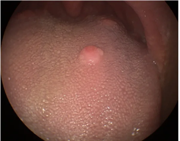
Fig. 1. Preoperative endoscopic finding shows a round, sessile mass in dorsal surface of the tongue
Fig. 2. The photomicrograph shows a well-circumscribed nod- ule composed of mature hyaline cartilage with typical chon- drocyte covered with non-keratinizing stratified squamous cell epithelium (H&E, X40)
Fig. 3. The photomicrograph shows small chondrocytes with a clear cytoplasm and round nuclei (H&E, X400)
small, spherical, erythematous, firm mass on the dorsal sur- face of the tongue, with a 1 cm diameter, and the overlying mucosa was slightly raised but intact(Fig. 1). The physical examinations of the other sites were unremarkable. There was no specific past medical history and family history.
And there were no previous trauma and inflammations in
this area.
The initial clinical diagnosis was pleomorphic adenoma, granuloma, and choristoma. Surgical excision was performed under local anaesthesia with proper margin. Surgical speci- mens were sent to pathologists and stained with H&E. This is composed of a piece of nodular mass, measuring 1.1 X 0.6 X 0.5 cm in size. The microscopic study of the tumor showed lobules of well-formed hyaline cartilage and sur- rounding fibrous tissue in the submucosa. There is no cellular atypia or abnormal mitosis(Fig. 2 and 3). Immunohistochemical stains revealed that chondrocytes and mature fat tissue show strong positive immunoreaction for S-100 protein. Whereas, there is no immunoreaction for cytokeratin and desmin, very low proliferating activity in Ki-67 stain(Fig. 4).
Wound healing was satisfactory, and vicryl wound su- tures were removed a week later. The postoperative course showed no recurrence and complication for 6 months.
Discussion
Oral cavity choristomas are extremely rare disease and mainly develop at foramen cecum.6) In the previous study, 39 cases of choristomas of the oral cavity were reported.
Foramen cecum and circumvallate papillae were the most common occurred site (36 cases) followed by the alveolar process and buccal vestibule (3 cases).1) In the past, chori- stoma was known as osteoma or chondroma. However, since 1913, 59 cases of choristomas were stated.7)
Although the cause of disease occurrence is uncertain and controversial, some hypotheses try to describe its origin.
First, embryologically tongue anterior two thirds is origi- nated from first branchial arch and posterior one third is originated from third branchial arch. Subsequently, tongue is combined around foramen cecum and probably branchial apparatus multipotency stem cell is regarded as origin of choristoma.8) Second, thyroid descends from foramen ce- cum during developmental process and undescended thyroid tissue could be origin of choristoma.8) Third, posterior one third of tongue is constantly stimulated by tongue movement caused by swallowing, pronunciation and such stimulations lead to calcium deposit and tissue hypertrophy.9)
Females occurred three to four times more than males and more than half of patients were twenties and thirties.8) Clinical symptoms of choristomas are globus sensation, dys-
- 45 - A
B
C
D
Fig. 4. The immunohistochemical stains of chondroid choristoma. The cartilage lobules are strong immunoreactive to S-100 protein (A, X100). Cytokeratin (epithelial marker, B, X100) and Desmin (C, X100) are not expressed in cartilage lobules. There is very low proliferating activity in Ki-67 stain (D, X100).
phagia, nausea, throat irritability, and snoring. However, symptom-free is most common.10) The diameter of tumor had a wide range from 4 to 25mm. But, patient's symptoms were not correlation with its diameter. Previous study sug- gested in patient with largest diameter 25 mm tumor had no symptom, whereas in patient with 7 mm tumor had globus sensation in throat.11)
Computed tomography scan analysis may be used to check tumor site and its relation to the anatomic structures to exactly plan surgery extent and well defined, round, ra- dio-opaque, and calcification lesion is detected.
Histological examination confirms definite diagnosis of choristomas. Histologically, choristoma is composed of car- tilaginous tissue surrounded by fibrous connective tissue with normal chondroblasts without atypia, mitosis, and necrosis.12) In previous study conducted by Park et al, the microscopic findings of choristoma located at lingual tonsil were consisted of mature bone tissue, covered with non-ker- atinizing stratifies squamous cell epithelium.15) By compar- ison, this case showed lobules of well-formed hyaline carti- lage and surrounding fibrous tissue in the submucosa. In
this regard, it can be found that chondroid choristoma has various histopathological appearance such as mature hyaline cartilage, bone and fat tissue. Chondroid choristoma re- sembles benign chondroma histologically, however, chori- stoma developed in locations that generally do not contain chondrocytes.
In addition, immunohistochemical study is crucial to rule out other disease. Choristoma has high positive S-100 pro- tein (marker of myoepithelial cell) immunoreactivity owing to cartilaginous tissue. There is a negative immunoreaction for cytokeratin (marker of epithelial cell), desmin (marker of muscle cell) and Ki-67 (marker of cellular proliferation).13) This examination can differentiate choristoma from thyro- glossal duct cyst, salivary gland neoplasm, fibroma, lipoma, giant cell tumor, and calcified lymph node.11) Especially ec- topic thyroid gland must be excluded if lesion is near the foramen cecum. Therefore, thyroid function test and thyroid scan must be conducted before surgery when lesion is lo- cated at posterior tongue.14) Chondrosarcoma could be ex- cluded according to cellular and nuclear morphology of the chondrocyte.11) The present case was simply distinguished
- 46 - as an encapsulated tumor of mature chondrocytes surround- ing by hyaline materials.
The treatment of chondroid choristomas of the tongue is surgical excision. Local recurrence is very rare but in- complete resection of the tumor or surrounding tissue has the possibility to develop new cartilaginous tissue.1) However, prognosis of choristoma is extremely favorable and long term follow up is not recommended.1)
In conclusion, oral cavity chondroid choristoma is very rare, but has a good clinical behavior. Surgical excision is a treatment of choice, and post-operative prognosis is ex- tremely good according to the previous study.
References
1) Chou LS, Hansen LS, Daniels TE. Choristomas of the oral cavity:
a review. Oral Surg Oral Med Oral Pathol. 1991;72:584-593.
2) Han JY, Han HS, Kim YB. Extraskeletal chondroma of the fallo- pian tube. J Korean Med Sci. 2002;17:276-278.
3) Sera H, Shimoda T, Ozeki S. A case of chondroma of the tongue.
Int J Oral Maxillofac Surg. 2005;34:99-100.
4) Norris O, Mehra P. Chondroma (cartilaginous choristoma) of the tongue: report of a case. J Oral Maxillofac Surg. 2012;70:643-646.
5) Gorini E, Mullace M, Migliorini L, Mevio E. Osseous chori- stoma of the tongue: a review of etiopathogenesis. Case Rep Otolaryngol. 2014;doi:10.1155/2014/373104.
6) Krolls SO, Jacoway JR, Alexander WN. Osseous choristomas
(osteomas) of intraoral soft tissues. Oral Surg Oral Med Oral Pathol. 1971;32(4):588-595.
7) Monserrat M. Osteome de la langue. Bull Soc Anat. 1913;88:
282-283.
8) Benamer MH, Elmangoush AM. Lingual osseous choristoma case report and review of literature. Libyan J Med 2007;2(1):46-48.
9) Toida M, Sugiyama T, Kato Y. Cartilaginous choristoma of the tongue. J Oral Maxillofac Surg. 2003;61:393-396.
10) Wesly RK, Zielinski RJ. Osteocartilaginouschoristoma of the tongue: clinical and histopathplogic considerations. J Oral Surg.
1978;36:59-61.
11) Supiyaphun P, Sampatanakul P, Kerekhanjanarong V, Chawakitchareon P, Sastarasadhit V. Lingual osseous chori- stoma: a study of eight cases and review of the literature. Ear Nose Throat J. 1998;77(4):316-325.
12) Pires FR, Abrahão AC, Cabral MG. Clinical, histological and im- munohistochemical features of ectomesenchymal chondromyx- oid tumor. Oral Surg Oral Med Oral Pathol Oral RadiolEndod.
2009;108:914-919.
13) Adegboyega PA, Qiu S. Immunohistochemical profiling of cyto- keratin expression by endometrial stroma sarcoma. Hum Pathol.
2008;39:1459-1464.
14) Andressakis DD, Pavlakis AG, Chrysomali E, Rapidis AD.
Infected lingual osseous choristoma. Report of a case and review of the literature. Med Oral Patol Oral Cir Bucal. 2008;13(10):
E627-632.
15) Jung KN, Park CH, Chun JH, Choi YH. Two cases of choristoma in base of the tongue. Korean J Head Neck Oncol. 2004; 20(1):
49-51.