A Comparative Study of Radiation Therapy Planning between Volumetric-Modulated Arc Therapy and
Three-Dimensional Conformal Radiotherapy in Nasopharyngeal Cancer
*Ji Sung Kim1*, Seok Ho Lee, MD1*, Seung Heon Lee, MD1, Hye-Young Kim, MSc1, Jin-Ho Choi, MSc1, Kyu Chan Lee, MD1, Dong Young Kim MD2
Department of Radiation Oncology,1 and Otolaryngology,2 Gachon University of Medical and Science, Inchon, Korea
비인두암의 방사선치료 시 삼차원입체조형 치료기법과 용적세기조절회전치료기법의 비교연구
*가천의과학대학교 의학전문대학원 방사선종양학교실,1 이비인후과학교실2
김지성1*·이석호1*·이승헌1·김혜영1·최진호1·이규찬1·김동영2
= 국 문 초 록 =
연구목적
비인두암 환자들을 대상으로 방사선치료 시 삼차원입체조형치료기법과 용적세기조절회전치료기법을 비교하고 이하 선을 포함한 정상조직 보호에 있어 그 차이점을 알아 보고자 본 연구를 시행하였다.
대상 및 방법
비인두암 환자 5명을 대상으로 치료계획용 CT(computed tomography)를 시행 후 삼차원입체조형방사선치료계획 과 용적세기조절회전치료계획을 시행하였다. 이를 바탕으로 얻은 선량분포, conformity index(CI) 그리고 선량체적 히 스토그램을 통해 손상위험장기(organ at risk)와 계획용표적체적(planning target volume)을 비교·분석하였다.
결 과
분석결과 이하선에 조사되는 평균선량이 용적세기조절회전치료계획에서는 43.9%로 삼차원입체조형치료계획에서의 89.4% 보다 유의하게(p=0.043) 감소하였다. 계획용표적체적 conformity index의 경우 용적세기조절회전치료계획 (CI=1.06)에서 삼차원입체조형치료계획(CI=2.55) 보다 유의하게(p=0.043) 향상된 결과를 보였다.
결 론
비인두암 환자에서 용적세기조절회전 치료계획 시 삼차원입체조형치료계획 보다 유의하게 이하선에 평균선량이 줄었 고 계획용 표적체적에 대한 conformity도 유의하게 향상되는 결과를 보였다. 본 연구가 적은 수의 환자를 대상으로 하였으나 용적세기조절회전치료기법을 시행 시 구강건조증의 발생을 줄일 수 있을 것으로 기대된다. 향후 더 많은 환자군을 대상으로 한 임상연구가 필요할 것으로 사료된다.
중심 단어:용적세기조절회전치료·삼차원입체조형치료·비인두암·이하선.
*Seok Ho Lee and Ji Sung Kim contributed equally to this study.
교신저자:이규찬, 405-760 인천광역시 남동구 구월동 1198 가천의과학대학교 의학전문대학원 방사선종양학교실 전화:(032) 460-3030·전송:(032) 460-3029·E-mail:[email protected]
Introduction
Xerostomia is the most common side effect and major cause of reduced quality of life following radiation therapy in head and neck cancer. In addition to its effects on sub- jective wellbeing, decreased saliva output causes alterations in speech, taste, difficulties with mastication and deglutition that create secondary nutritional deficiencies.1) To preserve salivary function after radiation therapy in head and neck cancer, it is critical to decrease the doses to parotid glands as much as possible.
Intensity-modulated radiotherapy(IMRT) has been draw- ing attention as a technique to reduce doses to normal tissues with resultant decreased complication rates after radiation therapy in head and neck cancer. Parotid gland sparing have been reported the decreased rate of xerostomia for patients treated with IMRT compared with three-dimensional con- formal radiotherapy(3D-CRT).1-4) Some studies reported va- rious tolerance dose of parotid gland to protect against xe- rostomia after IMRT.5) As well as parotid glands, doses to other organs at risk(OAR) such as spinal cord, eye balls and mandible should be limited under the tolerance doses.
Volumetric modulated arc therapy(VMAT) is a recently developed technique, which delivers intensity modulated ra- diation therapy to target using rotations of the gantry.6) This concept has been clinically implemented in the Eclipse treat- ment planning software(Varian Medical Systems, Palo Alto, CA, USA) as RapidArcTM. This technique is similar to to- motherapy in that a full 360 degree of beam directions are available for optimization but is fundamentally different in that the entire dose volume is delivered in a single or dou- ble rotation.7) Also, it delivers dose to the whole volume, ra- ther than slice by slice. During the delivery of VMAT, three parameters are simultaneously changed to achieve a inten- sity modulated conformal 3D dose delivery:1) gantry ro- tation speed, 2) shape of the treatment aperture using the movement of multileaf collimator leaves, and 3) dose rate.
The aim of the present study is to identify the potential do- simetric advantages of VMAT through comparing the cal- culated dose to target and various normal tissues especially, the parotid glands in 3D-CRT and VMAT planning.
Materials and Methods
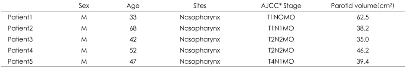
The 5 patients with nasopharyngeal cancer were included for the study. These patients were randomly selected from the list of patients with nasopharyngeal cancer that have been
receiving VMAT in 2010 at the department of Radiation oncology, Gachon University Gil hospital. The patients and tumor characteristics are shown in Table 1. For the VMAT planning, all patients were immobilized in supine position with customized aquaplast(thermoplastic) masks and target localization was accomplished using computed tomography (CT) simulation(Simens Medical Solutions, Germany). CT images were obtained by every 3mm slices. For each VMAT plan, corresponding 3D-CRT plan was generated for the comparison. The target volumes, doses, OAR volumes, and dose constraints were unchanged between the two plans. The Gross Tumor Volume(GTV) is defined as all known gross disease determined from clinical information, endoscopic finding, CT and magnetic resonance imaging(MRI). The gross extent of the tumor was outlined in conjunction with the other radiation oncologist. In our center, fusion the MRI im- ages along with the CT images was performed to more accu- rately define the GTV. The clinical target volume(CTV) was defined as the GTV plus areas considered to contain poten- tial microscopic disease. The margin between each GTV and its CTV was determined as a minimum value of 3mm except when the clivus was completely infiltrated with GTV and was adjacent to the brain stem. In those situations, the CTV margin was determined as small as 1mm. The CTV in- cludes the entire nasopharynx, retropharyngeal lymph nodes, clivus, skull base, pterygoid fossae, parapharyngeal space, inferior sphenoid sinus and posterior third of the nasal cavity and maxillary sinuses. The planning target volume(PTV) was defined as the CTV plus a minimum 5mm margin com- pensating for the variabilities of treatment set up and internal organ motion. Where the GTV or CTV is adjacent to the brain stem, where the PTV margin was defined as small as 1mm margin.
The PTV for the planning in VMAT was the same as the PTV in 3D-CRT planning. For the direct comparison with VMAT plan, the same dose prescription of 46Gy with 2.3Gy daily fractions to PTV was prescribed for each plan. How- ever, conventional fractionation of 2.3Gy per fraction daily was not usually used in the actual treatment using 3D-CRT, which was delivered in fraction sizes of 1.8Gy to 2.0Gy daily.
All plans were normalized to the mean dose of the PTV.
VMAT plans aimed to cover at least 100% of the PTV with a dose greater than 95% of the prescription dose. An over- dosage of up to maximum 107% was allowed to both clinical target volume(CTV) and PTV. For the OAR, the maximum dose were constrained to 25Gy for spinal cord, 25Gy for eye, 40Gy for mandible and 25Gy for parotid gland. Plann- ing software was the Varian Eclipse treatment planning sys-
tem(version 8.6.10) with 6 MV photon beams from Novalis Tx(Varian, USA) equipped with a High Definition MLC with 120 leaves with spatial resolution of 2.5mm at isocenter.
Plans for VMAT were optimized selecting a maximum dose rate of 600MU/min. The dose calculation algorithm was anisotropic analytical algorithm(AAA) photon dose cal- culation algorithm. The calculation grid was set to 2.5mm.
VMAT involves continuous variation of the instantaneous dose rate, according to varying multi-leaf collimator(MLC) leaf positions and gantry rotational speed to optimize the dose distribution. Details about VMAT optimization process have been published elsewhere.8) To minimize the contribution of tongue and groove effect during the arc rotation, the col- limator rotation in VMAT remains fixed to a value different from zero. For the study, the VMAT plans were optimized with double arc 360° for each plan.
In the technical detail for the 3D-CRT, patients were treated using 6 MV photon beams with customized blocks.
The upper neck field was planned with bilateral with or without anterior field(s) to a dose of 46Gy with 2.3Gy frac- tion for the comparison with VMAT plan. Lateral fields were prescribed to the midplane, while the boost plan dose was prescribed to the isodose line that completely encompassed the PTV. The lower neck was planned with both lateral or anterior with or without posterior field(s). However, the actual 3D-CRT was delivered in daily dose of 1.8Gy for 5 days per week and shrinking field technique was performed using electron beams to exclude the spinal cord beyond 45Gy.
To improve dose homogeneity, wedges were routinely used.
The 3D-CRT beam shape for each field was designed to ex- clude the eye, brain stem, spinal cord and parotid gland as much as possible without compromising the target volume.
Calculated doses and dose volume histogram(DVH) of PTV and OARs were compared between 3D-CRT and VM- AT plans. Conformity index for the PTV was also compared for nasopharyngeal cancer through dose comparison. This conformity index by RTOG was used in analysis. RTOG de- fined conformity index as volume of reference dose/target volume(Vm/TV).
For the statistical analysis, the Wilcoxon signed rank test
was performed for each parameters. Statistical significance was accepted when p-values were ≤0.05.
Results
1. Dose distribution
1) Planning target volume
The mean dose to the PTV was 100.5% with 3D-CRT and 103.5% with VMAT(p=0.465).
2) Organs at risk (1) Parotid glands
As shown in Table 2, average mean parotid gland dose de- creased from 89.4% with 3D-CRT to, 43.9% with VMAT (p=0.043). Average maximum dose to parotid gland was 104.8% with 3D-CRT and 93.5% with VMAT. In VMAT, mean dose reduction in parotid gland was 45.5% compared to 3D-CRT.
(2) Spinal cord
The average mean spinal cord dose was 58.2% with 3D- CRT and 30.2% with VMAT, which was not statistically sig- nificant(p=0.08) Average maximum cord dose was 103.3%
with 3D-CRT and 56.4% with VMAT(p=0.043).
(3) Eyes
Average maximum dose to eyes was 46% with 3D-CRT and 26.3% with VMAT. Average mean doses to eyes was 18.5% with 3D-CRT and 8.8%with VMAT(p=0.465). In the analysis of eyes, the datas of 4 patients were used. In other patients, eyes was not included in the RT field.
Table 2. Statistical analysis of PTV & OAR for 3D-CRT and VMAT 3D-CRT VMAT P-value*
PTV mean dose 100.5% 103.5% 0.465 Parotid mean dose 089.4% 043.9% 0.043 Spinal cord max dose 103.3% 056.4% 0.043 Eye mean dose 018.5% 008.8% 0.465 Mandible mean dose 066.2% 055.8% 0.893 The Wilcoxon matched-pair signed rank test is listed for 3D-CRT vs. VMAT
*:P value for wilcoxon signed rank test
Table 1. Patients’ characteristics
Sex Age Sites AJCC* Stage Parotid volume(cm2) Patient1 M 33 Nasopharynx T1NOMO 62.5
Patient2 M 68 Nasopharynx T1N1MO 38.2
Patient3 M 42 Nasopharynx T2N2MO 35.0
Patient4 M 52 Nasopharynx T2N2MO 46.2
Patient5 M 47 Nasopharynx T4N1MO 39.4
*:american joint committee on cancer
(4) Mandible
Average maximum dose to mandible was 106.7% with 3D-CRT and 107.2% with VMAT. Average mean doses to mandible was 58.5% with 3D-CRT and 58.6% with VMAT (p=0.893).
The analyzed results for PTV and OAR were summariz-
ed in Table 2.
2. Conformity index
Fig. 1 shows axial dose distributions of the patient 5 with nasopharyngeal cancer for 3D-CRT and VMAT technique.
The mean conformity index for VMAT and 3D-CRT were 1.06 and 2.55, respectively(p=0.043). The conformity in- dex for each patients is shown in Table 3.
3. Dose-volume histogram
Dose volume histograms comparing 3D-CRT and VMAT for patient 5 was shown in Figures(Fig. 2). The results in- cluded the histogram of PTV, parotid glands, spinal cord, eye and mandible. The DVHs of parotid gland in VMAT show the markedly lower OAR DVH compared with 3D-CRT.
Discussion
Extensive studies on clinical benefit of sparing of parotid glands in IMRT compared with 3D-CRT have been reported.
Significant reduction of patient- and observer-rated xeros- tomia, as well as other head and neck symptoms were reported comparing with 3D-CRT.9) The other study also reported that oral health-related quality of life was highly preserved in IMRT comparing 3D-CRT.10) Table 4 showed various tol- erance dose of parotid gland to protect against xerostomia
Table 3. Conformity index of PTV for VMAT and 3D-CRT
VMAT 3D-CRT
Patient 1 0.78 2.55 Patient 2 1.01 2.57 Patient 3 0.87 3.09 Patient 4 1.39 2.68 Patient 5 1.26 1.85 Mean conformity index 1.06 2.55
Table 4. Reports focusing on parotid tolerance dose after IMRT for head and neck cancer
Author No. of patients Follow up period(month) Treatment Parotid tolerance dose(Gy) Eisbruch et al.1) 88 12 3D-CRT*,IMRT† 260.
Chao et al.21) 41 06 3D-CRT,IMRT 320.
Saarilahti et al.22) 17 12 IMRT 25.5
Lee et al.5) 34 06 IMRT 27.5
*:3D-conformal radiation therapy, †intensity modulated radiation therapy Fig. 1. Axial dose distributions for the patient 5. The 95% isodose
lines show that VMAT(B) has better conformity of PTV than 3D- CRT(A).
A B
Fig. 2. The dose volume histogram for the patient 5. The DVHs of organs at risk such as parotid gland in VMAT(squares) show more sparing compared with 3D-CRT(triangles). In high dose area, dose coverages in eye(pink), spinal cord(yellow), and mandible (blue) show more sparing in VMAT compared with 3D-CRT.
Ration of total structure volume (%) 100 80 60 40 20
0
0 10 20 30 40 50 Dose (Gy)
Some structures are unapproved or rejected 0 21.739 43.478 65.217 86.956 108.69
Relative dose (%)
after IMRT.
VMAT has been investigated and compared with other te- chniques in a series of studies that included brain tumors, pro- state, head-and-neck, anal canal, and cervical uterine cancer, and other indications8,11-17) and some cases showed significant dosimetric improvements against other techniques.8,11-15,18)
Recently, the study comparing VMAT and 3D-CRT reported that the 3D-CRT revealed a poor PTV coverage in contrast to VMAT and IMRT in malignant glioma.19) However, in head and neck cancer, there is no study comparing 3D-CRT and VMAT techniques.
For the analysis of DVH between 3D-CRT and VMAT, mean parotid dose was used in our study. Previous studies on dose-response relationships and the effects of the volume of the irradiated glands on gland function have revealed that the mean parotid dose is the most important predictor for developing xerostomia.20-24) For the analysis of the eye and mandible, the mean dose was also used. These all struc- tures(parotid gland, eye, and mandible) are the parallel struc- tures, on the contrary, for spinal cord, maximum cord dose was used in the analysis because it is one of serial struc- tures. In the planning of VMAT, double arc techniques were applied to all patients to improve the homogeneity of the PTV dose distribution. There was a report that double arc techni- que offered improved target coverage with respect to con- ventional IMRT.15)
The present study resulted in that VMAT technique spared the OAR more than 3D-CRT dose. Especially, parotid glands showed statistically significant dose reduction in VMAT(p
=0.043). For the spinal cord, the statistical difference bet- ween VMAT and 3D-CRT also showed the significance(p
=0.043). Actually, 3D-CRT is not quite different from bi- lateral fields concerning to the sparing of parotid gland. The- refore, the sparing of parotid gland is not easier even in 3D-CRT. Due to the unsophisticated characteristics of 3D- CRT, we could predict that VMAT technique has better dosi- metrical advantages(especially for sparing the parotid gland) than 3D-CRT like in IMRT.
The conformity index was developed as an extension of section-by-section dosimetric analysis and dose-volume his- tograms and can be defined as an absolute value resulting from the relationship between tumor volume or a fraction of this volume and the volume delineated by an isodose or a fraction of this volume. It can also be defined by the ratio of an isodose with another isodose.25) The use of this type of tool could facilitate the choice of treatment and compa- risons of various treatment plans for 3D-CRT and VMAT.
In our study, the conformity index was 1.06 with VMAT and
2.55 with 3D-CRT, respectively. Because of sophisticated anatomic structures of the head and neck cancer, the results for conformity index showed better outcomes in VMAT than 3D-CRT. For the radiation therapy of nasopharyngeal cancer, the upper neck field was mostly planned with lateral fields.
The center structures such as spinal cord was irradiated in whole volume. It attributed to poor conformity index for 3D- CRT than VMAT.
In the results of dose distributions of these 5 patients using VMAT, low dose was irradiated in whole areas of head and neck including eye and mandible. The reason is due to the ad- vanced technique of VMAT that delivers a precisely sculpted 3D dose distribution with a double of 360 degree rotation of the gantry. To conclude the significance, more cases need to be added.
This study has several limitations. First, the number of pa- tients was too small for valid statistical analysis. Although the statistical difference in parotid gland, spinal cord and con- formity index were significant, the 5 patients were not suf- ficient sample sizes to support these results of statistical an- alysis. Further study enrolling more patients is needed. Se- cond, the actual 3D-CRT including shrinking field technique was not performed using electron beams to exclude the spinal cord. And, we adopted not the curative RT dose but dose of 46Gy with 2.3Gy fraction with the aim of comparison in this study. Generally, the prescribed dose for curative aim is more than 70Gy to the PTV in patients with nasopharyngeal can- cer. For these reasons, we cannot be sure this statistical sig- nificant difference in spinal cord comparing with 3D-CRT and VMAT although the statistical analysis for spinal cord shows the significant difference. Finally, despite of favora- ble results toward VMAT, only calculated dose to normal and target structures compared between two techniques. This study has a limitation that quality assurance for real patients or phantoms was not performed. Because the calculated dose doesn’t mean actual absorbed dose to the organ, further stu- dies including measurements in phantom is warranted.
Based on the results, we concluded that VMAT could im- prove locoregional control by escalating dose to targets and toxicity at current dose levels although further study is needed.
The present investigation was a preliminary study comparing 3D-CRT and the state-of-the art technique, VMAT and it would be useful data in clinical choice among various treat- ment techniques.
Conclusion
VMAT plan show a higher conformity index compared with
3D-CRT and calculated dose to parotid gland was also de- creased with VMAT compared to 3D-CRT in nasopharyn- geal cancer. Although these results were based on a rather small number of patients, the incidence of xerostomia is ex- pected to be decreased using VMAT technique. The clinical studies enrolling more patients will be necessary to draw the reliable results and clinical outcomes.
References
1) Eisbruch A, Ship JA, Dawson LA, Kim HM, Bradford CR, Terrell JE, et al. Salivary gland sparing and improved target irradiation by conformal and intensity modulated irradiation of head and neck cancer. World J Surg. 2003;27:832-837.
2) Pow EH, Kwong DL, McMillan AS, Wong MC, Sham JS, Leung LH, et al. Xerostomia and quality of life after intensity-modu- lated radiotherapy vs. conventional radiotherapy for early-stage nasopharyngeal carcinoma: Initial report on a randomized con- trolled clinical trial. Int J Radiat Oncol Biol Phys. 2006;66:981- 991.
3) Braam PM, Terhaard CH, Roesink JM, Raaijmakers CP. Intensity- modulated radiotherapy significantly reduces xerostomia com- pared with conventional radiotherapy. Int J Radiat Oncol Biol Phys. 2006;66:975-980.
4) Dijkema T, Terhaard CH, Roesink JM, Braam PM, van Gils CH, Moerland MA, et al. Large cohort dose volume response analysis of parotid gland function after radiotherapy: Intensity-modulated versus conventional radiotherapy. Int J Radiat Oncol Biol Phys.
2008;72:1101-1109.
5) Lee SH, Kim TH, Kim JY, Park SY, Pyo HR, Shin KH, et al.
Evaluation of parotid gland function following intensity modu- lated radiation therapy for head and neck cancer. Cancer Res Treat. 2006;38:84-91.
6) Otto K. Volumetric modulated arc therapy: IMRT in a single gan- try arc. Med Phys. 2008;35:310-317.
7) David Palma, Emily Vollans, Nakano S, Moiseenko V, Shaffer R, et al. Volumetric modulated arc therapy for delivery of prostate radiotherapy:comparison with intensity-modulated radiotherapy and three-dimentional conformal radiotherapy. Int J Radiat Oncol Biol Phys. 2008;72:996-1001.
8) Cozzi L, Dinshaw KA, Shrivastava SK, Mahantshetty U, Engineer R, Deshpande DD, et al. A treatment planning study comparing volumetric arc modulation with RapidArc and fixed field IMRT for cervix uteri radiotherapy. Radiother Oncol. 2008;89:180-191.
9) Vergeer MR, Doornaert PA, Rietveld DH, Leemans CR, Slotman BJ, Langendijk JA. Intensity-modulated radiotherapy reduces ra- diation-induced morbidity and imrpvoes health-related quality of life: results of a nonrandomized prospective study using a follow- up program. Int J Radiat Oncol Biol Phys. 2009;74:1-8.
10) Meirovitz A, Murdoch-Kinch CA, Schipper M, Pan C, Eisbruch A. Grading xerostomia by physicians or by patients after in- tensity-modulated radiotherapy of head and neck cancer. Int J
Radiat Oncol Biol Phys. 2006;66:445-453.
11) Clivio A, Fogliata A, Franzetti-Pellanda A, Nicolini G, Vanetti E, Wyttenbach R, et al. Volumetric arc modulated radiotherapy for carcinomas of the anal canal: A treatment planning comparison with fixed field IMRT. Radiother Oncol. 2009;92:118-124.
12) Fogliata A, Clivio A, Nicolini G, Vanetti E, Cozzi L. Intensity mo- dulation with photons for benign intracranial tumours: A plann- ing comparison of volumetric single arc, helical arc and fixed gan- try techniques. Radiother Oncol. 2008;89:254-262.
13) Kjaer-Kristoffersen F, Ohlhues L, Medin J, Korreman S. RapidArc volumetric modulated therapy planning for prostate cancer pa- tients. Acta Oncol. 2008;14:1-6.
14) Palma D, Vollans E, James K, Nakano S, Moiseenko V, Shaffer R, et al. Volumetric modulated arc therapy for delivery of pro- state radiotherapy: Comparison with intensity modulated radio- therapy and three-dimensional conformal radiotherapy. Int J Ra- diat Oncol Biol Phys. 2008;72:996-1001.
15) Vanetti E, Clivio A, Nicolini G, Fogliata A, Ghosh-Laskar S, Agarwal JP, et al. Volumetric arc modulated radiotherapy for carcinomas of the oro-pharynx, hypopharynx and larynx: A treat- ment planning comparison with fixed field IMRT. Radiother On- col. 2009;92:111-117.
16) Lagerwaard FJ, Meijer OW, van der Hoorn EA, Verbakel WF, Slotman BJ, Senan S. Volumetric modulated arc radiotherapy for vestibular schwannomas. Int J Radiat Oncol Biol Phys. 2009;
74:610-615.
17) Verbakel WF, Cuijpers JP, Hoffmans D, Bieker M, Slotman BJ, Senan S. Volumetric intensity modulated arc therapy versus con- ventional IMRT in head and neck cancer: A comparative plann- ing and dosimetric study. Int J Radiat Oncol Biol Phys. 2009;
74:252-259.
18) Verbakel WFAR, Senan S, Lagerwaard FJ, Hoffmans D, Slotman BJ. RapidArc vs. IMRT Planning: comparative Study with Dosi- metric Validation for Head and Neck, Glioma and Pancreas Cancer. Int J Radiat Oncol Biol Phys. 2008;72:596-597.
19) Wagner D, Christiansen H, Wolff H, Vorwerk H. Radiotherapy of malignant gliomas: Comparison of volumetric single arc te- chnique(RapidArc), dynamic intensity-modulated technique and 3D conformal technique. Radiother Oncol. 2009;93:593-596.
20) Eisbruch A, Ten Haken RK, Kim HM, Marsh LH, Ship JA. Dose, volume and function relationships in parotid salivry glands fol- lowing conformal and intensity modulated irradiation of head and neck cancer. Int J Radiat Oncol Biol Phys. 1999;45:577-587.
21) Chao KS, Deasy JO, Markman J, Haynie J, Perez CA, Purdy JA, et al. A prospective study of salivary function sparing in patients with head and neck cancers receiving intensity-modulated or three-dimensional radiation therapy: initial results. Int J Radiat Oncol Biol Phys. 2001;49:907-916.
22) Saarilahti K, Kouri M, Collan J, Hamalainen T, Atula T, Joensuu H, et al. Intensity modulated radiotherapy for head and neck can- cer: evidence for preserved salivary gland function. Radiother Oncol. 2005;74:251-258.
23) Amosson CM, Teh BS, Van TJ, Uy N, Huang E, Mai WY, et al.
Dosimetric predictors of xerostomia for head and neck cancer
patients treated with the smart(simultaneous modulated accele- rated radiation therapy) boost technique. Int J Radiat Oncol Biol Phys. 2003;56:136-144.
24) Franzen L, Funegard U, Ericson T, Henriksson R. Parotid gland function during and following radiotherapy of malignancies in the
head and neck. A consecutive study of salivary flow and patient discomfort. Eur J Cancer. 1992;28:457-462.
25) Feuvret L, Noel G, Mazeron JJ, Bey P. Conformity index: review.
Int J Radiat Oncol Biol Phys. 2006;64:333-342.