R E S E A R C H Open Access
Retrospective study on factors affecting the prognosis in oral cancer patients who
underwent surgical treatment only
Byeong-Guk Kim, Jun-Hwa Kim, Myung-In Kim, Jeong Joon Han, Seunggon Jung, Min-Suk Kook, Hong-Ju Park, Sun-Youl Ryu and Hee-Kyun Oh
*Abstract
Background: This study was performed to evaluate their 5-year survival rates and identify the factors affecting the prognosis of oral cancer patients who had undergone surgical treatment only.
Methods: Among 130 patients who were diagnosed with malignant tumor of oral, maxillofacial, and surgical treated in the Department of Oral and Maxillofacial Surgery at Chonnam National University Hospital within a period from January 2000 to December 2010, for 11 years, 84 patients were investigated who were followed up for more than 5 years after radical surgery; oral cancer is primary and received only surgical treatment. The survival rate according to gender, age, type and site of cancer, TNM stage, cervical lymph node metastasis and its stage, recurrence or metastasis, time of recurrence and metastasis, and differentiation were investigated and analyzed.
Results: Overall, 5-year survival rate in patients who received only surgical treatment was 81.2 %, and disease-specific 5-year survival rate was 83.1 %. The disease-specific 5-year survival rate based on TNM stage, metastasis of cervical lymph node, N stage, and presence of recurrence/metastasis was a significant difference ( p < 0.05). The disease-specific 5-year survival rate based on sex, age, type of tumor, primary site, and differentiation was not a significant difference ( p > 0.05).
Conclusions: These results suggest that good survival rate can be obtained with surgical treatment only, and stage of oral cancer, cervical lymph node metastasis and stage, recurrence or metastasis, time of recurrence, and metastasis have a significant effect on survival rate in oral cancer patients.
Keywords: Neoplasm metastasis, Oral cancer, Recurrence, Survival rate, TNM classification
Background
For the treatment of oral cancer, it is still controversial, but usually, surgical treatment is preferred in the initial oral cancer and the cases of progressed oral cancer like cervical lymph node metastasis or extracapsular spread have been performed with surgical treatment along with combination therapy and radiation therapy [1]. Recently, it has emerged as an important factor of treatment decisions, quality of life in addition to the possibility of oral cancer recovery [2].
Radiation therapy and chemotherapy in patients with oral cancer performed separately and also performed be- fore surgery or after surgery. Radiation therapy may be used for tongue cancer effectively but is performed limit- edly because of the influence on the adjacent normal tis- sues [3]. It induces side effects such as induction of malignant neoplasm, osteoradionecrosis (ORN), pronun- ciation disorders, dysphagia, dry mouth, and dental car- ies [4]. In addition, it has limited radiation therapy to perform radiation therapy again in the same site and it makes more complicated that there is a salvage treat- ment through surgery after radiotherapy [3]. Chemo- therapy is applied to advanced stage, extracapsular spread, recurrence or metastasis, and the case of pallia- tive therapy.
* Correspondence:[email protected]
Department of Oral and Maxillofacial Surgery, School of Dentistry, Dental Science Research Institute, Chonnam National University, 77, Yongbongro, Buk-Gu, Gwangju 500-757, South Korea
© 2016 Kim et al. Open Access This article is distributed under the terms of the Creative Commons Attribution 4.0 International License (http://creativecommons.org/licenses/by/4.0/), which permits unrestricted use, distribution, and reproduction in any medium, provided you give appropriate credit to the original author(s) and the source, provide a link to the Creative Commons license, and indicate if changes were made.
It is reported that the prognosis of oral cancer patients undergoing radiation therapy or combination therapy after surgical treatment is not significantly better than those who received only surgical treatment [4]. Perform- ing only surgical treatment is preferred since it prevents the side effects of chemotherapy and radiation therapy and obtains a good result. Wolfensberger et al. reported the disease-specific 4-year survival rate of 94 % and recur- rence or metastasis rate of 18 % in the 93 cases only through surgical procedures in oral cancer patients, and Lim et al. reported the disease-specific 5-year survival rate of 83 % and recurrence or metastasis rate of 21 % in the 76 cases [4, 5]. Liu et al. reported a 5-year survival rate of 72 % and recurrence or metastasis rate of 25 % in 72 cases [6].
There are a few reports on the factors that affect the prognosis of oral cancer patients. Massano et al. reported that TNM stage, extracapsular spread, resection margins of lesions, and the thickness of the tumor have high rele- vance to the prognosis of oral squamous carcinoma pa- tients; Rajapakshe et al. reported that factors which affect the prognosis and survival of oral squamous carcinoma patients are TNM staging, lymph node metastasis, and the status of the resection margin of lesions [7–9].
This study was performed to evaluate their 5-year survival rates and identify the factors affecting the prognosis of oral cancer patients who had undergone surgical treatment only.
Methods
1. Patients
a. Among oral cancer patients who have received the surgical treatment in the Department of Oral and Maxillofacial Surgery at Chonnam National University Hospital within a period from January 2000 to December 2010, for 11 years, 84 patients were investigated who were followed up for more than 5 years, had primary oral cancer, and received only surgical treatment.
2. Methods
(a) Examination of patients’ medical records The patient’s medical records were examined.
The clinical, pathological, and medical care information were collected retrospectively.
Biopsy for diagnosis, computed tomography (CT), whole body bone scan (WBBS), and positron emission computed tomography-computed tomog- raphy (PET-CT) findings of such were examined.
Overall survival rates, etc. were investigated after categorization referring to the classification table (AJCC cancer staging manual 7th edition) recommended by AJCC for the distribution of the survival status and location of the oral cavity of the patient [10].
(b) Surgical treatment
Patients with tumor-free dissection boundaries and who cannot get radiation treatment for can- cer underwent surgical treatment. Radical resec- tion was performed including a safety margin of 10 mm. Neck dissection was performed in 82 pa- tients among 84 patients, bilateral supraomo- hyoid neck dissection (SOHND) in 75 patients, ipsilateral SOHND in 5 patients, ipsilateral SOHND and opposite SND (Selective neck dis- section: level I only) in a patient, and bilateral modified radical neck dissection (MRND) in a patient. Direct closure was performed in 46 cases (55 %) of 84 cases and reconstruction in 38 cases (45 %). Local flap (16 %) for reconstruction was in 6 cases, and microvascular free flap was per- formed in 32 cases (84 %).
(c) Prognosis assessment of patients
The 5-year survival rate and the disease-specific 5-year survival rate were calculated. Factors affecting the prognosis of oral cancer patients, including gender and age of patients, type, stage and location of cancer, lymph node metastasis, stage of lymph node, recurrence and metastasis, time of recurrence and metastasis, and differentiation, were investigated.
Standard of classification table (AJCC cancer staging manual 7th edition) recommended by the AJCC is applied to TNM classification for staging of cancer [10].
(d) Statistical assessment
The survival rate was calculated using the Kaplan Meier method, and the log rank test was performed for significance test of the predicted factors that affect the prognosis. Each analysis was performed using the SPSS 20 (IBM, Chicago, Illinois, USA).
Results
1. Prognosis according to gender and age
Oral cancer patients receiving surgery treatment were 58 male and 26 female, and the rate was higher 2.3 times in men, and overall, 5-year survival rate was 81.2 %. The disease-specific 5-year survival rate according to gender (women 84.6 %, men 82.5 %) showed no significant difference, and results of log rank test showed that sex does not affect prognosis (Table 1).
According to the age distribution of oral cancer
patients, affected age was 60 or more (55 patients,
65 %). Disease specific 5-year survival rate by age
decreased slightly with age, but there was not
statistically significant difference, and the result of
log rank test showed that age does not affect the prognosis (Fig. 1).
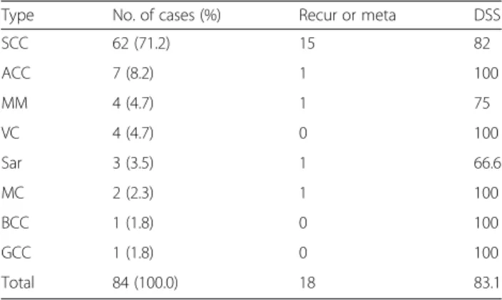
2. Prognosis according to type, site, and stage of cancer Squamous cell carcinoma among oral cancer was the most common with 62 cases (71.2 %); the disease-specific 5-year survival rate of these patients was 82.0 %. The disease-specific 5-year survival rate according to the type of oral cancer ranged from 100 to 0 %. Results of Kaplan Meier method and log rank test showed that the type of oral cancer does not affect prognosis (Table 2).
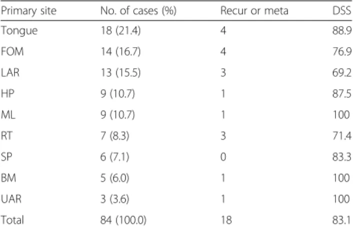
Areas most affected by oral cancer were anterior two-thirds of the tongue followed by floor of the mouth, inferior alveolar ridge. The disease-specific 5-year survival rate with the site of oral cancer ranged from 100.0 to 69.2 %, and results of log rank test showed that site of oral cancer does not affect prognosis (Table 3).
In the stage of patients, 25 people belong to stage I, 23 people to stage II, 16 people to stage III, and 20 people to stage IV, as stage progresses, the disease-specific 5-year survival rate decreases as follows: 96.0 % of patients belong to stage I, 90.9 % of patients to stage II, 86.7 % of patients to stage III, 57.1 % of patients to stage IV, and the p value is 0.003 by log rank test results, which showed the stage was significant factor in the survival rate (Fig 2).
3. Prognosis according to metastasis of cervical lymph node and stage of cervical lymph node
Table 1 Vital status and disease specific 5-year survival rate according to gender
Gender No.
of cases
Alive Dead DSS
TFNR TFAR AWPRM DTF DWC DPO
Male 58 38 5 4 4 5 2 82.5
Female 26 17 2 2 2 2 1 84.6
Total 84 55 7 6 6 7 3 83.1
ATFNR alive; tumor-free; no recurrence, ATFAR alive; tumor-free; after recur- rence, AWPRM alive; with persistent/recurrent/metastatic disease, DTF dead;
tumor-free, DWC dead; with cancer-primary/recurrent/metastatic, DPO dead;
postoperative, DSS disease specific 5-year survival rate
Fig. 1 Disease specific 5-year survival rate by age group (p = 0.093)
Table 2 Disease specific 5-year survival rate according to type of neoplasm
Type No. of cases (%) Recur or meta DSS
SCC 62 (71.2) 15 82
ACC 7 (8.2) 1 100
MM 4 (4.7) 1 75
VC 4 (4.7) 0 100
Sar 3 (3.5) 1 66.6
MC 2 (2.3) 1 100
BCC 1 (1.8) 0 100
GCC 1 (1.8) 0 100
Total 84 (100.0) 18 83.1
SCC squamous cell carcinoma, ACC adenoid cystic carcinoma, MM malignant melanoma, VC verrucous carcinoma, Sar sarcoma, MC mucoepidernoid carcinoma, BCC basal cell carcinoma, GCC ghost cell carcinoma, Recur recurrence, meta metastasis, DSS disease specific 5-year survival rate)
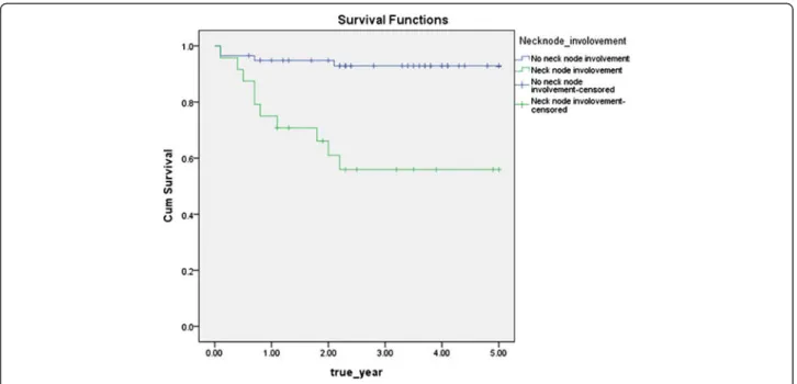
The disease-specific 5-year survival rate without cervical lymph node metastasis was significantly higher than that with cervical lymph node metastasis (93.2 vs 58.3 % (Fig 3)). As N stage progressed, the disease-specific 5-year survival rate significantly decreased (p < 0.05) (Fig 4).
4. Prognosis according to recurrence/metastasis or timing of recurrence/metastasis
The disease-specific 5-year survival rate according to recurrence or metastasis recurrence or metastasis was 89.1 % in the case without recurrence or metastasis and 63.2 % with recurrence or metastasis,
and whether or not, recurrence and metastasis were significant factors, since significant probabil- ity was 0.011 by log rank test results, which showed that recurrence and metastasis were significant factors in the survival rate (Fig 5).
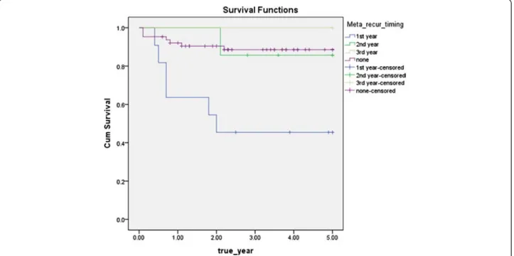
The survival rate varied according to the time of recurrence or metastasis of 19 patients who experienced postoperative recurrence or metastasis.
The disease-specific 5-year survival rate of patients who experienced recurrence or metastasis within 1 year after surgery was 45.5 %, within 1–2 years was 85.7 %, and within 2–3 years was 100 %, and significant probability was 0.002 by log rank test results, which showed that the time of recurrence
or metastasis after surgery was a significant factor in the survival rate (Fig 6).
Recurrence or metastasis occurs to all 18 patients (21.4 % of 84 patients), with local recurrence only occurring to 7 patients, regional recurrence only to 8 patients, local recurrence and regional recurrence to 1 patient, and with distant metastasis to 2 patients. One of 7 patients has local recurrence only, 3 of 8 patients with regional recurrence only, one patient with locoregional recurrence, and 1 of 2 patients with distant metastasis died (Table 4). It was most common that recurrence or metastasis occurred within 1 year to 10 (58 % of 18 patients) of 18 cases, within 1–2 years to seven cases (37 % of the 18 patients), within 2–3 years to one case (5 % of 18 patients),
Table 3 Disease specific 5-year survival rate according toprimary site of neoplasm
Primary site No. of cases (%) Recur or meta DSS
Tongue 18 (21.4) 4 88.9
FOM 14 (16.7) 4 76.9
LAR 13 (15.5) 3 69.2
HP 9 (10.7) 1 87.5
ML 9 (10.7) 1 100
RT 7 (8.3) 3 71.4
SP 6 (7.1) 0 83.3
BM 5 (6.0) 1 100
UAR 3 (3.6) 1 100
Total 84 (100.0) 18 83.1
Tongue anterior 2/3 of the tongue, FOM floor of mouth, LAR lower alveolar ridge, HP hard palate, ML mucosal lip, RT retromolar trigone, SP soft palate, BM buccal mucosa, UAR upper alveolar ridge
Fig. 2 Disease specific 5-year survival rate by TNM stage (p = 0.003)
and most recurrence or metastasis occurred within 2 years (95 %) after surgery.
5. Prognosis according to differentiation
Depending on the histopathological differentiation, the disease specific 5-year survival rate was 81.7 % (58 out of 73 patients survival) for the well differ- entiated type, 100.0 % (10 out of 10 patients sur- vival) for the moderately differentiated type, and
100.0 % (1 out of 1 patients survival) for the poorly differentiated type, and p value was 0.189 by log rank test results, which showed that the histo- pathological differentiation was not a significant factor in the survival rate (Fig 7).
Out of the 7 patients with local recurrence, 5 patients were T1-2 stage and 2 patients were T3-4 stage. Categorizing according to differentiation,
Fig. 3 Disease specific 5-year survival rate by positive neck node (p = 0.000)
Fig. 4 Disease specific 5-year survival rate by N stage (p = 0.000)
well differentiated type was 5 % (4 of 73 patients) and moderately differentiated type was 30 % (3 out of 10 patients).
Discussion
Radiotherapy or chemotherapy after the surgical proced- ure is largely determined by the histopathological find- ings of the lesion in the treatment decision of oral cancer [11, 12]. According to Brown et al., who reported
that the overall 5-year survival rate of surgical treatment and surgical treatment accompanied by postoperative radiotherapy was 71 and 54 %, respectively, for 193 pa- tients with oral squamous cell carcinoma of TNM stage I-II, good result can be obtained only through surgical treatment in the clean resection boundaries and the le- sion of low stage (Stage I-II) with low recurrence prob- ability [13]. In this study, the effect that sex, age, type of oral cancer, primary site, stage, cervical metastasis, stage
Fig. 5 Disease specific 5-year survival rate by recurrence / metastasis (p = 0.011)
Fig. 6 Disease specific 5-year survival rate by timing of recurrence/metastasis (p = 0.002)
of lymph node, metastasis depending on neck level, re- currence or metastasis, time of recurrence or metastasis, etc. has on survival rate was evaluated.
There are reports that showed excellent prognosis only through surgical treatment in oral cancer patients. Lim et al. reported a disease-specific 5-year survival rate of 83 % with only performing surgical procedure in 76 oral cancer patients [4]. Magge et al. reported that the prog- nosis of surgical treatment accompanied by postopera- tive radiotherapy compared to only surgical treatment did not improve [9]. In this study, disease-specific 5-year survival rate was 83.1 % only through surgical treatment for the oral cancer patients. This is similar when compared to the results reported in the other literature so far [14, 15].
In this study, with the gender distribution of oral cancer patients about 2.3:1 ratio (58 male, 26 female), the proportion of men was higher. The result was similar to gender distribution of the literature researched in Korea [4, 16]. There was no significant difference in the disease-specific 5-year survival rate by gender (male
82.5 %, female 84.6 %) as other reports (Liu et al., Roger et al.) [6, 17].
The effect that the age of oral cancer patients with surgical treatment has on prognosis has been controver- sial. Rogers et al. reported that as the age of the patient increase, disease-specific 5-year survival rate decreases, but Liu et al. reported that there was no significant dif- ferences statistically [6, 17]. In this study, year survival rate was slightly lower in the elderly after 50, but there was no significant difference.
With the result that squamous cell carcinoma patients only got surgical treatment, Lim et al. reported that 5- year survival rate was 83 % out of 76 patients, and Liu et al. reported that 5-year survival rate was 77 % out of 72 patients [4, 6]. In this study, the disease-specific 5- year survival rate was 82.0 % in the squamous cell carcinoma, melanoma, and sarcoma compared to the other tumor that showed a slightly lower survival rate, so the result was similar to the other literature [18–20], which was not statistically significant. The disease- specific 5-year survival rate based on type of tumor was not a significant difference.
Shah et al. reported that oral cancer showed another biological aspect according to primary site [21]. On the other hand, carcinomas on mucosal lip showed a good prognosis; carcinomas on anterior 2/3 of the tongue, floor of the mouth, and the lower alveolar ridge have high risk of metastasis to adjacent lymph nodes and showed a relatively poor prognosis. Rogers et al. re- ported that the disease-specific 5-year survival rate de- pending on primary site was 64–44 %, which was not statistically significant in the 489 oral cancer patients
Table 4 The number of cases and death of the patients whohad recurrence or meta
Recur or meta No. of cases (%) No. of death
Local recur only 7 (38.9) 1
Regional recur only 8 (44.4) 3
Locoregional recur 1 (55.6) 1
Distant meta 2 (11.1) 1
Total 18 (100.0) 6
Recur recurrence, meta metastasis, DSS disease specific 5-year survival rate
Fig. 7 Disease specific 5-year survival rate by histopathological differentiation (p = 0.189)
[17]. In this study, the disease-specific 5-year survival rate depending on primary site varied from 100.0 to 69.2 %, and there was no significant difference by the log rank test results.
Rajapakshe et al. and Geum et al. reported that TNM stage is the factor that has significant influence on the prognosis of oral cancer patients [8, 22]. In this study, as the stage increases, the disease-specific 5-year survival rate decreases (p = 0.003).
With the result only treated surgically for the 489 oral cancer patients, Rogers et al. reported that the disease specific 5-year survival rate (87 %) of the case without cervical lymph node metastasis was significantly higher than that of the case (54 %) with cervical lymph node metastasis [17]. In this study, the disease specific 5-year survival rate of the case (93.2 %) without cervical lymph node metastasis was significantly higher than that of the case (58.3 %) with cervical lymph node metastasis, which accorded with previous researches [4, 6, 17].
In study of Rogers et al., the disease-specific 5-year survival rate of N0, N1, and N2-3 stage was 87, 68, and 40 %, respectively [17]. In this study, the disease-specific 5-year survival rate according to cervical lymph node stage was 93.2 % for the N0 (60 patients), 66.7 % for the N1 (13 patients), 0 % for the N2a (a patient), 50.0 % for the N2b (8 patients), 100.0 % for the N2c (2 patients), and by the log rank test results, cervical lymph node stage had significant effects on oral cancer prognosis (p = 0.000).
Geum et al. reported that the disease-specific 5-year survival rate was 90.5 % for the patients without re- currence or metastasis and 30.0 % for the patients with recurrence or metastasis out of 37 oral cancer patients [22]. In this study, the disease-specific 5-year survival rate depending on recurrence or metastasis was 89.1 % for the case without recurrence or metas- tasis, was 63.2 % for the case with recurrence or me- tastasis, and by the log rank test results, recurrence or metastasis had an significant impact on oral cancer prognosis (p = 0.011).
Liu et al. reported that 72.2 % experienced a recurrence or metastasis after surgery within 2 years and 100 % did within 3 years out of patients with recurrence or metasta- sis [6]. In this study, 95 % experienced a recurrence or me- tastasis after surgery within 2 years and 100 % did within 3 years out of 18 cases with recurrence and metastasis, which was similar to report by Liu et al. [6].
Schwartz et al. reported that survival rate and progno- sis of patients who experienced recurrence or metastasis after 6 months of primary operation were satisfactory than those of within 6 months in the study for 350 oral squamous cell carcinoma patients [23]. The disease- specific 5-year survival rate of those who experienced re- currence or metastasis within 1 year after surgery was
45.5 %, within 1~2 years was 85.7 %, and within 2~3 years was 100 %. As the recurrence or metastasis occurred early, prognosis was significantly poor, in re- sults of the log rank test (p = 0.002).
Geum et al. reported that the disease-specific 5-year survival rate was 94.7 % for the well-differentiated type, 57.1 % for the moderately differentiated type, and 25.0 % for the poorly differentiated type related to survival rate of oral squamous cell carcinoma according to histo- pathological differentiation [22]. But Liu et al. reported that overall 5-year survival rate was 77.3 % for the well- differentiated type and 76.7 % for the moderately differ- entiated type, which was not statistically significant [6].
In this study, histologic differentiation did not have a significant impact on survival rate by the log rank test results.
Conclusions
These results suggest that good survival rate can be ob- tained with surgical treatment only, and stage of oral cancer, cervical lymph node metastasis and stage, recur- rence or metastasis, time of recurrence, and metastasis have a significant effect on survival rate in oral cancer patients.
Consent
The authors declare that they have no competing interests.
Abbreviations
ACC:adenoid cystic carcinoma; ATFAR: alive; tumor-free; after recurrence;
ATFNR: alive; tumor-free; no recurrence; AWPRM: alive with persistent/
recurrent/metastatic disease; BCC: basal cell carcinoma; DPO: dead;
postoperative; DSS: disease specific 5-year survival rate; DTF: dead;
tumor-free; DWC: dead; with cancer (primary/recurrent/metastatic);
GCC: ghost cell carcinoma; MC: mucoepidernoid carcinoma;
Meta: metastasis; MM: malignant melanoma; Recur: recurrence;
Sar: sarcoma; SCC: squamous cell carcinoma; VC: verrucous carcinoma.
Competing interests
The authors declare that they have no competing interests.
Authors’ contributions
BGK obtained the measurements and data and wrote the manuscript. JHK helped in obtaining the data. MIK helped in drafting the manuscript. JJH made substantial contributions to the analysis and interpretation of the data.
SGJ made substantial contributions to the analysis and interpretation of the data. HJP was involved in revising the manuscript. MSK participated in its design and coordination. SYR gave final approval of the manuscript to be published. HKO participated in its design and coordination and carefully reviewed and revised the manuscript. All authors read and approved the final manuscript.
Received: 3 November 2015 Accepted: 1 December 2015
References
1. Chen AY, Myers JN (2000) Cancer of oral cavity. Curr Probl Surg 37(10):633–731 2. Miloro M, Ghali GE, Larsen PE, Waite PD (eds) (2012) Peterson’s principles of
oral and maxillofacial surgery, 3rd edn. McGraw-Hill, Shelton, p 696 3. Omura K (2014) Current status of oral cancer treatment strategies: surgical
treatments for oral squamous cell carcinoma. Int J Clin Oncol 19(3):423–30
4. Lim YC, Choi EC (2008) Surgery alone for squamous cell carcinoma of the oral cavity: survival rates, recurrence patterns, and salvage treatment. Acta Otolaryngol 128:1132–1137
5. Wolfensberger M, Zbaeren P, Dulguerov P, Mu¨ller W, Arnoux A, Schmid S (2001) Surgical treatment of early oral carcinoma—results of a prospective controlled multicenter study. Head Neck 23:525e–30
6. Liu CH, Chen HJ, Wang PC, Chen HS, Chang YL (2013) Patterns of recurrence and second primary tumors in oral squamous cell carcinoma treated with surgery alone. Kaohsiung J Med Sci 29:554–559
7. Massano J, Regateiro FS, Janua’rio G, Ferreira A (2006) Oral squamous cell carcinoma: review of prognostic and predictive factors. Oral Surg Oral Med Oral Pathol Oral Radiol Endod 102:67–76
8. Rajapakshe RM, Pallegama RW, Jayasooriya PR, Siriwardena BS, Attygalla AM, Hewapathirana S et al (2015) A retrospective analysis to determine factors contributing to the survival of patients with oral squamous cell carcinoma.
Cancer Epidemiol 39(3):360–366
9. Magge KT, Myers EN, Johnson JT (2003) Radiation following surgery for oral cancer: impact on local control. Laryngoscope 113(6):933–935
10. Edge S, Byrd DR, Compton CC, Fritz AG, Greene FL, Trotti A (eds) (2010) AJCC cancer staging manual, 7th edn. Springer, Chicago, pp 29–40 11. Amdur RJ, Parsons JT, Mendenhall WM, Million RR, Stringer SP, Cassisi NJ
(1989) Postoperative irradiation for squamous cell carcinoma of the head and neck: an analysis of treatment results and complications. Int J Radiat Oncol Biol Phys 16:25–36
12. Dinshaw KA, Agarwal JP, Laskar SG, Gupta T, Shrivastava SK, Cruz AD (2005) Head and neck squamous cell carcinoma: the role of postoperative adjuvant radiotherapy. J Surg Oncol 91:48–55
13. Brown JS, Shaw JR, Bekiroglu F, Rogers SN (2012) Systematic review of the current evidence in the use of postoperative radiotherapy for oral squamous cell carcinoma. Br J Oral Maxillofac Surg 6:481–489 14. Hall J, Angele S (1999) Radiation, DNA damage and cancer. Mol Med
Today 5(4):157–164
15. Finlay PM, Dawson F, Robertson AG, Soutar DS (1992) An evaluation of functional outcome after surgery and radiotherapy for intraoral cancer. Br J Oral Maxillofac Surg 30:14–17
16. Lee JW, Kim JW, Kim CS (2008) A clinic-statistical study on cervical lymph node metastasis of oral squamous cell carcinoma. J Kor Oral Maxillofac Surg 34:594–601
17. Rogers SN, Brown JS, Woolgar JA, Lowe D, Magennis P, Shaw RJ et al (2009) Survival following primary surgery for oral cancer. Oral Oncol 45:201–211 18. Padhye A, D’souza J (2011) Oral malignant melanoma: a silent killer? J of Ind
Soci of Perio 4:425–428
19. Auluck A, Zhang L, Desai R, Rosin MP (2008) Primary malignant melanoma of maxillary gingiva—a case report and review of the literature. J Can Dent Assoc 74(4):367–371
20. Yamaguchi S, Nagasawa H, Suzuki T, Fujii E, Iwaki H, Takagi M et al (2004) Sarcomas of the oral and maxillofacial region: a review of 32 cases in 25 years. Clin Oral Investig 8:52–55
21. Shah J, Gil Z (2009) Current concepts in management of oral cancer—surgery. Oral Oncol 45:394–401
22. Geum DH, Roh YC, Yoon SY, Kim HG, Lee JH, Song JM et al (2013) The impact factors on 5-year survival rate in patients operated with oral cancer.
J Korean Assoc Oral Maxillofac Surg 5:207–216
23. Schwartz GJ, Mehta RH, Wenig BL, Shaligram C, Louis G (2000) Salvage treatment for recurrent squamous cell carcinoma of the oral cavity. Head Neck 22(1):34–41
Submit your manuscript to a journal and benefi t from:
7 Convenient online submission 7 Rigorous peer review
7 Immediate publication on acceptance 7 Open access: articles freely available online 7 High visibility within the fi eld
7 Retaining the copyright to your article
Submit your next manuscript at 7 springeropen.com