• 검색 결과가 없습니다.

Effect of Different Air Hole Diameters of the Inspiratory Muscle Trainer on the Rating of Perceived Exertion and Inspiratory Muscle Activity during Breathing Exercise

N/A
N/A
Protected

Academic year: 2021

Share "Effect of Different Air Hole Diameters of the Inspiratory Muscle Trainer on the Rating of Perceived Exertion and Inspiratory Muscle Activity during Breathing Exercise"

Copied!
7
0
0

로드 중.... (전체 텍스트 보기)

전체 글

(1)

Effect of Different Air Hole Diameters of the Inspiratory Muscle Trainer on the Rating of Perceived Exertion and Inspiratory

Muscle Activity during Breathing Exercise

Shin Areum, PT

1

· Kim Kisong, PT, Ph.D

1

Applied Kinesiology and Ergonomic Technology Laboratory, Dept of Physical Therapy, Graduate School of Yonsei University, Student

2ǂ

Dept of Physical therapy, College of Life and Health Science, Hoseo University, Professor

Abstract

Purpose : This study aims to investigate the rating of perceived exertion (RPE) and muscle activity of the inspiratory primary and accessory muscle during breathing exercise with different air hole diameters of the inspiratory muscle trainer (IMT).

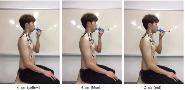
Methods : The Borg’s scale and surface electromyography (EMG) was used to collect data of the RPE and muscle activity of the inspiratory primary the external intercostal (EI) and diaphragm (DIA) and accessory muscles anterior scalene (AS), sternocleidomastoid (SCM), pectoralis major (PM), and upper trapezius (UT) muscles during breathing exercise with different air hole diameters (6 ㎜, 4 ㎜, and 2 ㎜) of the IMT in healthy young male subjects.

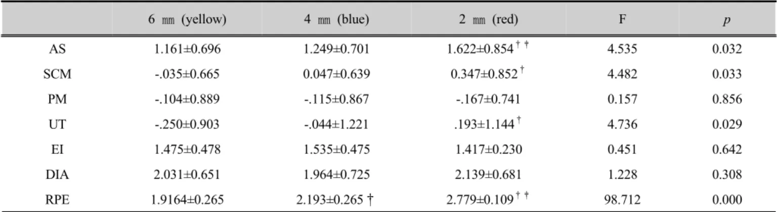
Results : The RPE and muscle activities of the AS, SCM, and UT are increased significantly in accordance to the decreasing diameter of air hole of air tip in IMT. However, there are no differences in the muscle activities of the PM, EI, and DIA based on differences of diameters of air hole of air tip in IMT.

Conclusion : The smaller the diametr of IMT air-hole, RPE and muscle activities of AS, SCM and UT were increased. Therefore, further study would be necessary to investigate the proper intensity and relaxation posture for the exercise protocol to strengthen the inspiratory primary muscles.

Key Words : different diameters of air tip of inspiratory muscle trainer, inspiratory primary and accessory muscle, muscle activity, rating of perceived exertion

ǂ

Corresponding author : Kim Kisong, [email protected]

2)

Received : April 25, 2019 | Revised : June 11, 2019 | Accepted : June 14, 2019

※ This research was supported by the Academic Research fund of Hoseo University in 2016 (20160296).

(2)

. Introduction

1. Background

Pulmonary diseases were classified as obstructive, restrictive, and other diseases (Watanabe et al., 2018).

Among the intervention methods performed for pulmonary rehabilitation, the inspiratory muscle trainer (IMT) developed for strengthening the inspiratory muscle of the patient has various types of resistance (Beaumont et al., 2018). According to the resistance method, the IMT were divided into a method of changing the air hole diameter of the air tip, a method of using elasticity of the spring, and a buoyancy device using the sound pressure in the exercise device (Lin et al., 2012). IMT device which was controlled through the size of the air hole diameter, has 6 different hole diameter size of the air tip ranging from 2 ㎜ to 7 ㎜.

IMT increased inspiratory muscle capacity, and improved exercise tolerance and perception of breathlessness during activities of daily living (Gosselink et al., 2011). Breathing exercise using IMT was usually applied once for 15~30 minutes, twice or three times a day, and the choice of the hole diameter size of the air tip to determine the resistance depends on the tolerance of the patient (Johnson et al., 1998). However, breathing exercises for strengthening inspiratory muscles often caused pain of trunk muscles and dyspnea. Pain and dyspnea during inspiratory muscle strengthening exercise can be considered as fatigue caused by overload due to resistance exceeding tolerance and exercise capacity of patient (Enoka & Duchateau, 2008).

Previous studies using IMT have focused only on change in clinical symptoms, such as forced vital capacity and quality of life after period of exercise (Riera et al., 2001).

However, no studies have examined the effect of air hole size change on the patient’s exercise intensity and the changes in muscle activity of the inspiratory main and accessary muscle during breathing exercise using different

air hole diameter of air tip of the IMT.

2. Purpose of study

The purpose of our study was to investigate the effect of air hole diameter size change of air tip on the rating of perceived exertion and muscle activity of the inspiratory primary and accessary muscle during breathing exercise using IMT in healthy young subjects. The hypothesis of this study was as follows: depending on the difference in diameter of air tip of the IMT, the rating of perceived exertion and muscle activity of the inspiratory primary main and accessary muscle would be different during breathing exercise using IMT.

. Methods

1. Participants

Sample size was determined with software of G*power

analysis. Necessary sample size of 14 was calculated

through a power of 0.80 and effect size of 0.37 (calculated

with a partial η₂of 0.121 from a pilot study with five

subjects) with a α level of 0.05. As a result, fifteen

healthy male subjects voluntarily participated in this study

(Table 1). Informed consent form was signed by individual

subjects after understanding the purposes and methods of

this present study. Exclusion criteria were as follows: 1)

subjects who complained of pain or discomfort during

breathing exercise with IMT, 2) subjects who were

diagnosed with musculoskeletal diseases or cardiorespiratory

disorders, 3) subjects who have been receiving recent

operations or taking any medications affecting breathing

patterns. Institutional review board of Hoseo University has

approved this study (104123-170904-HR-063-02).

(3)

Ethnicity Sex Age (y) Height (㎝) Weight (㎏) BMI (㎏/m

2

) FVC (L)

Korean Male 21.6±1.8 174.87±5.15 69.59±7.91 22.81±2.96 3.4±0.6

Table 1. Subject demographic characteristics and pulmonary functions

BMI; body mass index, FVC; forced vital capacity

2. Measurements

1) Forced vital capacity (FVC)

To measure the forced vital capacity, Desk-top Spirometer (Spirometer, Pony F/X, Italia) was used. In sitting position, the subjects inhaled as deep as possible and then exhaled maximally into the spirometer.

2) Borg’s perceived exertion scale

To measure the subject’s difficulty during strengthening exercise of inspiratory muscles, Borg’s perceived exertion scale was used. The Borg’s perceived exertion scale consists of 20 score from 6 score and the higher score means the harder perception.

3) Data collection

For surface electromyography (EMG) data collection and analysis, a Noraxon TeleMyo 2400 system (DTS EMG system, Noraxon, Inc, USA) and a Noraxon MyoResearch 1.06 XP software was used. The EMG signals were collected at 1000 ㎐ amplified, band-pass filtered at 20 and 450 ㎐, notch filtered at 60 ㎐ and processed into the root-mean-square values. EMG data were recorded from the anterior scalene (AS), sternocleidomastoid (SCM), pectoralis major (PM), upper trapezius (UT), external intercostal (EI), and diaphragm (DIA) on the dominant side. After shaving and rubbing the skin with alcohol, disposable Ag/AgCl surface electrodes were attached approximately 2 ㎝ apart and parallel to the each muscle fiber at the frequently referenced site (Chien et al., 2008; Cram, 1998); (1) AS, electrode was placed on the just posterior to and at a

slightly oblique angle relative to the SCM, just above the

clavicle and in the hollow triangle anterior to the UT; (2)

SCM, electrode was positioned slightly posterior from the

middle of the distance between the mastoid process and the

sternal notch; (3) PM, electrode was placed on the chest

wall at an oblique and angle toward the clavicle,

approximately 2 ㎝ inferior from the clavicle, just medial to

the axillary fold; (4) UT, electrode was positioned along the

ridge of the shoulder, slightly lateral to and one-half the

distance between the cervical spine 7 and the acromion; (5)

EI, electrode was placed on the second or third intercostal

space at the midclavicular line; (6) DIA, electrode was

positioned 7th or 8th intercostal space on the dominant side

of the body at the midclavicular line. Maximal voluntary

isometric contractions (MVIC) were collected to normalize

the EMG data from each muscle (AS, SCM, PM, and UT)

during 5 seconds. The position of each muscle for MVIC

was (1) for AS and SCM, the subject was positioned supine

with elbows flexed, hands beside the head, craniocervical

and cervical anterolateral flexion; (2) for PM, the subject

was positioned sitting on the table with elbow extended and

shoulder flexed at 90 °, rotated medially, horizontally

adducted toward the sternal end of the clavicle; (3) for UT,

the subject positioned sitting on the table with shoulder

abducted at 90 ° and cervical ipsilateral flexion and

contralateral rotation (McCreary & Provance, 2005). The

middle 3-s of MVIC was used for data analysis. The mean

value of two trials for each muscle was used for data

analysis. The mean EMG amplitude values for SCM, AS,

PM, UT were described as a percentage of MVIC

(%MVIC) (de Oliveira et al., 2008). The intraclass

correlation coefficient of EMG was 0.996 (95 % confidence

(4)

interval: 0.989-0.999) in this study.

3. Procedure

First, the subject who visited the laboratory listened to the purpose of the study and the experiment method while sitting comfortably in the chair. Then, primary investigator explained the method of Borg’s exertion scale. After the agreement of research participation, the subject took off the top and was attached electrodes on his body.

The subjects were instructed to breath in their normal habitual comfortable breathing style with IMT (Inspirayory

muscle trainer, Smiths Medical ASD. Inc, USA) in sitting position with 90 ° of hip, knee and ankle. The order of three difference air hole diameter of the air tips (yellow color for 6 ㎜, blue color for 4 ㎜, and red color for 2 ㎜) for IMT was randomized using the random number generator in Microsoft Excel (Excel, Microsoft Corp., USA) (Fig 1). After subjects adapted to breathe using IMT, subjects performed breathing exercise with IMT for 1 minute for the data collection. All EMG data during breathing exercises with IMT were recorded for 1 minute.

The middle 30 seconds were used for data analysis.

6 ㎜ (yellow) 4 ㎜ (blue) 2 ㎜ (red)

Fig 1. Breathing exercise with different diameter of IMT

4. Statistical analysis

SPSS version 25.0 (SPSS, IBM Inc, USA) was used for test the differences between conditions. After the results are changed as logit because of normal distribution, one way analysis of variance with repeated measured was employed for comparison for three difference diameters (yellow color, blue color, and red color) for IMT. In the case of significant differences between test positions, Bonferroni’s post hoc test was performed. In all analyses, p-values <0.05

were deemed to indicate statistical significance.

Ⅲ. Results

The muscle activities of the AS (F=4.535b, P=0.032), SCM (F=4.482b, p=0.033), UT (F=4.736b, p=0.029), and RPE (F=147.072b, p=0.001) are increased significantly in accord to decreasing diameter of air hole of air tip in IMT.

There are no differences in muscle activities of the PM

(5)

(F=0.157b, p=0.856), EI (F=0.342b, p=0.717), DIA (F=1.249b, p=0.319) in accord to differences of diameter of air hole of air tip in IMT. In Bonferroni’s post-hoc test, there are significant differences between 6 ㎜ and 2 ㎜ (p=0.007), and between 4 ㎜ and 2 ㎜ (p=0.016) in AS,

between 6 ㎜ and 2 ㎜ (p=0.014) in SCM, and between 6

㎜ and 2 ㎜ in UT (p=0.016), and between all matching of 6 ㎜ and 4 ㎜ (p=0.001), 6 ㎜ and 2 ㎜ (p=0.001), and 4

㎜ and 2 ㎜ (p=0.002) in RPE (Table 2).

6 ㎜ (yellow) 4 ㎜ (blue) 2 ㎜ (red) F p

AS 1.161±0.696 1.249±0.701 1.622±0.854

†‡

4.535 0.032

SCM -.035±0.665 0.047±0.639 0.347±0.852

4.482 0.033

PM -.104±0.889 -.115±0.867 -.167±0.741 0.157 0.856

UT -.250±0.903 -.044±1.221 .193±1.144

4.736 0.029

EI 1.475±0.478 1.535±0.475 1.417±0.230 0.451 0.642

DIA 2.031±0.651 1.964±0.725 2.139±0.681 1.228 0.308

RPE 1.9164±0.265 2.193±0.265† 2.779±0.109

†‡

98.712 0.000

Table 2. Logit comparisons among 3 different diameters of IMT (Mean ± standard deviation)

*significant differences among three diameters of IMT air-hole (p<0.05)

Post-hoc:

significant difference to 6 ㎜ (p<0.017),

significant difference to 4 ㎜ (p<0.017)

AS; anterior scalene, SCM; sternocleidomastoid, PM; pectoralis major, UT; upper trapezius, EI; external intercostal, DIA; diaphragm, RPE;

rating perceived exertion

Ⅳ. Discussion

The hypothesis of this study was that depending on the difference in diameter of the IMT air tip, the RPE and muscle activity of the inspiratory principal (DAI and EI) and inspiratory accessary muscle (AS, SCM, PM and UT) would be different during breathing exercise using IMT.

There were significant differences among the parameters in muscle activities of the AS, SCM, and UT and RPE.

Hence, our research hypothesis was partially supported.

As decreasing of air hole diameter of tip in IMT, the several muscle activities of the inspiratory accessory muscles (AS, SCM, and UT) were increasing significantly.

Previous results also showed significantly increased SCM activity during loading periods using IMT in chronic obstructive pulmonary disease (De Andrade et al., 2005).

These results could be caused by increasing resistance of

air flow (caused by decreasing of air hole diameter of tip in IMT) during inspiratory breathing phase (Dodd et al., 1984). The resistance of air flow increases muscular efforts to elevate upper chest wall and to obtain the amount of tidal volume in upper airway during inspiration.

There was no significant differences in muscle activities

of the inspiratory accessory muscle (PM). This result was

caused by no distal fixation of arm and no mechanical

advantage (block the role as an arm mover, and devote

itself on trunk elevator) for the PM as an inspiratory

accessory muscle (Vera-Garcia et al., 2010). Also there

were no significant differences in muscle activities of the

inspiratory primary muscles (EI and DIA) in accordance to

decreasing of air hole diameter of tip in IMT. These results

were caused by compensatory function of inspiratory

accessory muscles (AS, SCM, and UT) to overcome the

increasing resistance for primary muscles during inspiratory

(6)

phase, and might be caused by that resistance of mouth piece (through air hole of air tip in IMT) affect only the movement of upper chest wall (Corrêa & Bérzin, 2008).

However, the results of muscles (EI and DIA) could be investigated with further objective research, because our research did not adapt the %MVIC for the EI and DIA with comparable other muscles (AS, SCM, PM and UT).

As decreasing of air hole diameter of tip in IMT, RPE was increasing significantly. These result were caused by same reason of the muscle activities in inspiratory accessory muscles (AS, SCM, and UT). The breathing exercises for strengthening inspiratory muscles often cause pain of trunk muscles and dyspnea. These features might be caused by fatigable condition of the inspiratory accessory muscles (HajGhanbari et al., 2012). Hence, RPE could be an objective target point to prevent fatigue of inspiratory accessory muscles and respiratory burden. Moderate intensity of strengthening exercise with inspiratory muscle trainer might elicit favorable change in patients’ condition without unwanted effects (Romer et al., 2002).

There were several limitations in our research. The first, adapted sitting posture in this study is not favorable deep breathing posture after increasing ventilator demand condition. In previous studies, trunk leaning forward posture with arm support could be a recommendable posture to improve the function of inspiratory accessory muscles. As these mechanical advantages, chronic respiratory patients select trunk leaning forward posture with arm support for the relaxation and athletic players make these pose after running competition for resolution the hyper ventilator condition. Hence, several recommendable posture could be adapted for further research. The second, surface EMG could not be a suitable device to collect the EMG data of DIA. Previous studies have described this limitation in their research, therefore the data of DIA would be contaminated with artifact or not. Hence, further advanced design to collect EMG data of DIA would be adapted for the explanation of core muscular function.

Ⅴ. Conclusion

The decreasing the diameter of air-hole induces increasing muscle activities of the inspiratory accessory muscles. The exercise intensity for the chronic patients with dyspnea should be lower than patients without dyspnea.

Even with same condition of exercise intensity, the all-day using inspiratory muscles of chronic patients with dyspnea, could be faster fatigable than patients without dyspnea. The exercise intensity for the chronic patients with dyspnea should be adjusted to their cardiopulmonary symptoms at initial evaluation.

References

Beaumont M, Mialon P, Le Ber C, et al(2018). Effects of inspiratory muscle training on dyspnoea in severe COPD patients during pulmonary rehabilitation:

controlled randomised trial. Eur Respir J, 51(1), 1701107. Doi: 10.1183/13993003.01107-2017.

Chien MY, Wu YT, Chang YJ(2008). Assessment of diaphragm and external intercostals fatigue from surface EMG using cervical magnetic stimulation. Sensors, 8(4), 2174-2187.

Corrêa EC, Bérzin F(2008). Mouth breathing syndrome:

cervical muscles recruitment during nasal inspiration before and after respiratory and postural exercises on Swiss Ball. Int J Pediatr Otorhinolaryngol, 72(9), 1335-1343.

Cram JR(1998). Introduction to surface electromyography.

1st ed, Gaithersburg, Aspen Publishers, pp.279-351.

De Andrade AD, Silva TN, Vasconcelos H, et al(2005).

Inspiratory muscular activation during threshold®

therapy in elderly healthy and patients with COPD. J Electromyogr Kinesiol, 15(6), 631-639.

de Oliveira AS, de Morais Carvalho M, de Brum

DPC(2008). Activation of the shoulder and arm muscles

(7)

during axial load exercises on a stable base of support and on a medicine ball. J Electromyogr Kinesiol, 18(3), 472–479.

Dodd DS, Brancatisano T, Engel LA(1984). Chest wall mechanics during exercise in patients with severe chronic air-flow obstruction. Am Rev Respir Dis, 129(1), 33-38.

Enoka RM, Duchateau J(2008). Muscle fatigue: what, why and how it influences muscle function. J Physiol, 586(1), 11-23.

Gosselink R, De Vos J, van den Heuvel SP, et al(2011).

Impact of inspiratory muscle training in patients with COPD: what is the evidence?. Eur Resp J, 37, 416-525.

HajGhanbari B, Holsti L, Road JD, et al(2012). Pain in people with chronic obstructive pulmonary disease (COPD). Respir Med, 106(7), 998-1005.

Johnson PH, Cowley AJ, Kinnear WJM(1998). A randomized controlled trial of inspiratory muscle training in stable chronic heart failure. Eur Heart J, 19(8), 1249-1253.

Lin SJ, McElfresh J, Hall B, et al(2012). Inspiratory muscle

training in patients with heart failure: a systematic review. Cardiopulm Phys Ther J, 23(3), 29-36.

Kendall FP, McCreary EK, Provance PG, et al(2005).

Muscles: testing and function with posture and pain. 5th ed, Baltimore, Lippincott Williams & Wilkins, pp.165-203.

Riera HS, Rubio TM, Ruiz FO, et al(2001). Inspiratory muscle training in patients with COPD: effect on dyspnea, exercise performance, and quality of life.

Chest, 120(3), 748-756.

Romer LM, McConnell AK, Jones DA(2002). Effects of inspiratory muscle training upon recovery time during high intensity, repetitive sprint activity. Int J Sports Med, 23(5), 353-360.

Vera-Garcia FJ, Moreside JM, McGill SM(2010). MVC techniques to normalize trunk muscle EMG in healthy women. J Electromyogr Kinesiol, 20(1), 10-16.

Watanabe R, Shiraki M, Saito M, et al(2018). Restrictive

pulmonary dysfunction is associated with vertebral

fractures and bone loss in elderly postmenopausal

women. Osteoporos Int, 29(3), 625-633.

수치

Table 1. Subject demographic characteristics and pulmonary functions
Fig 1. Breathing exercise with different diameter of IMT
Table 2. Logit comparisons among 3 different diameters of IMT (Mean ± standard deviation)

참조

관련 문서

• 이명의 치료에 대한 매커니즘과 디지털 음향 기술에 대한 상업적으로의 급속한 발전으로 인해 치료 옵션은 증가했 지만, 선택 가이드 라인은 거의 없음.. •

Respiratory muscles training – Inspiratory muscle training (IMT) – Expiratory muscle training (Abd-FES) Respiratory muscles training. – Inspiratory muscle training (IMT)

The proposal of the cell theory as the birth of contemporary cell biology Microscopic studies of plant tissues by Schleiden and of animal tissues by Microscopic studies of

웹 표준을 지원하는 플랫폼에서 큰 수정없이 실행 가능함 패키징을 통해 다양한 기기를 위한 앱을 작성할 수 있음 네이티브 앱과

The meeting was attended by Assistant Foreign Minister for GCC Affairs, Ambassador, Nasser Al-Muzayyen, and Deputy Assistant Foreign Minister for the Office of the

“ Sheikh Nasser has a written message from HH the Amir, Sheikh Sabah Al-Ahmad Al-Jaber Al-Sabah to the Chinese President, Chi Gen Beng related to enhancing mutual

On his part, CEO of Express Roads Authority, Saud Al-Naqqi said that the heavy rains of the previous day led to clogging parts of the express

Kuwait will celebrate on Sunday the fourth anniversary of the UN honoring and proclamation of His Highness the Amir, Sheikh Sabah Al-Ahmad Al-Jaber Al-Sabah as