혈액투석환자에서 중심정맥 협착 치료
구동억
순천향대학교 의과대학 영상의학교실
Treatment of Refractory Central Vein Stenosis in Hemodialysis Patients
Dong Erk Goo
Department of Radiology, College of Medicine, Soonchunhyang University, Seoul, Korea
혈액투석환자에서 중심 정맥 협착은 흔한 합병증의 하나이며, 중 심 정맥관 삽입과 중심정맥 밸브 비후 등이 중요 원인이다. 실제 미 국의 통계를 보면 투석환자의 약 80%는 중심정맥관을 사용하여 투 석을 처음 시작한다. 중심 정맥 협착이 발생하면 팔과 얼굴에 부종이 발생하며, 가슴에는 많은 측부순환 정맥들을 관찰할 수 있다. 드물게 는 유방 확장이나 식도정맥류, 호흡곤란, 이명 뿐만 아니라 신경학적
증상도 동반할 수 있다[1].
중심정맥 협착 치료에서 반드시 고려해야 할 사항으로 무증상의 중심정맥 협착이다. 중심정맥은 말초정맥에 비하여 탄력적이며 또한 풍선확장술(percutaneous transluminal angioplasty, 이하 PTA) 후 탄성 반도가 흔하다. 혈관내 초음파(Intravenous ultrasound, 이하 IVUS) 검 사에서 중심정맥의 50% 이상에서 PTA 후 즉각적인 탄성반도가 나
Received: Oct 12, 2019, Revised: Nov 9, 2019, Accepted: Nov 15, 2019 책임저자 : 구동억
우 04401, 서울시 용산구 대사관로 59, 순천향대학교 의과대학 영상의학교실 Tel: 02-709-9077, Fax: 02-709-9066, E-mail: [email protected]
Central venous stenosis is one of the major problems in hemodialysis patients. Although balloon dilatation and stent placement have high a technical success rate, some patients have problems with limited response or frequently recurrence. The author investigated the intervention and surgical procedures that can be used when there are limitations to balloon dilation and stent placement for the central vein stenosis in hemodialysis patients. The stent graft placement for the central vein stenosis is one of the useful procedures. One article reported 67% patency rate at 1 year, which was higher than the stent procedure, with no difference of complications rate. Recently, some articles have reported drug eluting balloon procedure for the central vein stenosis, but it is very limited numbers. Procedural success was achieved in all cases and the primary patency rates at 6 months and 12 months were 93.8% and 31.2%, respectively. If central vein stenosis is no longer feasible, a method of reducing blood flow to the arteriovenous fistula (AVF) using banding procedure is also possible. When the blood flow was cut in half, most patients’ symptoms improved. Recently, the HeRO catheter provides a last chance to make arteriovenous graft (AVG) in patients who can no longer make dialysis access in the upper arm. This provided same dialysis quality but lower patency rate to the AVG. Several surgical bypass procedures are effective but invasive, and median assisted primary patency for axillo-femoral bypass was 197 days. Stent grafts and drug coating balloons appear to be good alternatives for refractory central vein stenosis. Reducting access flow is also a useful method, especially in the high flow AVF. HeRO catheter gives the upper arm the opportunity to make AVG one last time.
Refractory central vein stenosis can be thought to be managed to some extent with the appropriate procedure selection.
Key Words: Dialysis, Central vein stenosis, Percutaneous transluminal angioplasty
This is an Open Access article distributed under the terms of the Creative Commons Attribution Non-Commercial License (http://creativecommons.org/licenses/by-nc/4.0) which permits unrestricted non-commercial use, distribution, and reproduction in any medium, provided the original work is properly cited.
Copyright © The Korean Society for Dialysis Access | eISSN: 2635-8603
타나고, 이런 탄성반도로 인하여 스텐트 시술이 비교적 빈번하게 필 요하다. 하지만 PTA와 스텐트 시술은 잦은 재발을 유발하는데, 무증 상 중심정맥 협착을 중재 시술한 경우에도 빠른 속도로 재협착이 발 생하는 것으로 알려져 있다[2]. PTA 자체가 혈관 내막에 손상을 주어 neointimal hyperplasia를 가속화시키는 것으로 알려져 있기 때문에 이러한 무증상 중심정맥 협착 환자는 증상의 발생 여부를 관찰하면 서 추적하는 것이 추천된다.
중심정맥 협착에 대한 일차적 치료는 PTA와 스텐트 삽입술인데, 이들의 일차 개통률은 만족스럽지는 못하다. K/DOQI guideline에서 는 중심정맥 협착의 치료법으로 PTA를 우선적으로 권고하고 있으며 7090%의 높은 초기 성공률을 보고하고 있다. Cuthbert 등은[3] 1,030 명의 혈액투석환자 중 76명(7.4%)에서 중심정맥 협착이 발생하였으 며, 동 측에 중심정맥관을 삽입한 환자의 58%에서 협착이 발생하였 다고 보고하였다. 또한 45%에서는 완전 폐쇄 소견이며, 동정맥루 수 술 후 평균 24개월에 첫 번째 중심정맥 PTA를 시행하였다. 6, 12개 월 일차 보조 개통률(primary assisted patency)은 각각 87, 74%로 보 고하였지만 평균 7개월 후 재 시술이 필요하였고, 중심정맥관을 삽입 한 경우는 좀 더 짧은 개통률을 보였다. 한편, 고압력 풍선을 이용한 PTA에서 6개월 비보조 개통률(unassisted patency rate)은 60%, 12개 월 30%로 보고하여 고압력 풍선의 유용성을 주장하기도 한다[4].
스텐트는 탄성반도에 의한 협착이나 3개월 이내에 2번 이상의 재 협착, PTA 시술 실패가 발생하면 적응증이 된다. 스텐트는 탄성반도 협착에 즉각적인 효과를 나타내지만 역시 빈번한 재 협착은 PTA와 비슷하다. 자가팽창형 스텐트가 높은 기술적 성공률을 보이고 있어 탄성반도 병변에는 PTA 단독 시술보다는 자가팽창형 스텐트를 같 이 시술하는 것이 좀 더 좋은 결과를 보인다. PTA에 실패한 경우에 는 일반적으로 스텐트를 사용하지만, 일차적인 스텐트 설치(primary stenting)은 장기 개통률에 도움이 되지 않기 때문에 권고되지 않는 다. 스텐트의 일차, 이차 개통률은 제한적인데, 그 이유는 스텐트가 필요한 병변의 심한 혹은 반복적인 재 협착 특성 때문이다. 특히 PTA 에 실패하여 스텐트 삽입술을 한 경우에는 평균 138일, 19%의 1년 개통률을 보고하였다[5]. 따라서 PTA에 실패한 중심정맥 협착에서 스텐트는 짧은 기간 동안의 증상 호전에 도움이 되며, 단지 반복적인 확장술을 통해서만 장기 개통을 유지할 수 있다. Ronald 등[6]은 스 텐트 내 재 협착을 가진 중심정맥 협착환자에서 PTA 단독 치료 환자 와 PTA에 반응이 없는 경우 추가적인 스텐트 시술환자의 비교에서 6, 12개월 개통률이 38, 17%와 57, 32%로 스텐트 내 협착에서 풍선에 반응이 없는 경우 추가적인 스텐트 삽입술을 주장하였다.
최근에는 심장 리듬 장치(cardiac rhythm device)로 인한 중심정맥 협착을 자주 경험하게 된다. 이때 PTA는 안전하게 시술할 수 있는 방 법이지만, 6, 12개월 일차 개통률은 18%, 9%로 없는 경우와 비교하 면 매우 낮은 개통률을 보인다[7]. 이때 스텐트 시술이 필요한 경우에 는 심박동기(pacemaker) 제거 후 스텐트 시술을 하고 다시 설치해야 만 한다.
다음은 중심정맥 협착에 PTA와 스텐트 시술이 불충분한 경우, 혹 은 잦은 재발 때 선택할 수 있는 중재 시술 및 수술적 방법들에 대하
여 알아보고자 한다.
1. Sacrifice of the Access
중심 정맥 협착이나 폐쇄를 중재 시술이나 수술적으로 해결할 수 없는 경우에는 증상 완화를 위하여 동정맥루를 폐쇄하여야 한다. 오 래된 중심정맥 협착은 식도정맥류나 신경학적 합병증을 만들 수도 있기 때문이다. 동정맥루 폐쇄는 수술로 해결할 수도 있지만, 심한 부종으로 수술적 접근이 어려운 경우에는 코일 등을 이용하여 중재 시술로 폐쇄할 수도 있다.
2. Stent Graft
중심 정맥 협착에 대하여 stent graft 사용이 일부 보고되고 있다.
Jones 등[8]은 중심정맥 협착에 VIABAHN stent graft를 사용하여 1 년 개통률 67%, 1년 일차 보조 개통률(primary assisted patency) 94%
를 보고하였으며, 협착병변 보다는 폐쇄병변에서 개통률이 의미 있 게 짧았다고 하였다(p=0.05). 하지만 이것은 기존 bare stent에 비하여 높은 개통률이며 다른 합병증에서는 큰 차이를 보이지 않았다.
Vestandig 등[9]은 52예의 중심정맥 stent graft 시술에서 100% 성 공률과 60%, 40%, 28%의 6개월, 1년, 2년 계통률을 보고하였다. 이들 은 stent graft가 동정맥루 계통 연장에 도움이 됨을 보여주었지만, 시 술 후 83%의 환자에서 stent graft가 내경정맥이나 반대측 무명정맥의 입구를 덮고 있었다. 이러한 시술은 향후 내경정맥이나 반대측 동정 맥루를 형성하는데 방해가 될 수 있기 때문에 가능한 이러한 시술은 피할 것을 권고하였다(Fig. 1).
3. Collateral Vein PTA
중심 정맥 협착이 발생하면 다양한 경로의 측부순환 정맥이 발달 한다. 가장 흔한 측부순환 정맥은 쇄골하정맥 협착 때 요골측피부활 (cephalic arch)에서 외경정맥으로 발달하며, 그 밖에도 내경정맥과 갑 상샘하 정맥(inferior thyroid vein)을 통한 반대 측 중심정맥으로의 측 부순환이다. 특히 cephalic arch에서 외경정맥으로 발달한 측부순환 정맥은 길이가 짧으며 cephalic arch에서 접근하기도 용이하여 비교 적 쉽게 PTA를 할 수 있으며 증상도 호전시킬 수 있다. 이들 정맥을 PTA하는 것은 항상 가능한 것은 아니지만 측부순환 정맥을 이용한 PTA는 간단하면서 유용한 하나의 옵션이다[10] (Fig. 2).
4. Drug Eluting Balloon (DEB) PTA
동정맥 인조혈관(arteriovenous graft, 이하 AVG) 정맥문합부 협착 에 대한 DEB PTA 결과는 많이 보고되었지만 중심 정맥 협착에 대 한 DEB PTA 결과 보고는 많지 않다. 40명의 동정맥루(arteriovenous fistula, 이하 AVF)와 AVG 환자의 중심 정맥 협착에 대한 연구에서 중재 시술없이 유지된 기간(intervention free period)은 DEB는 179일, 일반적인 풍선확장술(convention balloon)은 124.5일로 통계적으로 의 미가 있다는 보고가 있다[11]. Hongsakul 등은 16명의 중심 정맥 재 협착 환자에서 67 mm DEB double balloon PTA에서 100% 시술 성 공률과 93.8%, 31.2%의 6, 12개월 개통률을 보고하였다. 또한 DEB의
평균 개통률은 9개월인 반면, convention balloon의 평균 개통률은 2.5 개월로 보고하였지만 환자수가 너무 적은 한계점이 있다[12]. 향후 중심정맥에 대한 DEB PTA와 관련된 더 많은 연구가 필요하다.
5. HeRO Catheter
최근에는 수술용 그라프와 중심정맥관을 결합한 기구로 HeRO catheter가 중심정맥 협착에 이용되고 있다. 중심정맥 협착으로 새로 운 동정맥루를 만들 수 없는 경우에 먼저 중재 시술로 HeRO catheter 를 중심정맥에 삽입 후, 수술용 그라프를 동맥과 연결 후 그라프와 중심정맥관을 연결하여 새로운 동정맥루를 만드는 것이다. 이것은 중심정맥 협착 때문에 일반적인 방법으로 더 이상 상완에 동정맥루 를 만들 수 없는 경우 최후로 AVG를 만들 수 있는 하나의 옵션으로 생각할 수 있다. 여러 보고에서 평균 1년 일차 개통률은 21.9%, 감염 율은 10.1%로 보고하였다. 메타분석에서는 투석의 적정성과 감염률 은 AVG와 비슷하였고, 평균 2.21회/년 중재 시술이 필요하여 하지 AVG의 1.17회/년 보다는 높았다. 하지만 터널형 중심정맥관에 비하 여는 낮은 감염률을 보고하였다[13]. 아직 국내에서는 이용할 수 없 다.
6. Flow Reduction in the Central Vein Stenosis 특히 brachiocephalic fistula인 경우 radiocephalic fistula에 비하 여 과혈류가 문제가 되는 경우가 흔하다. brachiocephalic fistula는 시간이 지나면서 점차 혈류가 많아지면서, 심장에 부담이 되기도 하
며, 투석용 정맥에는 동맥류성 확장과 압력증가, 지혈이 되지 않는 등의 부작용이 발생한다. 또한 과혈류는 중심 정맥 협착을 더욱 악화 시키는 역할을 하기도 한다. 이러한 중심 정맥 협착에서 PTA나 스텐 트 삽입술이 효과가 없는 경우에는 단순히 동정맥루 혈류를 줄이는 것만으로도 증상완화에 도움이 된다. 한 연구에서 22명의 교정이 불 가능한 중심 정맥 협착환자에서 동정맥루 혈류를 밴딩 시술로 1640 ml/min에서 820 ml/min으로 줄여서 20명에서 증상 완화가 있었다고 보고하였다[14]. 교정이 불가능한 중심정맥 협착에서 혈류를 줄이는 간단한 시술만으로도 많은 환자에서 증상 완화를 이룰 수 있음을 보 여주고 있다(Fig. 3).
7. 외과적 우회술
중심 정맥 협착에 대한 외과적 우회술은 여러 가지 방법이 있 다. 그라프를 이용한 우회술이 가장 일반적이며, jugular vein transposition도 이용할 수 있다. 반대쪽 중심정맥으로 우회할 수도 있 으며, 동 측의 대퇴정맥으로 그라프를 이용한 우회도 가능하다. 중심 정맥 협착에 그라프를 이용한 axillofemoral bypass 수술에서 median assisted primary patency는 197일이었다[15]. 이 방법은 중재 시술로 치료할 수 없는 중심정맥 협착에서 동정맥루를 유지하면서 증상을 완화할 수 있는 선택이지만 침습적인 단점이 있다. 본원에서는 지난 10년간 71명의 중심정맥 협착 우회술을 시행하였으며, 평균 9.3개월 의 1차 개통률을 보였고 이들은 이후 지속적인 중재 시술이 필요하 였다(Fig. 4). 중심정맥 협착환자에서 PTA, 스텐트 시술, 외과적 우회
A B C
Fig. 1. A 60-year-old female patient with left upperarm graft fistula. (A) Fistulogram shows total occlusion of stent placed left innominate vein 5 months ago. After stent graft (12×60 mm) placement (B), (C) 8 months follow up fistulogram shows only minimal in-stent intimal hyperplasia.
A B
Fig. 2. A 54-year-old male patient with right brachio-cephalic fistula.
(A) Note complete occlusion of subclavian vein (arrow) with colla- teral flow from cephalic arch to external jugular vein. (B) After 8 mm balloon PTA at the collateral vein, note the decreased filling of the collateral venous drainage.
술은 모두 반복적인 재 협착이 발생하며 결국 반복적인 인터벤션 시 술이 필요함을 지적하였다[16]. Edwards 등[17]은 14명의 중심정맥 협착이 있는 환자에서 첫 번째 갈비뼈 절제, 쇄골 절제와 외과적 우 회술을 동시에 시행하여 90%의 증상 완화와 71.4%의 1년 일차 보조 개통률(primary assisted patency)를 보고하였다.
8. 기타
치료가 힘든 중심정맥 협착에서 그 밖의 선택으로는 대퇴정맥관, 요추정맥관, 대퇴정맥을 이용한 AVG, 복막투석이 있다. 이들의 1년 일차 보조 개통률(primary assisted patency)는 2356%로 보고되고 있 다[18]. 그 밖에도 translumbar, transhepatic, transrenal IVC access를
시도하기도 하며[19], 측부순환 정맥을 통한 중심정맥관 삽입도 보고 되기도 한다[20].
결 론
PTA나 스텐트 시술에 반응이 없는 중심정맥 협착은 투석환자에 서 상완에 어떤 종류의 동정맥루도 사용할 수 없는 상황이 발생할 수 있다. 이것은 반복적이고 오랜 기간 투석용 중심정맥관을 사용한 경 우 흔히 볼 수 있다. 상완에서 동정맥루를 포기하는 경우에는 대퇴 정맥을 이용한 동정맥루를 만들어야 하며, 이 때 일부 환자에는 심한 거부 반응을 보이기도 한다. 따라서 우리는 다양한 다른 방법으로 중 심정맥 협착을 치료해 볼 가치가 있다. 다만 성공적인 재개통술 후에 도 많은 환자에서 개통을 유지하기 위해서는 반복적인 시술이 필요 하다. Stent graft와 DEB PTA가 기존 방법에 비하여 좀 더 좋은 개통 률을 보이지만 아직 광범위한 연구가 좀 더 필요해 보인다. 그리고 혈류가 많은 동정맥루는 단순히 혈류를 줄이는 것만으로도 증상 완 화를 이룰 수 있기 때문에 시도해 볼 만하다.
REFERENCES
1. Herzig DW, Stemer AB, Bell RS, et al. Neurological sequelae from brachiocephalic vein stenosis. J Neurosurg.
2013; 118(5): 1058-62.
2. Levit RD, Cohen RM, Kwak A, et al. Asymptomatic central venous stenosis in hemodialysis patients. Radiology. 2006;
238(3): 1051-6.
3. Cuthbert GA, Lo ZJ, Kwan J, et al. Outcomes of Central Venoplasty in Haemodialysis Patients. Ann Vasc Dis. 2018 sep 25; 11(3): 292-7.
4. Buriánková E, Köcher M, Bachleda P, et al. Endovascular treatment of central venous stenoses in patients with dialysis
A B
Fig. 3. A 64-year-old male patient with right radio-cephalic fistula. Patient had surgical bypass from the right axillary vein to the jugular vein for the subclavian vein occlusion, but still complained of arm swelling. (A) Collateral veins are still persistent in the upperarm after bypass surgery. (B) After banding procedure at the distal cephalic vein (arrow), fistulogram shows completely disappeared collateral veins.
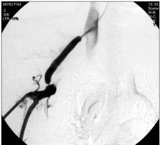
08763 F/54 :3 :5/8 LTR: 10%
13: 53 Scene W-B:
W-C:
X:
Y:
Fig. 4. A 54-year-old female patient with surgical bypass from the axillary vein to the internal jugular vein. Fistulogram shows short segmental stenosis at the both graft anastomosis site.
shunts. Biomed Pap Med Fac Univ Palacky Olomouc Czech Repub. 2003; 147(2): 203-6.
5. Maya ID, Saddekni S, Allon M. Treatment of refractory central vein stenosis in hemodialysis patients with stents.
Semin Dial. 2007; 20(1): 78-82.
6. Ronald J, Davis B, Guevara CJ, et al. Treatment of central venous in-stent restenosis with repeat stent deployment in hemodialysis patients. J Vasc Access. 2017; 18(3): 214-9.
7. Asif A, Salman L, Carrillo RG, et al. Patency rates for angioplasty in the treatment of pacemaker-induced central venous stenosis in hemodialysis patients: results of a multi- center study. Semin Dial. 2009; 22(6): 671-6.
8. Jones RG, Willis AP, Jones C, et al. Long-term results of stent-graft placement to treat central venous stenosis and occlusion in hemodialysis patients with arteriovenous fistulas. J Vasc Interv Radiol. 2011; 22(9): 1240-5.
9. Verstandig AG, Berelowitz D, Zaghal I, et al. Stent grafts for central venous occlusive disease in patients with ipsilateral hemodialysis access. J Vasc Interv Radiol. 2013; 24(9):
1280-7.
10. Ahmad I. Salvage of arteriovenous fistula by angioplasty of collateral veins establishing a new channel. J Vasc Access.
2007; 8(2): 123-5.
11. Kitrou PM, Papadimatos P, Spiliopoulos S, et al. Paclitaxel- Coated Balloons for the Treatment of Symptomatic Central Venous Stenosis in Dialysis Access: Results from a Randomized Controlled Trial. J Vasc Interv Radiol. 2017;
28(6): 811-7.
12. Hongsakul K, Bannangkoon K, Rookkapan S, et al.
Paclitaxel-Coated Balloon Angioplasty for Early Restenosis
of Central Veins in Hemodialysis Patients: A Single Center Initial Experience. Korean J Radiol. 2018; 19(3): 410-6.
13. Maqsood MH, Rubab K.Cureus. Quality of Life of Patients Using the Hemodialysis Reliable Outflow (HeRO) Graft in Hemodialysis. Cureus. 2019; 11(1): e3915.
14. Jennings WC, Miller GA, Coburn MZ, et al. Vascular access flow reduction for arteriovenous fistula salvage in symptomatic patients with central venous occlusion. J Vasc Access. 2012; 13(2): 157-62.
15. Grimm JC, Beaulieu RJ, Sultan IS, et al. Efficacy of axillary- to-femoral vein bypass in relieving venous hypertension in dialysis patients with symptomatic central vein occlusion. J Vasc Surg. 2014; 59: 1651-6
16. Kundu S. Central venous disease in hemodialysis patients:
prevalence, etiology and treatment. J Vasc Access. 2010;
11(1): 1-7.
17. Edwards JB, Brooks JD, Wooster MD, et al. Outcomes of venous bypass combined with thoracic outlet decompression for treatment of upper extremity central venous occlusion. J Vasc Surg Venous Lymphat Disord. 2019; 7(5): 660-4.
18. Aitken E, Jackson AJ, Kasthuri R, et al. Bilateral central vein stenosis: options for dialysis access and renal replacement therapy when all upper extremity access possibilities have been lost. J Vasc Access. 2014; 15(6): 466-73.
19. Shin JH. Hemodialysis access salvage techniques in patients with exhausted access. J Korean Dial Access 2018; 1(1):
21-7
20. Kaminski R, Strozecki P, Kosinski A, et al. Left superior intercostal vein as the last resort for hemodialysis vascular access. J Vasc Access. 2016; 17(1): e5-6