Received: 2013.1.30. Revised: 2013.4.12. Accepted: 2013.5.7.
Corresponding author: Young Min Choi
Department of Obstetrics and Gynecology, The Institute of Reproductive Medicine and Population, Medical Research Center, Seoul National University College of Medicine, 101 Daehak-ro, Jongno-gu, Seoul 110-744, Korea
Tel: +82-2-2072-2385 Fax: +82-2-762-3599 E-mail: [email protected]
Articles published in Obstet Gynecol Sci are open-access, distributed under the terms of the Creative Commons Attribution Non-Commercial License (http://creativecommons.
org/licenses/by-nc/3.0/) which permits unrestricted non-commercial use, distribution, and reproduction in any medium, provided the original work is properly cited.
Copyright © 2013 Korean Society of Obstetrics and Gynecology
Introduction
Polycystic ovary syndrome (PCOS) is one of the most common causes of endocrine dysfunction in women of reproductive age with a prevalence that ranges from 4% to 7% [1,2].
Metabolic disturbances such as visceral obesity, hypertension, dyslipidemia, insulin resistance, and glucose intolerance are well-recognized clinical features of this syndrome. These fac- tors, which are cluster in patients with PCOS, are also closely related to atherosclerosis.
Carotid intima-media thickness (CIMT) has been widely
Carotid intima-media thickness in mainly non-obese women with polycystic ovary syndrome and age-matched controls
Jin Ju Kim 1,2 , Young Min Choi 1,3 , Jin Hwa Kang 2,4 , Kyu Ri Hwang 1,5 , Soo Jin Chae 6 , Sun Mie Kim 1,2 , Seung Yup Ku 1,3 , Seok Hyun Kim 1,3 , Jung Gu Kim 1 , Shin Yong Moon 1,3
1
Department of Obstetrics and Gynecology, Seoul National University College of Medicine;
2Healthcare System Gangnam Center, Seoul National University Hospital;
3The Institute of Reproductive Medicine and Population, Medical Research Center, Seoul National University College of Medicine;
4
Department of Radiology, Seoul National University Hospital;
5Seoul National University Boramae Medical Center;
6Department of Obstetrics and Gynecology, Maria Fertility Hospital, Seoul, Korea
Objective
Metabolic disturbances are well-recognized clinical features of polycystic ovary syndrome (PCOS). Carotid intima-media thickness (CIMT) has been widely used as a surrogate marker of atherosclerosis and cardiovascular disease (CVD).
CIMT in women with PCOS has been investigated in many studies, but there has been only one report in the Korean population. The aim of the present study was to compare the presence of subclinical atherosclerosis in young untreated Korean women with PCOS and age-matched controls, specifically by measuring their CIMT.
Methods
CIMT was measured by one radiologist in 56 PCOS patients and 56 controls. To compare the CIMT according to PCOS phenotypes, women with PCOS were divided into two subgroups according to the presence of hyperandrogenism.
Results
Although PCOS patients were more obese and had higher blood pressure and insulin resistance index than the age- matched controls, the CIMT was not different between the two groups (0.49 ± 0.09 mm in PCOS patients vs. 0.50 ± 0.11 mm in controls, respectively, p = 0.562). When the CIMT in the control group was compared with hyperandrogenic and non-hyperandrogenic PCOS groups, also no significant differences were found.
Conclusion
Despite the significant differences in some vascular risk factors between women with PCOS and controls, PCOS patients did not have a significantly higher CIMT (even in the hyperandrogenic subgroups). Although our study did not show the increased risk of subclinical atherosclerosis in PCOS patients, the role of CIMT continues to be investigated considering the importance of screening and monitoring CVD risk factors in women with PCOS.
Keywords: Atherosclerosis; Carotid intima-media thickness; Insulin resistance; Polycystic ovary syndrome http://dx.doi.org/10.5468/ogs.2013.56.4.249
pISSN 2287-8572 · eISSN 2287-8580
used as a surrogate marker of atherosclerosis and cardiovas- cular disease (CVD) events [3-8]. The association between PCOS and CIMT has been investigated in many studies [9-13], but there has been only one report in the Korean population:
in 24 women with PCOS and 16 matched controls, mean CIMT was significantly higher in PCOS group than controls (0.57 ± 0.12 mm vs. 0.49 ± 0.11 mm, respectively, P = 0.004) [14]. The aim of the present study was to compare the pres- ence of premature atherosclerosis in young untreated Korean women with PCOS and age matched controls, specifically by measuring their CIMT.
Materials and methods
1. Subjects
Fifty-six women with PCOS (range, 18 to 40 years) were recruited using the Rotterdam criteria [15]. Clinical hyper- androgenism (HA) was defined as a modified Ferriman and Gallwey score (mF-G score) of 6 or greater and biochemical HA was defined as follows: total testosterone>0.68 ng/mL, free testosterone >1.72 pg/mL, and free androgen index (FAI)
>5.36 [16,17]. To determine the distribution of the different PCOS phenotypes, patients with PCOS were divided into two subgroups according to the presence of HA. All women with PCOS were screened to exclude hyperprolactinemia and thy- roid dysfunction. Serum 17-hydroxyprogesterone (OHP) was also measured, and if the serum 17-OHP level was over 2 ng/
mL, a repeat test was performed during the early morning fol- licular phase. The patients who showed continuous elevation of 17-OHP were excluded from the study group.
A total of 56 age-matched (±1 year) premenopausal wom- en served as controls, and the match ratio was 1 to 1. Control women visited Seoul National University Hospital as part of a group check-up for work and lacked specific health problems.
All controls had regular (21 to 35 day) menstrual cycles, a mF-G score <6, and all received a transvaginal or transrectal pelvic ultrasound examination to evaluate ovarian morphol- ogy and were excluded if PCOS morphology was identified.
None of the patients with PCOS and controls had taken combined oral contraceptives, lipid-lowering agents or insulin sensitizer. The Institute Review Board (IRB) for human research of Seoul National University Hospital approved this project (IRB number: H-0807-031-250) and written informed consent was obtained from each woman.
2. Clinical and biochemical measurements
Clinical variables, such as body weight, height, waist circum- ference, and blood pressure were assessed in all subjects.
Using radioimmunoassay (RIA) (Siemens, Los Angeles, CA, USA), serum levels of total testosterone, free testosterone and sex hormone-binding globulin (SHBG) were measured in all patients with PCOS and in a subset of controls (n=14) whose blood samples were taken during the follicular phase of the menstrual cycle. FAI was calculated as total testosterone/
SHBG ×100, and the values for testosterone were converted from ng/mL to nmol/L using the following index proposed by the manufacturer: 1 ng/mL = 3.467 nmol/L. The intra-assay and inter-assay coefficients of variation were 4.0% to 11.0%
and 5.9% to 12.0% for total testosterone, and 4.0% to 17%
and 8.0% to 18.3% for free testosterone, respectively.
In all subjects, after 12-hour overnight fast, fasting plasma glucose (FPG) (hexokinase method), total cholesterol (cho- lesterol oxidase-N-[3-sulfopropyl]-3-methoxy-5-methylaniline [HMMPS] method), triglycerides (glycerol-3-Phosphatase oxidase–HMMPS glycerol blanking), high density lipoprotein (HDL)-cholesterol (selective elimination method) and low den- sity lipoprotein (LDL)-cholesterol (selective elimination method) were measured (Wako Pure Chemical Industries Ltd., Osaka, Japan). Circulating highly sensitive C-reactive protein (hs-CRP) was measured using a latex turbidimetric immunoassay with a sensitivity of 0.01 mg/dL (Wako Pure Chemical Industries Ltd., Japan). Fasting insulin levels were measured using RIA (Bio- Source Europe S.A., Nivelles, Belgium). The homeostatic model for insulin resistance was calculated by glucose (mg/dL) × insu- lin (μU/mL)/405, and HOMA
βcell(%) was calculated as follows:
(20×fasting insulin)/(fasting glucose-3.5).
Although transducer frequency is best between 8 to 12
MHz [4], CIMT measurement was conducted using a high-res-
olution 7.5-MHz phased-array transducer (Vivid 7 Cardiovas-
cular Ultrasound, GE Healthcare, Milwaukee, WI, USA) by one
radiologist (K.J.H.) who was blinded to the patients’ clinical
profiles. Depth and gain were optimized to reduce noise, and
to get best image, the operator manipulated transducer for
ultrasound beam is perpendicular to the intima-media struc-
ture. Both common carotid arteries were explored in B-mode
and intra-assay variation was <10%. The posterior carotid
wall at 1 cm of the common carotid bulb was imaged and
CIMT was estimated by visual assessment of the distance be-
tween the lumen/intima and intima/adventicia interphases in
longitudinal frame. Each left and right carotid artery IMT was
calculated as the average of three recordings, and the mean CIMT, which was calculated from the bilateral CIMT values, was used as the outcome variable.
3. Statistical analysis
Deviation of the data from a normal distribution was exam- ined through visual inspection of quantile-normal plots and/or the Shapiro-Wilk test of normality. The data are shown as the mean ± standard deviation or median value with the range. If Gaussian distribution was achieved by natural logarithmic or square root transformation, the data are shown as geometric means and 95% confidence intervals (95% CI). Continuous parameters were compared using Student’s t or Mann-Whitney U test. Univariate regression analyses were conducted with CIMT as a dependent variable and traditional CVD risk factors
and serum androgens as independent variables. All data analy- ses were performed using the Statistical Package for the Social Sciences software ver. 19.0 (IBM SPSS, Somers, NY, USA), and statistical significance was set at two-sided P-values <0.05.
Power calculations were performed using the G-power ver.
3.1.5 software (http://www.psycho.uni-duesseldorf.de/ab- teilungen/aap/gpower3). Given the specified sample size (56 PCOS patients and 56 controls), the power to detect a mean CIMT difference 0.5 mm (an α value of 0.05) was 0.75.
Results
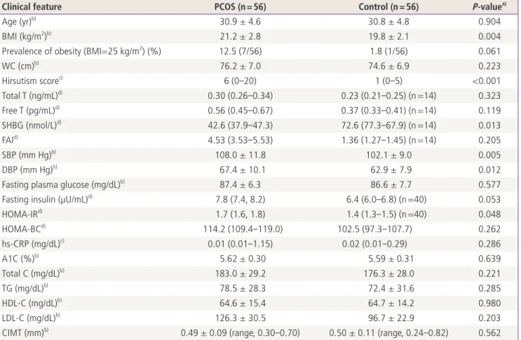
Clinical and biochemical characteristics of the subjects are shown in Table 1. Women with PCOS and controls were same
Table 1. Clinical features of the patients with PCOS and matched controls
Clinical feature PCOS (n = 56) Control (n = 56) P-value
a)Age (yr)
b)30.9 ± 4.6 30.8 ± 4.8 0.904
BMI (kg/m
2)
b)21.2 ± 2.8 19.8 ± 2.1 0.004
Prevalence of obesity (BMI=25 kg/m
2) (%) 12.5 (7/56) 1.8 (1/56) 0.061
WC (cm)
b)76.2 ± 7.0 74.6 ± 6.9 0.223
Hirsutism score
c)6 (0-20) 1 (0-5) <0.001
Total T (ng/mL)
d)0.30 (0.26-0.34) 0.23 (0.21-0.25) (n =14) 0.323
Free T (pg/mL)
d)0.56 (0.45-0.67) 0.37 (0.33-0.41) (n =14) 0.119
SHBG (nmol/L)
d)42.6 (37.9-47.3) 72.6 (77.3-67.9) (n =14) 0.013
FAI
d)4.53 (3.53-5.53) 1.36 (1.27-1.45) (n =14) 0.205
SBP (mm Hg)
b)108.0 ± 11.8 102.1 ± 9.0 0.005
DBP (mm Hg)
b)67.4 ± 10.1 62.9 ± 7.9 0.012
Fasting plasma glucose (mg/dL)
b)87.4 ± 6.3 86.6 ± 7.7 0.577
Fasting insulin (µU/mL)
d)7.8 (7.4, 8.2) 6.4 (6.0-6.8) (n =40) 0.053
HOMA-IR
d)1.7 (1.6, 1.8) 1.4 (1.3-1.5) (n =40) 0.048
HOMA-BC
d)114.2 (109.4-119.0) 102.5 (97.3-107.7) 0.262
hs-CRP (mg/dL)
c)0.01 (0.01-1.15) 0.02 (0.01-0.29) 0.286
A1C (%)
b)5.62 ± 0.30 5.59 ± 0.31 0.639
Total C (mg/dL)
b)183.0 ± 29.2 176.3 ± 28.0 0.221
TG (mg/dL)
b)78.5 ± 28.3 72.4 ± 31.6 0.285
HDL-C (mg/dL)
b)64.6 ± 15.4 64.7 ± 14.2 0.980
LDL-C (mg/dL)
b)126.3 ± 30.5 96.7 ± 22.9 0.203
CIMT (mm)
b)0.49 ± 0.09 (range, 0.30-0.70) 0.50 ± 0.11 (range, 0.24-0.82) 0.562
PCOS, polycystic ovary syndrome; BMI, body mass index; WC, waist circumference; T, testosterone; SHBG, sex- hormone binding globulin; FAI, free androgen index; SBP, systolic blood pressure; DBP, diastolic blood pressure; HOMA-IR, homeostatic model assessment for insulin resistance;
HOMA-BC, homeostatic model assessment for beta cell capacity; hs-CRP, high-sensitivity C-reactive protein; A1C, hemoglobin A1c; C, cholester- ol; TG, triglyceride; HDL-C, high-density lipoprotein cholesterol; LDL-C, low-density lipoprotein cholesterol; CIMT, carotid intima media thickness.
a)
P-values were analysed by Student’s t test or Mann-Whitney U test except the prevalence of obesity (Fisher’s exact test);
b)Data are shown
as means ± standard deviations;
c)Medians (ranges);
d)Geometric means with 95% confidence intervals.
in age (30.9 vs. 30.8, P = 0.904). Although mean BMI of the PCOS patients was significantly higher than that of the con- trols (21.2 kg/m
2vs. 19.8 kg/m
2, respectively, P = 0.004), most (87.5%) of the women with PCOS in the present study were not obese. The PCOS group had significantly higher blood pressure, HOMA-IR as well as lower SHBG levels than the controls, but there were no significant differences in serum androgen, lipid profiles, fasting glucose and A1C levels be- tween the two groups.
The CIMT ranged from 0.30 to 0.70 mm in women with PCOS and from 0.24 to 0.82 mm in controls. Despite the significant differences in some vascular risk factors between women with PCOS and controls, the mean CIMT was not dif- ferent between the two groups (0.49 ± 0.09 mm in PCOS pa- tients vs. 0.50 ± 0.11 mm in controls, respectively, P = 0.562).
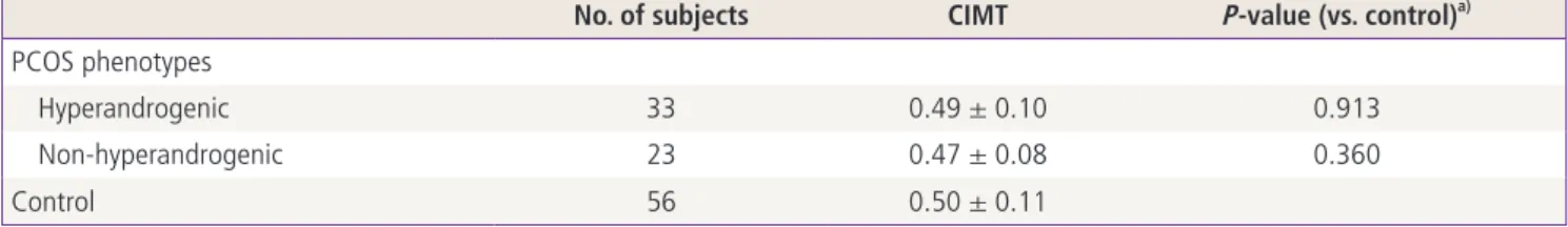
When the CIMT in the control group was compared with hyperandrogenic and non-hyperandrogenic PCOS groups, there were also no significant differences between the groups (Table 2). The univariate linear correlations between CIMT and common atherogenic factors or androgenic parameters are presented in Table 3. CIMT was not correlated with any of the above parameters in all subjects as well as the subset of PCOS patients.
Discussion
We have presented a case-control study of the CIMT, a marker of subclinical atherosclerosis, in women with PCOS and age- matched controls. Although young Korean PCOS patients Table 2. Comparison of CIMT between the control group and hyperandrogenic and non-hyperandrogenic PCOS patients
No. of subjects CIMT P-value (vs. control)
a)PCOS phenotypes
Hyperandrogenic 33 0.49 ± 0.10 0.913
Non-hyperandrogenic 23 0.47 ± 0.08 0.360
Control 56 0.50 ± 0.11
CIMT, carotid intima media thickness; PCOS, polycystic ovary syndrome.
a)