https://doi.org/10.4174/astr.2017.92.5.355 Annals of Surgical Treatment and Research
Clinical outcomes of laparoscopic radiofrequency ablation of single primary or recurrent hepatocellular carcinoma (≤3 cm)
Byung Gon Na, Jong Man Kim, Dong Kyu Oh, Kyo-Won Lee, Tae-Wook Kang1, Gyu-Seong Choi, Min Woo Lee1, Choon Hyuck David Kwon, Hyun Chul Lim1, Jae-Won Joh
Departments of Surgery, 1Radiology, Samsung Medical Center, Sungkyunkwan University School of Medicine, Seoul, Korea
INTRODUCTION
Surgical resection for hepatocellular carcinoma (HCC) has been a beneficial treatment option for long-term survival. Early HCC has been the preferred candidate for resection, because the 5-year disease-free survival rate is approximately 50%.
Although small HCC (<2 cm) has shown diverse outcomes in terms of clinicopathologic features after surgical resection, it is con sidered to have a good prognosis [1,2].
However, surgical resection is not always mandatory for HCC.
HCC typically develops in multiple locations, and can appear in combination with liver cirrhosis and other comorbidities.
Radiofrequency ablation (RFA) is a novel local treatment for preserving the hepatic parenchyma and preventing liver failure after surgical resection [3]. RFA exhibits similar outcomes to those of surgical resection in terms of overall and disease-free survival, especially for very-early-stage HCC by the Barcelona Clinic Liver Cancer classification [4,5]. In addition, compared to surgical resection, RFA is associated with a shorter hospital stay and a lower complication rate [6]. However, when the HCC is located in a subcapsular lesion, it is likely to rupture during ablation, while when it is adjacent to organs and vessels, RFA can damage these organs. Although there are many techniques to facilitate successful percutaneous RFA, the risk of organ Purpose: Percutaneous radiofrequency ablation (RFA) for hepatocellular carcinoma (HCC) has some limitations such as poor sonic window and injury to adjacent organs. The laparoscopic approach has been suggested as an alternative option.
The aim of this study was to show the safety and efficacy of laparoscopic RFA for single, small (≤3 cm), and primary or recur rent HCC that is not suitable for percutaneous RFA or surgical resection.
Methods: We reviewed the cases of 37 patients (32 men and 5 women, mean age 61 ± 8.1 years) who underwent lapar- oscopic RFA for single, small HCC (≤3 cm) that was unsuitable for percutaneous RFA or surgical resection.
Results: The technical success rate was 94.6% and 34 patients (95%) had no complications. There were no conversions to open RFA and no operative mortality. The primary effectiveness rate 1 month after the procedure was 100%. The overall recurrence rates at 3, 6, 12, and 24 months after the laparoscopic RFA were 8.1%, 14.4%, 25%, and 35.7%, respectively.
The local tumor progression rate was 4.2% at 6 months and 8.7% at 9 months.
Conclusion: Laparoscopic RFA is a safe and effective treatment for HCC cases that are unsuitable for percutaneous RFA.
[Ann Surg Treat Res 2017;92(5):355-360]
Key Words: Hepatocellular carcinoma, Laparoscopy, Catheter ablation, Local neoplasm recurrence
Reviewed January February March April May June JulyAugust September October November December
Received August 8, 2016, Revised October 20, 2016, Accepted November 28, 2016
Corresponding Author: Jong Man Kim
Department of Surgery, Samsung Medical Center, Sungkyunkwan University School of Medicine, 81 Irwon-ro, Gangnam-gu, Seoul 06351, Korea
Tel: +82-2-3410-1719, Fax: +82-2-3410-0040 E-mail: [email protected]
Copyright ⓒ 2017, the Korean Surgical Society
cc Annals of Surgical Treatment and Research is an Open Access Journal. All articles are distributed under the terms of the Creative Commons Attribution Non- Commercial License (http://creativecommons.org/licenses/by-nc/4.0/) which permits unrestricted non-commercial use, distribution, and reproduction in any medium, provided the original work is properly cited.
injury and tumor seeding still exists.
The purpose of this study was to examine the clinical outcomes of laparoscopic RFA in patients with single, small (≤3 cm), primary or recurrent HCC that was unsuitable for percutaneous RFA. We also analyzed the local tumor progression rate after the laparoscopic procedure.
METHODS
Patient selection
This was a single-center descriptive study that was based on a retrospective analysis of RFA for HCC via open or laparoscopic surgery. Between January 2011 and April 2015, a total of 91 patients with HCC underwent a RFA procedure at Samsung Medical Center. If a patient had a resectable Child-Pugh class A tumor, surgical resection was recommended as first-line treatment and local treatment as an alternative option. RFA was only performed if the patient expressed a preference for less invasive treatment. This retrospective study protocol was approved by our Institutional Review Board and waived the requirement for informed consent.
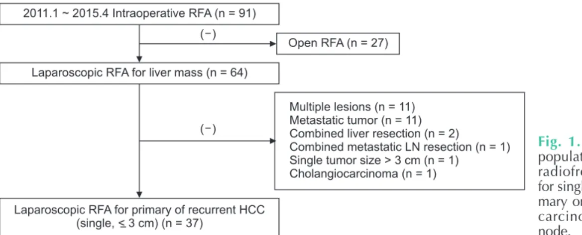
RFA was performed via open surgery in 27 patients, whereas a laparoscopic approach was used in 64. Of these, 37 patients who had a single and small (≤3 cm) HCC were included in this study. Preoperative planning ultrasonography (US) established that they were not eligible for percutaneous RFA for the following reasons: (1) poor sonic window to delineate the tumor because of the depth and location of the tumor (n = 21, 56.8%);
(2) adjacent organs close to the tumor, such as the diaphragm, heart, gallbladder, stomach, and colon (n = 7, 18.9%); or (3) adjacent major vessels, such as inferior vena cava, portal vein, hepatic vein, and collaterals (n = 9, 24.8%). Cases with multiple HCC lesions were excluded because of the difficulty of assessing the technical success of laparoscopic RFA for individual tumors (Fig. 1).
Laparoscopic RFA procedure
All patients underwent scheduled surgery under general anesthesia. A 12-mm laparoscopy trocar was placed in a subumbilical incision and two 5-mm trocars were inserted in the subcostal area bilaterally to handle the liver. After the dissection of ligaments around the liver and adhesive tissue, another 12-mm trocar was placed in the upper middle quadrant of the abdomen for the US probe (Aloka Inc., Tokyo, Japan). Once the target tumor was considered to be exposed, intraoperative US was performed so the lesion could be confirmed by the surgeon and radiologist. The tumor size and the distance between the abdominal wall and index tumor were then measured to determine the puncture site. A radiologist inserted the electrode percutaneously to the correct position in the index tumor. RFA was conducted using internally cooled electrode systems with generators (Cool-tip RF System, Covidien, Mansfield, MA, USA; or VIVA RFA System, STARmed, Goyang, Korea) by 1 of 2 radiologists with sufficient experience in RFA. The 15-G or 17-G electrode and the single- (Proteus RF Electrode, STARmed) or multiple-separable-electrode (Octopus electrode, STARmed) were deployed by a radiologist. It was difficult to achieve a sufficient ablation margin when there was a poor sonic window due to echogenic bubbles after initial ablation with a single electrode. The multiple separable electrodes were applied using overlapping ablation techniques to overcome such shortcomings. Ablative treatment consisted of one or more cycles on the tumor lesion to accomplish complete ablation. The RFA energy was transmitted for 6–12 min per ablation. On completion of the procedure, we cauterized the tract during electrode removal to prevent postoperative bleeding.
Follow-up
For evaluation of postoperative outcomes and complications, all patients underwent CT after the procedure on the operative day. The ablation margin secured was at least 5 mm. If a residual lesion was detected on CT, additional RFA was attempted via a
2011.1 ~ 2015.4 Intraoperative RFA (n = 91)
Open RFA (n = 27)
Laparoscopic RFA for liver mass (n = 64)
Multiple lesions (n = 11) Metastatic tumor (n = 11) Combined liver resection (n = 2)
Combined metastatic LN resection (n = 1) Single tumor size > 3 cm (n = 1)
Cholangiocarcinoma (n = 1)
Laparoscopic RFA for primary of recurrent HCC (single, < 3 cm) (n = 37)
( ) ( )
Fig. 1. Flowchart of the study pop ulation. Patients undergoing radio frequency ablation (RFA) for single, small (≤3 cm), and pri- mary or recurrent hepa to cellular car cinoma (HCC). LN, lymph node.
laparoscopic or percutaneous approach on the following day. For therapeutic efficacy, technical success was defined as complete ablation of the index tumor on follow-up imaging after the procedure. Once complete ablation was achieved, the primary effectiveness rate was evaluated 1 month after initial discharge, and tumor markers such as serum α-FP and protein induced by vitamin K absence/antagonism II were also measured. The follow-up policy was described in a previous study [2,7]. Local tumor progression was defined as the appearance of enhanced tumor around the ablation zone on follow-up CT. New lesions, as represented by intrahepatic metastases, were defined as newly developed tumors away from the ablation zone on CT.
Statistical analysis
Descriptive analysis was performed for all variables.
Categorical variables are shown as frequencies/percentages.
Continuous variables are expressed as mean ± standard devia-
tion for normally distributed data and median (inter quartile range) for nonnormally distributed data. Overall sur vival curves and cumulative recurrence curves were analyzed by the Kaplan- Meier method. A P-value <0.05 was considered statistically significant. All analyses were performed using IBM SPSS ver.
18.0 (IBM Co., Armonk, NY, USA).
RESULTS
Baseline characteristics
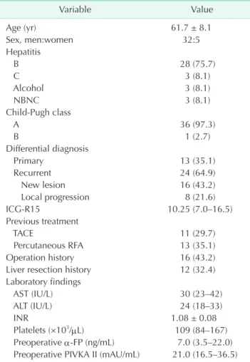
The patients included 32 men and 5 women, with a mean age of 61 ± 8.1 years (range, 44–84 years). Twenty- eight patients had cirrhosis with hepatitis B, 3 patients had cirrhosis due to hepatitis C, 3 patients had alcoholic cirrhosis, and the remaining 3 had non-B and non-C cirrhosis. Most of the patients (97.3%) were classified as Child-Pugh class A.
Thirteen patients had a solitary primary HCC lesion, whereas 24 had recurrent lesions. In the recurrent HCC cases, 16 le- sions were new and 8 were local progression recurrence during the follow-up period after previous treatment for HCC.
The previous treatments for HCC before laparoscopic RFA included transarterial chemoembolization (TACE; 29.7%) and percutaneous RFA (35.1%). Sixteen patients had undergone prior operations: appendectomy (n = 1), cholecystectomy (n = 2), stomach surgery (n = 1), or liver resection (n = 12). The patients with a history of previous liver resection had undergone right hemihepatectomy (n = 4), extended right hemihepatectomy (n = 2), extended left hemihepatectomy (n = 1), left lateral sectionectomy (n = 3), or wedge resection (n = 2) (Table 1). The mean tumor size was 15 mm (range, 10–19.5 mm). Six tumor nodules were located in the posterior sections, S6 (n = 4) and S7 (n = 2) (Table 2).
Perioperative outcomes
The mean durations of surgery and of RFA were 150 and 18 minutes, respectively. Twenty-two patients underwent a combined procedure, involving cholecystectomy (n = 7) Table 1. Demographic characteristics of 37 patients
Variable Value
Age (yr) 61.7 ± 8.1
Sex, men:women 32:5
Hepatitis
B 28 (75.7)
C 3 (8.1)
Alcohol 3 (8.1)
NBNC 3 (8.1)
Child-Pugh class
A 36 (97.3)
B 1 (2.7)
Differential diagnosis
Primary 13 (35.1)
Recurrent 24 (64.9)
New lesion 16 (43.2)
Local progression 8 (21.6)
ICG-R15 10.25 (7.0–16.5)
Previous treatment
TACE 11 (29.7)
Percutaneous RFA 13 (35.1)
Operation history 16 (43.2)
Liver resection history 12 (32.4)
Laboratory findings
AST (IU/L) 30 (23–42)
ALT (IU/L) 24 (18–33)
INR 1.08 ± 0.08
Platelets (×103/μL) 109 (84–167)
Preoperative α-FP (ng/mL) 7.0 (3.5–22.0) Preoperative PIVKA II (mAU/mL) 21.0 (16.5–36.5) Values are presented as mean ± standard deviation, number (%), or median (interquartile range).
NBNC, non-B non-C; ICG-R15, indocyanine green retention rate at 15 minutes; TACE, transarterial chemoembolization; RFA, radio- frequency ablation; INR, international normalized ratio; PIVKA II, protein induced by vitamin K absence/antagonism II.
Table 2. The size and location of hepatocellular carcinomas ablated in the study
Variable Value
Tumor size (mm) 15 (10–19.5)
Tumor location
Segment 2 3 (8.1)
Segment 3 6 (16.2)
Segment 4 11 (29.7)
Segment 5 2 (5.4)
Segment 6 4 (10.8)
Segment 7 2 (5.4)
Segment 8 9 (24.3)
Values are presented as mean (range) or number (%).
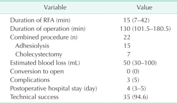
or adhesiolysis (n = 15). The mean blood loss during the operation was 50 mL (range, 30–100 mL). There were no cases of conversion to open RFA and no operative deaths. Thirty-four patients (95%) had no complications, whereas complications were detected in 3 cases by CT performed on the day of surgery.
One patient had gastric wall injury from thermal ablation, as shown by edematous change of the wall adjacent to the ablated lesion. The second patient had subcutaneous emphysema, and the third had right pneumothorax. These patients had no symptoms or signs related to the complications and improved without further treatment. The median duration of hospitalization was 4 days (range, 3–5 days).
The technical success rate was 94.6% in this study. Technical failure occurred in 2 patients, as shown by follow-up CT. One patient had a history of right hem-hepatectomy, percutaneous RFA, and TACE. During the laparoscopic RFA procedure, advanced adhesion precluded any further dissection after detection of a lesion that was mistaken for the target index
tumor. The target lesion was not ablated and the patient was subsequently scheduled for the second laparoscopic RFA. In the other patient, a daughter nodule located medially to the target index tumor was ablated. The residual tumor was found on follow-up CT, and percutaneous RFA was applied to the target tumor. The primary effectiveness rate was 100% 1 month after the procedure. None of the cases were converted to open surgery (Table 3).
Tumor recurrence rate
The median follow-up period for all patients was 13.7 months (range, 3–28 months). During follow-up, tumor recurrence was observed in 10 patients (27%). Three of the 13 patients (23%) with primary HCC and 7 of the 24 patients (29%) with recurrent HCC experienced recurrence. The overall recurrence rates 3, 6, 12, and 24 months after laparoscopic RFA were 8.1%, 14.4%, 25%
and 35.7%, respectively (Fig. 1). The 10 cases of tumor recurrence involved new intrahepatic lesions (n = 8) or local tumor progression (n = 2). Local tumor progression was observed in 7%
of patients with primary HCC and 4% of those with recurrent HCC. There were no patients with both new lesions and local tumor progression. The local tumor progression rate was 4.2%
at 6 months and 8.7% at 9 months during the follow-up period (Fig. 2).
DISCUSSION
In this study, we showed that laparoscopic RFA of single, small (≤3 cm), recurrent or primary HCC has a low rate of perioperative morbidity (5%) and no associated mortality.
The 2-year local tumor progression rate and intrahepatic distant recurrence rate were 7.3% and 30%, respectively. The postoperative hospital stay after RFA was short (3.6 ± 1.3 days) in our study compared with that reported in another study [8].
This result may be attributed to the low rate of morbidities and Table 3. The perioperative outcomes of laparoscopic radio-
fre quency ablation
Variable Value
Duration of RFA (min) 15 (7–42)
Duration of operation (min) 130 (101.5–180.5)
Combined procedure (n) 22
Adhesiolysis 15
Cholecystectomy 7
Estimated blood loss (mL) 50 (30–100)
Conversion to open 0 (0)
Complications 3 (5)
Postoperative hospital stay (day) 4 (3–5)
Technical success 35 (94.6)
Values are presented as median (interquartile range) or number (%) unless otherwise indicated.
RFA, radiofrequency ablation.
Cumulativerecurrence
0 25
Time (mo) 0
1.0
0.8
0.6
0.4
0.2
20 15
10 5
Localtumorprogression
0 25
Time (mo) 0
1.0
0.8
0.6
0.4
0.2
20 15
10 5
Fig. 2. Cumulative recurrence rates and local tumor progression rates after laparoscopic radiofrequency ablation for single, small (≤3 cm), and primary or recurrent hepatocellular carcinoma.
mild complications, such as Clavien-Dindo class I. Although 2 cases had technical failure, there was no case of incomplete ablation at the 1-month follow-up evaluation.
Herbold et al. [8] evaluated the outcomes of laparoscopic RFA for HCC in 34 consecutive patients from 2002 to 2008. They performed laparoscopic RFA instead of percutaneous RFA when the tumor was located within 1 cm of the liver capsule or in the dome of the liver. Laparoscopic RFA was also indicated when the size of the tumor to be treated was larger than 25 mm. The mean tumor size was 32.1 mm. During a mean follow-up period of 36.9 months, the local tumor recurrence rate was 61.8%.
The investigators found no perioperative mortality within 30 days and no surgical complications, except for 1 patient with a transitory decrease in liver function and ascites.
Birsen et al. [9] analyzed the 90-day morbidity and mortality of 910 patients who underwent a total of 1,207 laparoscopic RFA procedures for 2,890 malignant liver tumors between 1996 and 2012. In this large study, they found that laparoscopic RFA was associated with a complication rate of 4% (n = 50) and a mortality of 0.4% (n = 5). A total of 5 patients needed laparotomy because of postoperative bleeding from the liver (n
= 4) or mesentery (n = 1). In all cases of bleeding from the liver, the target lesions were located in the right posterior section.
Since these tumors required the longest needle insertion tract, there was a greater likelihood of injuring vessels.
Other studies related to RFA have reported a morbidity ranging between 0% and 34.7%, and mortality between 0% and 5.2%, regardless of approach [10,11].
Our results regarding morbidity and mortality are similar to those of the above-mentioned studies. However, all cases with postoperative morbidity improved spontaneously without any treatment. Furthermore, none of the patients developed postoperative bleeding or mortality. Cauterization of the needle tract during withdrawal could prevent postoperative parenchymal bleeding, especially in cases involving the right posterior section.
The procedure-related complications are mainly visceral damage, intraperitoneal bleeding, and tumor seeding. Kasugai et al. [12] showed that small vessel adhesion around the liver due to previous open surgery is a risk factor for bleeding during procedures. Yeung et al. [13] reported a case of delayed colonic perforation diagnosed on the eighth day after RFA.
They suggested percutaneous RFA for lesions, especially those within 1 cm of the colonic wall, and for patients with a history of abdominal surgery. A laparoscopic or open approach has also been suggested, because the colon can safely be mobilized away from the target lesion. Kang et al. [14] assessed factors affecting the technical failure of percutaneous RFA using artificial ascites in 113 patients with or without a history of previous HCC treatment. There was some risk of injury to the adjacent organ with conventional percutaneous RFA. Consequently, they tried
to create artificial ascites around the target lesion via catheter to avoid thermal damage from the RFA. The technical success rate for the formation of artificial ascites was 84.1%. However, this rate decreased to 55.8% in patients with previous hepatic resection. The multivariate study showed that a history of hepatic resection was a significant independent predictive factor affecting the technical failure of artificial ascites forma- tion. In our study, of the 16 patients (43.2%) with a prior history of abdominal operation, 12 (32.4%) had a history of liver resection. However, in spite of this history, 11 of these patients underwent a technically successful laparoscopic ablation, despite adhesions in the operative field. The laparoscopic RFA approach can substitute for the artificial ascites of percutaneous RFA and enable sufficient mobilization and dissection around the liver to achieve accurate ablation of the target lesion without injury to adjacent organs and vessels.
Laparoscopic US is useful for RFA. Asahina et al. [15]
suggested that mandatory use of laparoscopic US could pre- vent thermal damage to intrahepatic vessels and bile ducts.
Furthermore, effective tumor ablation without a “heat sink”
effect could be possible. In our practice, intraoperative US played a pivotal role in determining the width and depth of the tumor and the most satisfactory placement of needles around the target lesion. All procedures should begin with an appropriate arrangement of electrodes along the contour of the lesion, without direct puncture of a tumor. Efficient overlapping ablation can then be performed safely, without rupture and dissemination of the tumor. Consequently, laparoscopic US can render accurate ablation of a target lesion possible and lead to a higher rate of technical success, which reached 94.6% in our practice.
The portal and hepatic veins are main routes for the dissemination of HCC. The tumor can invade the portal branches and spread tumor emboli over the neighboring branches of the same segment [16]. Local tumor progression after RFA may be caused by insufficient ablation of the primary tumor or the presence of tumor venous dissemination in the adjacent regions of the liver. Considering tumor dissemination, surgical resection is fundamental treatment for HCC. Accord- ingly, RFA must be alternative treatment to patient not suitable for resection.
The benefits of minimally invasive surgery made laparoscopic approach applicable to RFA. Topal et al. [17] indicated that laparoscopic RFA (n = 61), compared with open RFA (n = 12), was associated with lower intraoperative blood loss, shorter duration of operation, fewer postoperative complications, and shorter postoperative hospital stay in subgroup analysis that included small tumor (≤3 cm) cases after exclusion of cases with simultaneous and/or hepatic resection. In consideration of technical aspect, open RFA may be treatment option to the cirrhotic patient with larger tumor (>3 cm).
In this study, there were some limitations. First, it was not a prospective study. There was also a selection bias related to baseline liver disease, previous treatment, and liver function.
Second, our study could not show that the comparison of laparoscopic RFA with open RFA because of small cases. This study was focused on the description of laparoscopic RFA. More data from multiple centers and with long-term follow-up after laparoscopic RFA will be needed to validate this approach.
In conclusion, as described above, our practice of laparoscopic RFA for single, small (≤3 cm), and primary or recurrent HCC was associated with a low rate of complications and mortality.
Furthermore, a similarly low rate of tumor recurrence has been shown in other studies. Laparoscopic US can facilitate safe and efficient laparoscopic RFA in spite of adhesions. Consequently, laparoscopic RFA is appropriate for certain HCC cases in which percutaneous RFA is not indicated.
CONFLICTS OF INTEREST
No potential conflict of interest relevant to this article was reported.
1. Yamashita Y, Tsuijita E, Takeishi K, Fujiwara M, Kira S, Mori M, et al. Pre- dic tors for microinvasion of small hepa- to cellular carcinoma ≤ 2 cm. Ann Surg Oncol 2012;19:2027-34.
2. Kim JM, Hyuck C, Kwon D, Joh JW, Lee JH, Paik SW, et al. Protein induced by vitamin K antagonist-II (PIVKA-II) is a reliable prognostic factor in small hepatocellular carcinoma. World J Surg 2013;37:1371-8.
3. Kang TW, Kim JM, Rhim H, Lee MW, Kim YS, Lim HK, et al. Small hepatocellular carcinoma: radiofrequency ablation ver- sus nonanatomic resection: propensity score analyses of long-term outcomes.
Radiology 2015;275:908-19.
4. Feng K, Yan J, Li X, Xia F, Ma K, Wang S, et al. A randomized controlled trial of radiofrequency ablation and surgical re- sec tion in the treatment of small hepa to- cellular carcinoma. J Hepatol 2012;57:794- 802.
5. Lee SD, Han HS, Cho JY, Yoon YS, Hwang DW, Jung K, et al. Safety and efficacy of laparoscopic radiofrequency ablation for hepatic malignancies. J Korean Surg Soc 2012;83:36-42.
6. Chen MS, Li JQ, Zheng Y, Guo RP, Liang HH, Zhang YQ, et al. A prospective ran- dom ized trial comparing percutaneous
lo cal ablative therapy and partial hepa tec- tomy for small hepatocellular carcinoma.
Ann Surg 2006;243:321-8.
7. Kim JM, Kang TW, Kwon CH, Joh JW, Ko JS, Park JB, et al. Single hepatocellular car cinoma ≤ 3 cm in left lateral segment:
liver resection or radiofrequency ablation?
World J Gastroenterol 2014;20:4059-65.
8. Herbold T, Wahba R, Bangard C, Demir M, Drebber U, Stippel DL. The laparoscopic approach for radiofrequency ablation of hepa to cellular carcinoma: indication, tech ni que and results. Langenbecks Arch Surg 2013;398:47-53.
9. Birsen O, Aliyev S, Aksoy E, Taskin HE, Akyuz M, Karabulut K, et al. A critical an aly sis of postoperative morbidity and mortality after laparoscopic radio fre- quency ablation of liver tumors. Ann Surg Oncol 2014;21:1834-40.
10. Kong WT, Zhang WW, Qiu YD, Zhou T, Qiu JL, Zhang W, et al. Major complications after radiofrequency ablation for liver tumors: analysis of 255 patients. World J Gastroenterol 2009;15:2651-6.
11. Mulier S, Mulier P, Ni Y, Miao Y, Dupas B, Marchal G, et al. Complications of radio- fre quency coagulation of liver tumours.
Br J Surg 2002;89:1206-22.
12. Kasugai H, Osaki Y, Oka H, Kudo M, Seki T;
Osaka Liver Cancer Study Group. Severe complications of radiofrequency ablation therapy for hepatocellular carcinoma: an an alysis of 3,891 ablations in 2,614 pa- tients. Oncology 2007;72 Suppl 1:72-5.
13. Yeung YP, Hui J, Yip WC. Delayed colo nic perforation after percutaneous radio fre- quency ablation of hepatocellular car ci- noma. Surg Laparosc Endosc Percutan Tech 2007;17:342-4.
14. Kang TW, Lee MW, Hye MJ, Song KD, Lim S, Rhim H, et al. Percutaneous radio- frequency ablation of hepatic tumours:
fac tors affecting technical failure of arti- fi cial ascites formation using an angio- sheath. Clin Radiol 2014;69:1249-58.
15. Asahina Y, Nakanishi H, Izumi N. Lapar- os copic radiofrequency ablation for hepa- to cellular carcinoma. Dig Endosc 2009;21:
67-72.
16. Hasegawa K, Kokudo N, Imamura H, Matsuyama Y, Aoki T, Minagawa M, et al.
Prognostic impact of anatomic resection for hepatocellular carcinoma. Ann Surg 2005;242:252-9.
17. Topal B, Hompes D, Aerts R, Fieuws S, Thijs M, Penninckx F. Morbidity and mortality of laparoscopic vs. open radio- fre quency ablation for hepa tic malig- nancies. Eur J Surg Oncol 2007;33:603-7.