Veterinary Science
http://dx.doi.org/10.4142/jvs.2013.14.4.467
Received: 7 Dec. 2012, Revised: 16 Feb. 2013, Accepted: 19 Feb. 2013
Original Article
*Corresponding author: Tel: +82-2-880-1263; Fax: +82-2-874-2738; E-mail: [email protected]
ⓒ 2013 The Korean Society of Veterinary Science.
This is an Open Access article distributed under the terms of the Creative Commons Attribution Non-Commercial License (http://creativecommons.org/licenses/by-nc/3.0) which permits unrestricted non-commercial use, distribution, and reproduction in any medium, provided the original work is properly cited.
Development of a novel enzyme-linked immunosorbent assay to detect anti-IgG against swine hepatitis E virus
Won Jung Lee, Min Kyoung Shin, Seung Bin Cha, Han Sang Yoo*
Department of Infectious Diseases, College of Veterinary Medicine, Seoul National University, Seoul 151-742, Korea
Swine hepatitis E virus (HEV) is widespread throughout pigs in both developing and industrialized countries. This virus is an important zoonotic agent and a public concern worldwide. Infected pigs are asymptomatic, so diagnosing swine HEV relies on detection of the virus or antibodies against the virus. However, several obstacles need to be overcome for effective and practical serological diagnosis. In this study, we developed an enzyme-linked immunosorbent assay (ELISA) that used a purified recombinant capsid protein of swine HEV. The potential clinical use of this assay was evaluated by comparing it with a commercial kit (Genelabs Technologies, Diagnostics, Singapore). Results of the ELISA were highly correlated with those of the commercial kit with a sensitivity of 97% and specificity of 95%. ROC (receiving operator characteristic) analysis of the ELISA data produced a value of 0.987 (95% CI, 0.977∼
0.998, p < 0.01). The cut-off value for the ELISA was also determined using negative pig sera. In summary, the HEV-specific ELISA developed in the present study appears to be both practical and economical.
Keywords: diagnosis, ELISA, swine hepatitis E virus
Introduction
Hepatitis E virus (HEV) is a significant global public health issue because this virus causes an enterically transmitted form of viral hepatitis in humans [7,18]. The mortality rate associated with hepatitis E is less than 1% in the general population, but can reach up to 28% in pregnant women [18]. HEV infection is endemic in developing countries [genotype 1 (Asia and Africa), genotype 4 (Asia), or genotype 2 (Mexico and Africa)] in areas with poor sanitation and hygiene standards, and causes medium- to large-sized waterborne epidemics with
sporadic cases of acute hepatitis [3,16]. In developed countries (genotype 3), viral infection is primarily found among travelers who have visited disease-endemic regions [4,16,25]. HEV may also be more prevalent than previously thought in industrialized countries [5].
The occurrence of zoonotic infection in non-endemic areas has been reported more frequently, and the incidence of chronic HEV infection in transplant recipients in industrialized countries is rising [19,22]. In addition, the number of animal species infected with HEV is increasing worldwide and now includes pigs, chickens, and several wild species [16]. Among these, pigs are particularly important zoonotic reservoirs because swine HEV is genetically similar to the human strain, and much information is available about HEV infection transmitted by infected pigs [8,15]. Presently, the seroprevalence of anti-HEV IgG is 39.5% and 80% in individual pigs and swine herds, respectively [14]. The prevalence of anti-HEV antibodies in the adult Korean population is about 20% with a higher prevalence among older individuals [23]. These data represent a significant increase compared to that previously reported [2,4]. Thus, the concern about zoonotic infection is increasing [18,20].
Although several diagnostic methods have been
developed and used to identify hepatitis E virus infection,
establishment of new assays with superior performance
characteristics such as enhanced efficacy and
cost-effectiveness is required [1]. Commercial kits are
available for identifying HEV based on the detection of
short ORF2 and ORF3 fragments of genotypes 1 and 2
[21]. These assays detect anti-HEV antibodies in human
sera or plasma but they might have lower sensitivity for
identifying infections with genotype 3 strains, the most
prevalent genotype among swine and humans in
industrialized countries [14]. Various reports have
indicated that commercial assays may also fail to detect
specific antibodies in sera from patients with proven HEV genotype 3 infection [6,14,27]. Furthermore, commercial ELISA kits are also very expensive for routine testing and the secondary antibody for the kits needs to be substituted with an anti-pig antibody to detect swine HEV. These considerations suggest the need for more sensitive, specific, simple, standardized, and low-cost assays to detect swine HEV. In the present study, an ELISA was developed that uses a recombinant capsid protein, which is the most immunogenic portion of the HEV. The efficacy of this ELISA was evaluated using pig sera obtained in the field.
Materials and Methods Viral RNA and cDNA synthesis
All reagents for isolating viral RNA and PCR amplification were purchased from Invitrogen (USA). Pig sera were obtained from jugular vein of pigs in Korean pig farms and isolated with centrifugation at 750 × g for 10 min. HEV viral RNA was purified from pig sera using Trizol LS reagent (Invitrogen) according to the manufacturer’s instructions. The viral RNA was eluted with a total volume of 30 μL RNase-free water and stored at −70
oC until further analysis. cDNA was synthesized using an external reverse primer specific for the capsid gene of HEV and M-MLV (moloney murine leukemia virus) Reverse Transcriptase. Briefly, 10 μL of viral RNA and 1 μL of external reverse primer (2 pmol/μL) were mixed, incubated at 70
oC for 10 min, and chilled at 4
oC for 5 min. This mixture was added to 4 μL of 5× First Strand Buffer that contained 2 μL of 0.1 M DTT (dithiothreitol), 1 μL of 10 mM dNTP mixture, 1 μL of RNase free water, 0.5 μL of RNaseOUT RNA inhibitor, and 0.5 μL of M-MLV Reverse Transcriptase. The samples were incubated at 37
oC for 1.5 h, 70
oC for 10 min, and 4
oC for 5 min before being stored at −20
oC.
PCR and cloning the capsid gene
Two-step PCR amplification was performed using cDNA made from HEV isolated from swine serum as previously described [2]. The nested forward primer sequence was slightly modified for cloning: 5´-CACCAACCCTCT CTTGCCTCT-3´. The 724 bp PCR product corresponding to the capsid gene was cloned into the Champion pET-100/
TOPO vector (Invitrogen, USA) in TOP10 competent Escherichia (E.) coli cells (Invitrogen) according to the manufacturer’s protocol. The sequence of the resulting construct was confirmed with automatic-dye-terminator DNA sequencing (ABI Prism 377 L; Applied Biosystems, USA). The cloned plasmid was used to transform BL21 Star E. coli cells (Invitrogen) for expression.
Expression and purification of the recombinant capsid protein
Bacteria containing the cloned capsid gene were grown by adding 100 mL of seed culture to 1 L of LB broth containing 100 µg/mL ampicillin and cultured at 37
oC for 1.5 h with shaking at 200 rpm. Next, 1 mM isopropyl- β-D-thiogalactoside (IPTG; Duchefa Biochemie, The Netherlands) was added and culturing was continued for 5.5 h with periodic mixing. The cells were harvested by centrifugation at 750 × g for 20 min at 4
oC, and resuspended in 40 mL of 10 mM imidazole lysis buffer (20 mM Tris, 500 mM NaCl, 8 M urea, 10 mM imidazole, and 1 mM β-mercaptoethanol, pH 8.0 in distilled water). The cells were lysed with a repeated freeze-thaw process. The lysate was purified using an His-spin Trap (GE Healthcare, UK) with the 10 mM imidazole lysis buffer and a 500 mM imidazole elution buffer (20 mM Tris, 500 mM NaCl, 8 M urea, 500 mM imidazole, and 1 mM β-mercaptoethanol, pH 8.0 in distilled water). The concentration of the purified recombinant capsid protein (6.3 mg/mL) was measured with a BCA Protein Assay Kit (Pierce, USA).
Monoclonal antibody production
The HEV capsid protein was expressed after cloning the ORF2 gene (481∼1,200 bp) into a pQE-30 UA vector (Invitrogen) as previously described [2]. The expressed protein was purified and used to immunize a BALB/c mouse by injecting the protein with Freund’s incomplete adjuvant twice at a 2-week interval. Next, the inguinal lymph node was isolated and fused with SP2/0 Ag14 myeloma cells to produce monoclonal antibody. The hybridoma clone producing a monoclonal antibody specific for HEV was selected by performing an ELISA.
3F9 cell clone (IgG2b) was selected and used for the present study.
SDS-PAGE and Western blot analysis
The purified recombinant capsid protein was separated by 12% sodium dodecyl sulfate-polyacrylamide gel electrophoresis (SDS-PAGE) and stained with Coomassie blue. Western blot analysis was performed by electrotransferring the separated proteins from the SDS-PAGE gel onto an iBlot Gel Transfer Stack nitrocellulose membrane (Invitrogen). The membrane was incubated with mouse anti-swine HEV monoclonal antibodies at a 1 : 5,000 dilution and an alkaline phosphatase (AP)-conjugated goat anti-mouse IgG (H+L; Bethyl Laboratories, USA) at a 1 : 2,000 dilution.
Antibody binding was detected using an AP conjugate
substrate kit (Bio-Rad, USA). The purified recombinant
protein was immunoblotted with pig serum that had been
determined to be HEV-positive with a commercial ELISA kit
(Diagnostics; Genelabs Technologies, Singapore) at a 1 : 1,000
dilution and an alkaline phosphatase-conjugated rabbit
anti-pig IgG (H+L; Bethyl Laboratories) at a 1 : 2,000 dilution.
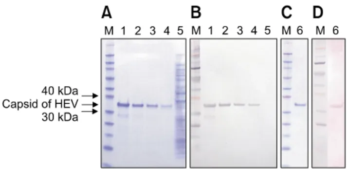
Fig. 1. Purification of the recombinant capsid protein of swine HEV. The recombinant capsid protein was analyzed by SDS-PAGE (A and C) and Western blotting with an anti-HEV monoclonal antibody (B). Western blotting with HEV-positive pig serum was also performed (D). Lane M: prestained protein molecular mass marker (kDa), lanes 1-4, and 6: induced capsid protein, lane 5: uninduced capsid protein.
The blots were developed with the AP conjugate substrate kit.
Optimized ELISA analysis of purified capsid protein using pig sera collected in the field
Optimal working dilutions of the recombinant capsid protein, pig serum, and horseradish peroxidase (HRP) conjugate were determined by checkerboard titration. The optimal concentration of the purified protein was verified using pig sera known to be positive or negative for swine HEV. Briefly, wells in the first lane of the ELISA plates were coated with 10 µg/mL of the recombinant protein in PBS and serially diluted two-fold. Aliquots of pig sera diluted two-fold (from 1 : 100 to 1 : 3,200) in antibody diluent [PBS containing 0.1% Triton X-100 (PBST) and 5% horse serum]
were dispensed into the wells of the plates. HRP-conjugated goat anti-pig IgG (Bethyl Laboratories) in antibody diluent was added at a dilution of 1 : 1,000 or 1 : 2,000.
A total of 235 field pig sera samples collected in Korea were tested using a commercial HEV ELISA kit (Diagnostics; Genelabs Technologies) and HRP-conjugated rabbit anti-pig IgG (Sigma-Aldrich, USA) according to the manufacturer’s instructions. The same sera were also analyzed with the ELISA developed in the present study using the recombinant protein. Briefly, 96-well ELISA plates were coated with 250 ng/well of the purified HEV capsid protein in PBS and incubated overnight at 4
oC. The ELISA plate was then washed three times with 250 µL of PBST and blocked with 250 µL of PBS containing 10%
horse serum at 37
oC for 1 h. After washing three times, 100 µL of pig sera diluted 1 : 100 in antibody diluent were added to each well and the plate was incubated at 37
oC for 1 h. The plates were again washed three times with PBST, and then 100 µL of HRP-conjugated goat anti-pig IgG (Bethyl Laboratories) diluted 1 : 2,000 in PBST was added. The plates were subsequently incubated for 1 h at 37
oC. After washing three times with PBST, 100 µL of an HRP substrate solution was added to each well and the plate was incubated for 15 min at room temperature. Absorbance was read at an adjusted optical density (OD) of 405 nm using an Emax Precision Microplate Reader (MDS Analytical Technologies, USA). The cut-off value was determined as previously reported using negative pig sera [10]. All samples were assayed in triplicate.
Statistical analysis
The sensitivity, specificity, efficiency, and Youden index values were calculated to evaluate the diagnostic accuracy of the novel ELISA. The following formulae were used to calculate the ELISA sensitivity, specificity, efficiency, and Youden index values: Sensitivity = no. of true positive/(no.
of true positive + no. of false negative); Specificity = no. of true negative/(no. of true negative + no. of false positive);
Efficiency (%) = [(no. of true positive + no. of true negative)/(no. of true positive + no. of false positive + no.
of true negative + no. of false negative)] × 100; Youden index = sensitivity + specificity – 1. The Youden index measures the probability of correct classification that is invariant to prevalence [6]. Values for the area under the receiving operator characteristic (ROC) curve of the developed ELISA were evaluated using the Statistical Package for the Social Sciences, 12.0 (SPSS, USA) at a 95% confidence interval (CI) [9,12].
Results
Expression and purification of the recombinant capsid protein
The identity of the cloned gene was confirmed as the swine HEV capsid gene by sequencing of the gene. The swine HEV isolate belonged to genotype 3 and the sequence (GenBank accession No. 01-18934D2, AF466681; 01-19248-3, AF466660) was the same. The capsid protein was purified via elution with an imidazole gradient, and yielded a purified capsid protein concentration of 6.3 mg/mL (maximum). SDS–PAGE analysis demonstrated that the recombinant capsid protein had an approximate molecular mass of 35 kDa, which was slightly higher compared to the software-predicted molecular weights (Fig. 1A). However, Western blot analysis using the anti-HEV monoclonal antibody revealed the shifted band to be the recombinant capsid protein with monoclonal antibody (Fig. 1B and C) and known positive pig serum (Fig. 1D).
ELISA optimization using the recombinant capsid protein
Concentrations of the recombinant capsid protein along
with dilution ratios of the field-collected pig sera and
conjugate were optimized using a checkerboard titration
assay with pig sera confirmed to be HEV-positive or
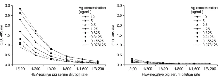
Fig. 2. Determination of the optimal antigen concentration (recombinant capsid protein) for the developed ELISA. A two-fold dilution series of the recombinant capsid protein was performed to identify the optimum concentration of the recombinant protein, and a titration experiment was performed using pig sera already known to be HEV-positive. Results of the checkerboard titration analysis demonstrated that the most optimal and reliable results were obtained when each microplate well was coated with 1.25 µg/mL of the capsid protein.
Fig. 4. Correlation between the results of the developed ELISA and a commercial kit for 235 field-collected pig sera samples.
After the 235 pig sera were tested with the HEV-specific commercial ELISA kit, the same 235 samples were subjected analysis with the developed ELISA test. The correlation ratio (R
2) between the developed ELISA and the commercial kit results was 0.717.
Fig. 3. Receiver operating characteristics (ROC) analysis of the developed ELISA. The area under the ROC curve (AUC) of the developed ELISA was 0.987 (95% CI, 0.977∼0.998; p < 0.001).
The blue line represents the test curve and the green line corresponds to the non-informative test curve. Sensitivity and specificity of the developed ELISA were 97% and 95%, respectively, when the optimal cut-off OD value was 0.6.
-negative with a commercial ELISA kit. The optimal concentration of recombinant capsid protein used to coat the ELISA plate was 1.25 µg/mL using a 1 : 100 dilution. This produced the highest positive/negative ratio for the standard checkerboard titration. The largest differences between absorbance of the positive and negative control sera samples were obtained by the addition of a 1 : 2,000 dilution of the conjugate to wells coated with the recombinant capsid protein under the optimized conditions (Fig. 2).
ELISA quantification of the purified capsid protein using field-collected pig sera
The 235 pig sera samples collected in the field were analyzed with a commercial HEV-specific ELISA kit.
Results of this analysis were compared to those from the ELISA developed in this study using the recombinant capsid protein. The ROC analysis revealed that the area under the curve for the developed ELISA was 0.987 (95%
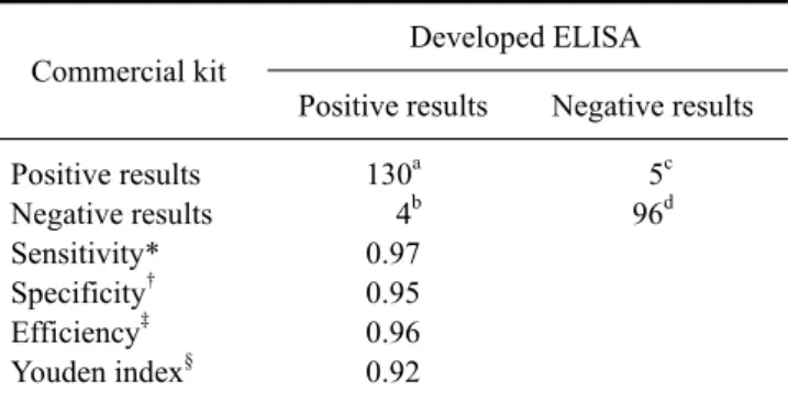
CI, 0.977∼0.998; p < 0.001; Fig. 3), and the sensitivity and specificity were 97% and 95%, respectively (Table 1).
The cut-off value (0.607) was determined using 47 negative
Table 1. Results for the ELISA assay developed for the detection of swine HEV in pig sera collected in the field
Commercial kit Developed ELISA
Positive results Negative results Positive results
Negative results Sensitivity*
Specificity
†Efficiency
‡Youden index
§130
a4
b0.97 0.95 0.96 0.92
5
c96
dOptimal cut-off values were determined using 47 HEV-negative pig sera samples. A cut-off value was identified according to a previous study [8]. HEV-positive (188) and -negative (47) pig sera were used to analyze the ELISA sensitivity and specificity. *Sensitivity = a/(a + b), †Specificity = d/(c + d), ‡Efficiency = (a + d)/(a + b + c + d),
§Youden index = Se + Sp−1.