Korean Circulation Journal
Introduction
Ischemic heart disease is complex and often involves changes in the epicardial coronary arteries and myocardial microvasculature.
Cardiac troponin is a useful tool that may help the clinician in di- agnosing myocardial infarction.1) Recent studies have shown that
Print ISSN 1738-5520 • On-line ISSN 1738-5555
Association between Cardiac Troponin Level and Coronary Flow Reserve in Patients without Coronary Artery Disease: Insight
from a Thermodilution Technique Using an Intracoronary Pressure Wire
Kyungil Park, MD
1,2, Minkwan Kim, MD
2, Young-Rak Cho, MD
1,2, Jong-Sung Park, MD
1,2, Tea-Ho Park, MD
1,2, Moo Hyun Kim, MD
1,2, and Young-Dae Kim, MD
1,21Regional Cardiocerebrovascular Center, Dong-A University Hospital, Busan,
2Division of Cardiology, Department of Internal Medicine, Dong-A University College of Medicine, Busan, Korea
Background and Objectives: Cardiac troponins are associated with increased mortality, even among patients with no coronary artery dis- ease. Elevated cardiac troponin levels are frequently observed in patients without significant coronary lesions, although the mechanism underlying this finding is unclear. The aim of our study was to evaluate the association between the levels of cardiac troponin and coronary flow reserve (CFR).
Subjects and Methods: We evaluated serum cardiac troponin-I in 19 patients (9 female; age 61.9±10.9 year-old). All patients had an ejec- tion fraction >40% and angiographically normal coronary arteries. Simultaneous measurements of fractional flow reserve (FFR), the index of microcirculatory resistance (IMR), and CFR measurements using an intracoronary temperature- and pressure-sensing guidewire under basal conditions and during maximal hyperemia were performed in three vessels: the left anterior descending artery (LAD), left circumflex artery (LCX) and right coronary artery (RCA).
Results: All patients were followed for a median of 13 months. FFR, IMR, and CFR measurements were performed successfully in all sub- jects. Mean CFRs of LAD, LCX, and RCA were 1.98±1.20, 2.75±2.11, and 4.44±2.51, respectively. Mean IMRs of LAD, LCX and RCA were 33.28±
18.78, 29.11±26.70, and 30.55±23.65, respectively. There was a poor correlation between CFR and troponin-I values in each vessel. In se- lecting the lowest value of CFR in each patient as the corresponding value, the lowest CFR was not associated with troponin-I levels (r=
-0.219, p=0.367).
Conclusion: In patients without significant coronary lesions, the correlation between CFR and troponin-I level was not significant using a thermodilution technique. Further study of a larger population with longer-term follow-up may be needed to more fully understand micro- vascular dysfunction. (Korean Circ J 2014;44(3):141-147)
KEY WORDS: Myocardiol coronary flow reserve; Troponin; Vascular resistance; Microvessels.
Received: January 8, 2014 / Revision Received: March 25, 2014 / Accepted: April 4, 2014
Correspondence: Young-Dae Kim, MD, Regional Cardiovascular Center, Dong-A University Hospital, Division of Cardiology, Department of Internal Medi- cine, Dong-A University College of Medicine, 26 Daesingongwon-ro, Seo-gu, Busan 602-714, Korea
Tel: 82-51-240-5959, Fax: 82-51-242-5852, E-mail: [email protected]
• The authors have no financial conflicts of interest.
This is an Open Access article distributed under the terms of the Creative Commons Attribution Non-Commercial License (http://creativecommons.org/licenses/
by-nc/3.0) which permits unrestricted non-commercial use, distribution, and reproduction in any medium, provided the original work is properly cited.
cardiac troponins can detect myocardial injury precisely and may predict short- and long-term mortality, even among those with no significant obstructive coronary artery disease, because high cardi- ac troponin levels have been associated with anatomical and func- tional conditions that place patients at risk of future cardiovascular events.2-4)
Functional impairment in the coronary microcirculation is thought to be a major pathway in the development of myocardial injury in normal epicardial vessels.5)6) Coronary flow reserve (CFR) provides information on both epicardial and microvascular resistance.7) In the absence of epicardial artery disease, CFR is an important index of coronary microcirculatory function. Recently, coronary thermodilu- tion-derived CFR has been developed, which permits the simulta- neous assessment of CFR and fractional flow reserve (FFR) using a single coronary pressure wire.8)9)
Elevation of cardiac troponin is frequently observed in patients without significant coronary lesions, although the mechanism under- lying this finding remains unclear.10) The aim of our study was to eval- uate the association between the levels of cardiac troponin and CFR.
Subjects and Methods
Study population
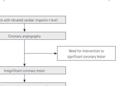
We studied 19 patients with acute chest pain and a lone elevated troponin-I level. None of the patients had significant coronary ste- noses (>30% diameter stenosis). Patients were excluded if they had a history of myocardial infarction in the epicardial vessel in the previous 12 months, a Thrombolysis in Myocardial Infarction (TIMI) flow of less than grade 3, severe renal impairment (estimated Glo- merular Filtration Rate <30 mL/min), acute inflammatory illness, chronic atrial fibrillation, left ventricular (LV) ejection fraction <40%, previous coronary artery bypass surgery, or significant valvular heart disease. Echocardiography was used to rule out concomitant hyper- trophic or dilated cardiomyopathy in all patients. Fig. 1 shows the study design.
The study protocol was approved by the institutional review board of our hospital. Written informed consent was obtained from all pa- tients.
Troponin-I assay
Troponin-I was measured using an Abbott AxSYM analyzer (Abbott Laboratories, Abbott Park, IL, USA) by mass immunoassay with a normal upper limit of 0.3 ng/mL, as specified by the manufacturer.
According to the manufacturer’s recommendations and based on our analyses, a definite elevation of troponin-I was defined as a tro- ponin-I level higher than 0.3 ng/mL.11) The 99th percentile of the URL was calculated to be 0.3 ng/mL, based on a sample of 100 healthy patients from the hospital’s catchment area. Detectable elevation was defined as between 0.10 and 0.30 ng/mL.11) Study participants were divided into two groups, based on the troponin-I level.
Catheterization protocol
Coronary angiography was performed with a femoral artery ap- proach. At the beginning of the procedure, heparin (5000 units) was routinely administered intra-arterially. Coronary lesions were as- sessed by visual estimation as well as with quantitative coronary angiography. The provocation test was conducted with ergonovine in all patients after identifying insignificant lesions during the an- giogram.
After the injection of intracoronary nitroglycerin (200 µg), simul- taneous measurements of FFR and CFR were performed for all ar- teries. A 6 Fr or 7 Fr coronary guiding catheter without side holes was used to engage the selected coronary artery. A 5 Fr sheath was placed in the right femoral vein for the injection of adenosine. A 0.014 coronary temperature and pressure-sensing guidewire (Pres- sureWire Certus, St. Jude Medical, MN, USA) was calibrated for the pressure recording, and then equalized with the aortic pressure in the guiding catheter. Next, the wire was advanced to the distal third part of the artery. For the induction of maximal hyperemia, intrave- nous adenosine was administered via the right femoral vein (140 mg/kg/min).
Physiological measurements
Fractional flow reserve, index of microcirculatory resistance (IMR), and CFR were measured under maximal hyperemia, induced by ade- nosine. FFR was calculated as the mean distal coronary pressure di- vided by the mean aortic pressure during hyperemia. For the mea- surement of CFR, basal coronary flow under basal conditions was determined by intracoronary administration of 3 mL of room-tem- perature saline, three times in succession manually (3 mL/s). Then, maximal hyperemia was induced, and three additional room tem- perature saline boluses of 3 mL were administered intracoronarily for the determination of peak coronary flow, presented as peak mean transit time. During saline injection, careful attention was given to the position of the guiding catheter and the distal sensor.
CFR was calculated automatically from the ratio of the mean transit
Patients with elevated cardiac troponin-I level
Coronary angiography
Insignificant coronary lesion
Follow-up Measure coronary flow reserve &
fractional flow reserve for 3 vessels
Need for intervention to significant coronary lesion
Fig. 1. Study design.
times during hyperemia and baseline. Adenosine was turned off for 5 minutes after physiological measurements for each artery and the following vessels were measured in sequence: left anterior de- scending artery (LAD), left circumflex artery (LCX), and right coro- nary artery (RCA). After the evaluation of FFR and CFR, IMR was cal- culated. In all vessels, a simplified method of calculating IMR was used, as follows:
IMR=Pd×Tmn
where Pd and Tmn are the mean hyperemic distal coronary pressure and hyperemic transit time, respectively.
Statistical analyses
Data are expressed as means±standard deviations for continuous variables and as numbers (%) for categorical variables, as appropri- ate. Comparisons of mean values were performed using Student’s or the paired t-test. For comparisons of discrete variables, the χ2 test was used. Correlations between CFR and FFR variables were ana- lyzed using a Spearman correlation analysis. CFR was compared with troponin-I by linear regression analysis. A p<0.05 was consid- ered to indicate statistical significance.
Results
Baseline characteristics
In total, 19 patients with TIMI 3 flow at baseline angiography were enrolled. The baseline characteristics of all patients are presented in Table 1. The mean age was 61.9±10.9 years, and nine (47.4%) of the patients were women. None of the patients had previous coronary revascularization, such as stenting or coronary bypass surgery. The overall population was classified into definite and detectable ele- vation according to troponin-I level. Clinical characteristics of two groups, based on the troponin-I concentration, are shown in Table 2.
Of the 19 patients, 15 had a definite elevation in troponin-I, with a median level of 2.34 ng/mL.
Physiological outcomes
The study was completed successfully according to the protocol in all populations. In total, 52 arteries were studied in 19 patients with- out evidence of significant coronary artery lumen obstruction. In all study arteries, FFR values were above 0.8.
Coronary flow reserve and IMR were available from 50 arteries (87.7% of all vessels). The main reasons for not obtaining adequate data were chest discomfort after adenosine infusion and small ves- sel size. The CFR of LAD, LCX, and RCA were 1.98±1.20, 2.75±2.11 and 4.44±2.51, respectively (Table 1). There was a significant differ-
ence in CFR between LAD and RCA (p=0.003). However, no signifi- cant difference was observed between the three vessels in IMR measurements. Coronary physiological data are outlined in Table 2.
There was no difference with regard to CFR, FFR, or IMR between the two groups (Table 2). Fig. 2 shows a poor correlation between CFR and troponin-I value in each vessel. Even in selecting the lowest value of CFR in each patient as the corresponding value, the lowest CFR was not associated with the troponin-I level (Fig. 3).
Additionally, the correlation between IMR and troponin-I was not significant in LAD (r=0.378, p=0.111), LCX (r=0.468, p=0.068), or RCA (r=0.044, p=0.881) (Fig. 4). However, in selecting the highest value of IMR in each patient as the corresponding value, the highest IMR was significantly higher in patients with definite elevation com- pared with detectable elevation (Table 2). Among physiological vari- ables, only the highest IMR correlated with troponin-I concentration Table 1. Baseline characteristics of study population
Variable Overall (n=19)
Age (years) 61.9±10.9
Female (%) 9 (47.4)
Diabetes mellitus (%) 6 (31.6)
Arterial hypertension (%) 9 (47.4)
Hyperlipidemia (%) 1 (5.3)
Current smoking (%) 6 (31.6)
Left ventricular ejection fraction (%) 52±11
Laboratory measurements
Hemoglobin (g/dL) 12.8±1.8
Platelet (×109/L) 250.1±116.9
Creatinine (mg/dL) 0.8±0.2
Total cholesterol (mg/dL) 180.2±51.7
LDL-C (mg/dL) 108.3±47.9
Cardiac troponin-I (ng/mL) 2.74±5.21
Physiological measurements Coronary flow reserve
Left anterior descending artery 1.98±1.20
Left circumflex artery 2.75±2.11
Right coronary artery 4.44±2.51
Fractional flow reserve
Left anterior descending artery 0.89±0.05
Left circumflex artery 0.96±0.05
Right coronary artery 0.96±0.04
Index of microvascular resistance
Left anterior descending artery 33.28±18.78
Left circumflex artery 29.11±26.70
Right coronary artery 30.55±23.65
Values are shown as means±standard deviations or n (%). LDL-C: low den- sity lipoprotein-cholesterol
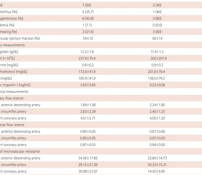
Table 2. Clinical and physiological data in patients with definite and detectable elevation of cardiac troponin-I
Variable Definite elevation (n=14) Detectable elevation (n=5) p
Age (years) 60.4±11.1 66.2±10.0 0.30
Female (%) 7 (50) 2 (40) 0.56
Diabetes mellitus (%) 5 (35.7) 1 (40) 0.48
Arterial hypertension (%) 6 (42.9) 3 (60) 0.44
Hyperlipidemia (%) 1 (7.1) 0 (0.0) 0.74
Current smoking (%) 3 (21.4) 3 (60) 0.15
Left ventricular ejection fraction (%) 54±10 50±14 0.62
Laboratory measurements
Hemoglobin (g/dL) 13.2±1.8 11.4±1.3 0.06
Platelet (×109/L) 231.9±70.4 302±201.8 0.89
Creatinine (mg/dL) 0.8±0.2 0.9±0.3 0.34
Total cholesterol (mg/dL) 172.6±41.8 201.0±76.4 0.57
LDL-C (mg/dL) 100.4±41.9 136.0±76.3 0.66
Cardiac troponin-I (ng/mL) 3.63±5.85 0.23±0.06 0.001
Physiological measurements Coronary flow reserve
Left anterior descending artery 1.89±1.08 2.24±1.60 0.96
Left circumflex artery 2.83±2.29 2.40±1.25 0.99
Right coronary artery 4.51±2.71 4.05±1.20 0.93
Fractional flow reserve
Left anterior descending artery 0.90±0.05 0.87±0.06 0.64
Left circumflex artery 0.95±0.05 0.97±0.03 0.90
Right coronary artery 0.97±0.03 0.94±0.05 0.24
Index of microvascular resistance
Left anterior descending artery 34.36±17.85 22.80±14.73 0.14
Left circumflex artery 29.15±21.39 20.33±15.31 0.23
Right coronary artery 30.08±21.07 14.50±4.95 0.43
Lowest CFR per patient 1.44±0.69 2.24±1.60 0.64
Highest IMR per patient 46.64±21.68 22.80±14.73 0.04
Values are shown as means±standard deviations or n (%). LDL-C: low density lipoprotein-cholesterol, CFR: coronary flow reserve, IMR: index of microvas- cular resistance
Fig. 2. Scatterplot of troponin-I versus CFR in LAD (A), LCX (B), and RCA (C). CFR: coronary flow reserve, LAD: left anterior descending artery, LCX: left cir- cumflex artery, RCA: right coronary artery.
25 20 15 10 5 0
25 20 15 10 5 0
25 20 15 10 5 0
CFR in LAD CFR in LCX CFR in RCA
Troponin-I Troponin-I Troponin-I
2.0 4.0 6.0 8.0 10.0 2.0 4.0 6.0 8.0 10.0 2.0 4.0 6.0 8.0 10.0 r=-0.089
p=0.718
r=-0.103 p=0.704
r=0.286 p=0.322
A B C
(Fig. 5).
Clinical outcomes
Clinical follow-up was available in all patients. At the 12-month cli- nical follow-up, all patients remained asymptomatic and free of car- diac events.
Discussion
The main finding of our investigation is that the CFR, using a th- ermodilution technique, is poorly associated with troponin-I level in patients with troponin-I elevation, but having no significant epicar- dial coronary artery stenosis. In published articles, cardiac biomarker measurement has been demonstrated be a statistically significant independent predictor of intermediate and long-term outcomes for cardiac events.2-4) Furthermore, minor elevations in troponin levels below the 99th percentile of the URL have been associated with
mortality and adverse cardiac events.11) Thus, understanding their mechanism will be important in patients with normal coronary ar- teries. Different pathogenic mechanism are responsible for tropo- nin-I elevation in patients with insignificant coronary lesions. Im- paired coronary microcirculation3)12) is one that can contribute to myocardial ischemia in patients with angiographically normal coro- nary arteries, and can be aggravated by microcirculatory stagnation, direct impairment, vasoconstriction, and thrombosis.13)
Coronary flow reserve is a commonly used index for coronary mi- crovascular function. The concept of CFR, introduced 50 years ago by Coffman and Gregg,7) provides a method for describing the ca- pacity of the coronary circulation to conduct maximal hyperemic blood flow. Previous studies have shown that the presence of an abnormal CFR is related to a worse outcome in various disorders,14)15) and CFR provides significant data on the improvement in myocardial function in patients with previous myocardial infarction.16)17) It has also been demonstrated that the severity of impairment is correlated Fig. 4. Scatterplot of troponin-I versus IMR in LAD (A), LCX (B), and RCA (C). IMR: index of microcirculatory resistance, LAD: left anterior descending artery, LCX: left circumflex artery, RCA: right coronary artery.
25 20 15 10 5 0
25 20 15 10 5 0
25 20 15 10 5 0
IMR in LAD IMR in LCX IMR in RCA
Troponin-I Troponin-I Troponin-I
20 40 60 80 100 20 40 60 80 100 20 40 60 80 100 r=0.378
p=0.111
r=0.468 p=0.068
r=0.044 p=0.881
A B C
Fig. 3. Correlation between the lowest CFR value measured by thermodilu- tion and cardiac troponin-I (CFR: horizontal axis; cardiac troponin-I: verti- cal axis). CFR: coronary flow reserve.
25
20
15
10
5
0
Lowest CFR
Troponin-I
1.0 2.0 3.0 4.0 5.0 6.0 r=-0.219 p=0.367
Fig. 5. Correlation between the highest IMR value measured by thermodi- lution and cardiac troponin-I (IMR: horizontal axis; cardiac troponin-I: ver- tical axis). IMR: index of microcirculatory resistance.
25
20
15
10
5
0
Highest IMR
Troponin-I
20 40 60 80 100 r=0.459 p=0.048
with cardiac markers.18) Thus, CFR has been considered to be a mark- er of coronary microvascular response. However the studies have some limitations. First, CFR assessment was performed only in the LAD, leading to caution in interpreting the data for general microvas- cular function, although it did not seem to have any influence.14)15)18) Second, CFR was measured by transthoracic echocardiography, in which the accuracy to detect coronary flow is influenced by image quality and angle dependence of Doppler velocities.14)15)17)
We tried to measure CFR in three vessels in each patient, because the value of LAD CFR may not be completely appropriate for iden- tifying a reduced CFR response. Also, an invasive method was used for the assessment of CFR to overcome non-invasive methodologi- cal limitations. However, our trial showed that CFR did not correlate with troponin-I concentrations. This finding differs from studies of stable patients with minor or normal LV function.19) This result may be due to hemodynamic dependence of CFR and submaximal hy- peremia with inadequate adenosine infusion.20)21) CFR is influenced by hemodynamics and the coronary microcirculation.20) Hyperemic flow, which is linearly related to coronary perfusion pressure, de- pends on total coronary resistance, whereas baseline flow is influ- enced by several factors, including myocardial oxygen demand and vasomotor tone. Additionally, the accuracy of CFR can be limited in a submaximal hyperemic condition.21)
The present study also showed that the CFR value was signifi- cantly lower in the LAD than in the RCA. Because CFR is a summed response of both the epicardial and microvascular flows, a normal CFR indicates that both the epicardial and minimally achievable mi- crovascular bed resistances are low and normal.21) When abnormal, CFR does not indicate which component is affected. Thus, CFR is the best available tool to assess the microcirculation in the absence of epicardial coronary narrowings. In our study, the mean FFR in LAD was 0.89, lower than the results of LCX and RCA. These results may suggest that most of the subjects likely had worse coronary ath- erosclerosis in the LAD. This finding also may be due to submaxi- mal hyperemia with inadequate adenosine infusion, because CFR measurements in each patient were attempted first in the LAD.
Recently, IMR was reported to be more useful in evaluating the microcirculation than CFR.22-24) The present study demonstrated that the highest IMR in each patient correlated with the troponin-I level.
This may suggest that the coronary microcirculation is impaired in patients with troponin-I elevation, and IMR can be a territory-spe- cific physiological parameter. IMR seems to be helpful in the func- tional assessment of coronary microvasculature.
This study has several limitations. The first major limitations were possible selection bias and the small sample size without objective references. Second, there was no control group with which to com- pare the CFR value of negative troponin-I patients. Invasive physio-
logical measurements were only performed in patients with tropo- nin-I elevation. Third, as the mean age of our patients was 62, most of them likely had some degree of coronary atherosclerosis. Even though we excluded patients with significant coronary artery dis- ease, the influence of an atherosclerotic component in the results could not be excluded. Fourth, clinical diagnoses in the population were heterogeneous. Finally, our study could not establish the clini- cal relevance of CFR due to the small sample size.
In conclusion, the correlation between CFR, using a thermodilu- tion technique, and troponin-I levels was not significant in patients with no significant coronary artery disease. CFR may be limited to estimating the microcirculation. Further studies of larger popula- tions with longer-term follow-up are needed to more fully under- stand microvascular dysfunction.
Acknowledgments
This study was supported by a grant from the Korean Society of Cardiology (2011), Republic of Korea.
References
1. Thygesen K, Alpert JS, Jaffe AS, et al. Third universal definition of myo- cardial infarction. J Am Coll Cardiol 2012;60:1581-98.
2. Leonardi S, Thomas L, Neely ML, et al. Comparison of the prognosis of spontaneous and percutaneous coronary intervention-related myocar- dial infarction. J Am Coll Cardiol 2012;60:2296-304.
3. Agewall S, Giannitsis E, Jernberg T, Katus H. Troponin elevation in coro- nary vs. non-coronary disease. Eur Heart J 2011;32:404-11.
4. Eggers KM, Lagerqvist B, Venge P, Wallentin L, Lindahl B. Persistent car- diac troponin I elevation in stabilized patients after an episode of acute coronary syndrome predicts long-term mortality. Circulation 2007;116:
1907-14.
5. Leung DY, Leung M. Non-invasive/invasive imaging: significance and assessment of coronary microvascular dysfunction. Heart 2011;97:
587-95.
6. Camici PG, Crea F. Coronary microvascular dysfunction. N Engl J Med 2007;356:830-40.
7. Coffman JD, Gregg DE. Reactive hyperemia characteristics of the myo- cardium. Am J Physiol 1960;199:1143-9.
8. De Bruyne B, Pijls NH, Smith L, Wievegg M, Heyndrickx GR. Coronary thermodilution to assess flow reserve: experimental validation. Circu- lation 2001;104:2003-6.
9. Barbato E, Aarnoudse W, Aengevaeren WR, et al. Validation of coronary flow reserve measurements by thermodilution in clinical practice. Eur Heart J 2004;25:219-23.
10. Eggers KM, Lind L, Ahlström H, et al. Prevalence and pathophysiological mechanisms of elevated cardiac troponin I levels in a population-based sample of elderly subjects. Eur Heart J 2008;29:2252-8.
11. van den Bos EJ, Constantinescu AA, van Domburg RT, Akin S, Jordaens LJ, Kofflard MJ. Minor elevations in troponin I are associated with mor-
tality and adverse cardiac events in patients with atrial fibrillation.
Eur Heart J 2011;32:611-7.
12. Layland JJ, Whitbourn RJ, Burns AT, et al. The index of microvascular resistance identifies patients with periprocedural myocardial infarc- tion in elective percutaneous coronary intervention. Heart 2012;98:
1492-7.
13. Merx MW, Weber C. Sepsis and the heart. Circulation 2007;116:793- 802.
14. Nakanishi K, Fukuda S, Shimada K, et al. Prognostic value of coronary flow reserve on long-term cardiovascular outcomes in patients with chronic kidney disease. Am J Cardiol 2013;112:928-32.
15. Cortigiani L, Rigo F, Gherardi S, et al. Coronary flow reserve during di- pyridamole stress echocardiography predicts mortality. JACC Cardio- vasc Imaging 2012;5:1079-85.
16. Beleslin B, Ostojic M, Djordjevic-Dikic A, et al. The value of fractional and coronary flow reserve in predicting myocardial recovery in patients with previous myocardial infarction. Eur Heart J 2008;29:2617-24.
17. Løgstrup BB, Høfsten DE, Christophersen TB, et al. Association between coronary flow reserve, left ventricular systolic function, and myocardial viability in acute myocardial infarction. Eur J Echocardiogr 2010;11:
665-70.
18. Takashio S, Yamamuro M, Izumiya Y, et al. Coronary microvascular dys- function and diastolic load correlate with cardiac troponin T release measured by a highly sensitive assay in patients with nonischemic
heart failure. J Am Coll Cardiol 2013;62:632-40.
19. Sicari R, Rigo F, Cortigiani L, Gherardi S, Galderisi M, Picano E. Additive prognostic value of coronary flow reserve in patients with chest pain syndrome and normal or near-normal coronary arteries. Am J Cardiol 2009;103:626-31.
20. Ng MK, Yeung AC, Fearon WF. Invasive assessment of the coronary microcirculation: superior reproducibility and less hemodynamic de- pendence of index of microcirculatory resistance compared with cor- onary flow reserve. Circulation 2006;113:2054-61.
21. Kern MJ, Lerman A, Bech JW, et al. Physiological assessment of coro- nary artery disease in the cardiac catheterization laboratory: a scientif- ic statement from the American Heart Association Committee on Di- agnostic and Interventional Cardiac Catheterization, Council on Clinical Cardiology. Circulation 2006;114:1321-41.
22. Lim HS, Yoon MH, Tahk SJ, et al. Usefulness of the index of microcircu- latory resistance for invasively assessing myocardial viability immedi- ately after primary angioplasty for anterior myocardial infarction. Eur Heart J 2009;30:2854-60.
23. Oh JH, Kim C, Ahn J, et al. The relationship between microcirculatory resistance and fractional flow reserve in patients with acute Myocar- dial infarction. Korean Circ J 2013;43:534-40.
24. Fearon WF, Shah M, Ng M, et al. Predictive value of the index of micro- circulatory resistance in patients with ST-segment elevation myocardi- al infarction. J Am Coll Cardiol 2008;51:560-5.