Yonsei Med J http://www.eymj.org Volume 52 Number 1 January 2011 113
The Correlation between Increased Serum Concentrations of Interleukin-6 Family Cytokines and Disease Activity
in Rheumatoid Arthritis Patients
Soo-Jin Chung, Yong-Jin Kwon, Min-Chan Park, Yong-Beom Park, and Soo-Kon Lee
Division of Rheumatology, Department of Internal Medicine, Institute for Immunology and Immunologic Disease, Brain Korea 21 Project for Medical Science, Yonsei University College of Medicine, Seoul, Korea.
Received: November 16, 2009 Revised: March 5, 2010 Accepted: March 17, 2010
Corresponding author: Dr. Soo-Kon Lee, Division of Rheumatology, Department of Internal Medicine, Institute for Immunology and Immunologic Disease, Brain Korea 21 Project for Medical Science, Yonsei University College of Medicine, 250 Seongsan-ro, Seodaemun-gu, Seoul 120-752, Korea.
Tel: 82-2-2228-1947, Fax: 82-2-393-6884 E-mail: [email protected]
∙ The authors have no financial conflicts of interest.
© Copyright:
Yonsei University College of Medicine 2011 This is an Open Access article distributed under the terms of the Creative Commons Attribution Non- Commercial License (http://creativecommons.org/
licenses/by-nc/3.0) which permits unrestricted non- commercial use, distribution, and reproduction in any medium, provided the original work is properly cited.
Purpose: This study was performed to determine whether the serum concentra- tions of interleukin (IL)-6 family cytokines are elevated in patients with rheuma- toid arthritis (RA) and to investigate the relationship between IL-6 family cytokine levels and disease activity in RA patients. Materials and Methods: We obtained serum samples from 40 patients with RA and 40 age- and sex-matched healthy controls, and we assessed the clinical parameters of disease activity, including the 28-joint disease activity score (DAS28) and C-reactive protein (CRP) levels. Se- rum samples from five patients with high disease activity (DAS28 > 5.1) were also collected at the eighth week of treatment. Serum concentrations of IL-6, IL-11, and leukemia inhibitory factor (LIF) were measured using an enzyme-linked im- munosorbent assay (ELISA). Results: Serum concentrations of IL-6 family cyto- kines, including IL-6, IL-11, and LIF, were significantly elevated in patients with RA compared to those of healthy controls. Although there was no significant rela- tionship between IL-6 family cytokine levels and DAS28, the IL-6 levels of pa- tients with RA showed a significant correlation with CRP levels. After eight weeks of medical treatment in patients with high disease activity, a decrease in DAS28 was associated with a significant decrease in the serum concentrations of IL-6 and IL-11. Conclusion: The serum concentrations of IL-6 family cytokines were sig- nificantly elevated in patients with RA, and they decreased with medical treat- ment. These findings suggest a possible role for IL-6 family cytokines in the pathogenesis of RA.
Key Words: Rheumatoid arthritis, interleukin-6 family cytokines, interleukin-6, interleukin-11, leukemia inhibitory factor
INTRODUCTION
Rheumatoid arthritis (RA) is a chronic disease that causes inflammation mainly in
the synovium and produces destruction and deformity of the joints. The etiology
of RA remains unclear, but it is known to be associated with genetic and environ-
mental factors.
1cal Center between November 2007 and January 2008, and who fulfilled the American College of Rheumatology (ACR) 1987 revised criteria for the classification of RA.
17Forty age- and sex-matched healthy adults without any evi- dence of chronic inflammatory disease served as the con- trols. This study was approved by Ethics Committee of our institute, and all study subjects provided their signed in- formed consent.
At baseline, we calculated the 28-joint disease activity score (DAS28) using the number of joints with tenderness or swelling and the erythrocyte sedimentation rate (ESR),
18and we measured the severity of pain reported by each pa- tient using the pain visual analogue scale (VAS).
19Based on the DAS28, the patients were subdivided into four groups as follows: remission (DAS28 ≤ 2.6), mild (2.6 < DAS28 ≤ 3.2), moderate (3.2 < DAS28 ≤ 5.1) and severe (5.1 <
DAS28).
20In patients whose DAS28 exceeded 5.1, a fol- low-up assessment of DAS28 and blood sampling were conducted to examine whether the serum levels of cyto- kines were correlated with disease activity.
At the time of clinical assessment for disease activity, blood samples were collected for the measurement of levels of C-reactive protein (CRP) and IL-6 family cytokines.
Measurement of IL-6, IL-11 and LIF levels
Serum samples were collected intravenously and stored in -20°C after centrifugation (2,500×g for 10 minutes at 4°C) until analysis.
The serum concentration of IL-6 was measured using a commercial enzyme-linked immunosorbent assay (ELISA) kit (R&D Systems Inc., Minneapolis, MN, USA). One hun- dred microliters each of serum sample and assay diluent were placed in each well of a 96-well plate coated with a monoclonal mouse IgG against IL-6. This mixture was in- cubated for two hours at room temperature, and each well was aspirated and washed four times with wash buffer.
Subsequently, 200 μL of conjugate solution was placed into each well and the reaction was performed for four hours at room temperature. Again, each well was washed four times with wash buffer. Following this, 200 μL of substrate solu- tion, which was prepared with equal amounts of stabilized hydrogen peroxide (H
2O
2) and tetramethylbenzidine, was added for a 20-minute reaction under dark conditions. The reaction was quenched by the addition of 50 μL stop solu- tion (2N H
2SO
4). Within 30 minutes, the optical density was measured at a wavelength of 450 nm using the Spectra Max 340 (Molecular Device Co., Sunnyvale, CA, USA).
Various proinflammatory cytokines, such as tumor ne- crotic factor (TNF)-α, interleukin (IL)-1β, interferon (IFN)-γ, and IL-6, are increased in the synovial tissue or sy- novial fluid of patients with RA.
2,3Increased levels of pro- inflammatory cytokines lead to the proliferation of synovial tissue, and thereby cause damage in the articular cartilage and bone destruction in the adjacent area.
4,5In particular, IL-6 is a cytokine with various functions. When IL-6 is ac- tivated, acute inflammatory responses such as fever or ane- mia are induced. IL-6 promotes the proliferation of B cells and thus is involved in the production of the rheumatoid factor.
6Recently, RA has been observed to be associated with high levels of IL-6 in the synovial membrane and se- rum.
7,8This led to the speculation that IL-6 plays a patho- genic role in RA. Recently, an IL-6 receptor antagonist, to- cilizumab, was developed and showed clinical efficacy in the treatment of RA.
9-11IL-6 family cytokines consist of IL-6, IL-11, leukemia inhibitory factor (LIF), oncostatin M, ciliary neurotrophic factor (CNTF), and cardiotrophin-1 (CP-1). Recent studies suggest that some of the IL-6 family cytokines such as IL- 6, IL-11, and LIF could be related with RA. IL-6 family cy- tokines such as IL-11 and LIF share glycoprotein 130 as a re- ceptor for signal transduction, which promotes the secretion of acute reactive proteins.
12Previous studies have shown that IL-6 family cytokines were detected at high concentra- tions in the synovial membrane and synovial fluid of pa- tients with RA, suggesting a role in the pathogenesis of RA.
However, there is a debate on whether serum levels of IL-6 family cytokines are positively correlated with RA disease activity.
13-16In the current study, we sought to examine whether se- rum levels of IL-6 family cytokines are increased in pa- tients with RA and whether the increased levels are signifi- cantly correlated with RA disease activity. We compared the serum concentrations of IL-6 family cytokines in pa- tients with RA and those in normal controls and then inves- tigated the correlation between serum levels of IL-6 family cytokines and the clinical parameters that indicate the dis- ease activity of RA.
MATERIALS AND METHODS
Subjects and clinical assessment
This study was conducted in 40 patients who had visited
the Division of Rheumatology at Yonsei University Medi-
Yonsei Med J http://www.eymj.org Volume 52 Number 1 January 2011 115
the RA patients were 4.2 ± 3.3 (range: 0-10) and 3.9 ± 1.3 (range: 1.29-6.50), respectively. Patients were divided into four groups according to DAS28: five in the remission group (DAS28 ≤ 2.6), nine in the mild group (2.6 < DAS28
≤ 3.2), 19 in the moderate group (3.2 < DAS28 ≤ 5.1), and seven in the severe group (5.1 < DAS28).
The mean age of the healthy controls was 53 ± 12.3 years (range: 28-76 years), and the control group was comprised of 12 males and 28 females.
A comparison of cytokine levels at baseline
The serum concentration of IL-6 was 41.76 ± 20.28 pg/mL (range: 18.0-109.1 pg/mL) in the patient group and 6.56 ± 5.33 pg/mL (range: 0-16.55 pg/mL) in the control group.
The serum concentration of IL-11 was 378.32 ± 230.31 pg/
mL (range: 45-900 pg/mL) in the patient group and 102.43
± 101.72 pg/mL (range: 0-375 pg/mL) in the control group.
The serum concentration of LIF was 57.44 ± 33.82 pg/mL (range: 1.3-120.3 pg/mL) in the patient group and 10.38 ± 7.46 pg/mL (range: 0-25.8 pg/mL) in the control group. Se- rum concentrations for all three IL-6 family cytokines were significantly elevated in the patient group compared with those of the control group (p < 0.001) (Fig. 1).
Relationship of baseline cytokine levels to disease activity At baseline, serum concentrations of IL-6 showed a signifi- cantly positive correlation with CRP levels (r = 0.45, p = 0.007) (Fig. 2A), but not with DAS28 (r = 0.28, p = 0.102), or pain VAS (r = 0.04, p = > 0.807). Serum concentrations of IL-11 and LIF were not significantly correlated with CRP levels, DAS28, or pain VAS (Table 2). The IL-6 fami- ly cytokines showed significant correlations with each other at baseline (Fig. 2B, C and D).
In subgroup analysis, serum concentrations of IL-6 were The serum concentration of IL-6 was determined based on
a standard concentration curve. The correlation coefficient (r) of the standard concentration curve was 0.990.
The IL-11 and LIF ELISA kits were purchased from R&D Systems, and serum concentrations of each cytokine were determined using similar methods. The respective correlation coefficients (r) were 0.993 and 0.999 for IL-11 and LIF.
Statistical analysis
Data analyses were performed with SAS version 9.1 (SAS Institute Inc., Cary, NC, USA). All of the descriptive vari- ables were expressed as the mean ± standard deviation. The intergroup analysis of cytokine levels was performed using the Student’s t-test and Chi-square test. The correlations among the concentrations of each cytokine, DAS28, pain VAS, and CRP level were tested using Pearson’s correla- tion test. The subgroup analyses of serum cytokine concen- trations from the four disease activity subgroups were per- formed using one-way ANOVA and Tukey’s post-hoc analyses. Eight weeks after treatment, the follow-up mea- surement of cytokine levels was performed using a Paired t-test. For all tests, a p value less than 0.05 was considered statistically significant.
RESULTS
Clinical characteristics of the study subjects
The demographic and clinical data of the subjects are shown in Table 1. The mean age of the 40 patients with RA was 54.7 ± 14.4 years (range: 19-83 years), and the patient group was comprised of 13 males and 27 females. The mean dis- ease duration from symptom onset was 63.6 ± 95.2 months (range: 2-360 months). The mean pain VAS and DAS28 of Table 1. Clinical Characteristics of the Study Subjects
Character RA patients (n = 40) Healthy controls (n = 40) p*
Age (yrs) 54.7 ± 14.4 53.0 ± 12.3 NS
Sex (male/female) 13 / 27 12 / 28 NS
Disease duration (months) 63.6 ± 95.2 - -
DAS28 3.9 ± 1.3 - -
Remission (DAS28 ≤ 2.6) (n) 5 - -
Mild (2.6 < DAS28 ≤ 3.2) (n) 9 - -
Moderate (3.2 < DAS28 ≤ 5.1) (n) 19 - -
Severe (5.1 < DAS28) (n) 7 - -
VAS 4.2 ± 3.3 - -
RA, rheumatoid arthritis; DAS28, disease activity score 28; VAS, visual analogue scale; NS, not significant.
*The p values were calculated using Student’s t-test and the Chi-square test.
Yonsei Med J http://www.eymj.org Volume 52 Number 1 January 2011 116
Fig. 1. A comparison of serum concentrations of IL-6, IL-11, and LIF between the two groups. Serum concentrations of IL-6, IL-11, and LIF were significantly higher in the patient group than in the control group. IL, interleukin; LIF, leukemia inhibitory factor.
Fig. 2. The correlations among serum concentrations of IL-6, IL-11, LIF, and CRP level. (A) Serum concentrations of IL-6 showed a signifi- cant positive correlation with CRP levels (r = 0.45, p = 0.007) in RA patients. (B) Serum concentrations of IL-6 showed significant correla- tions with serum concentrations of IL-11. (C) Serum concentrations of IL-6 showed significant correlations with serum concentrations of LIF. (D) Serum concentrations of IL-11 showed significant correlations with serum concentrations of LIF. IL, interleukin; LIF, leukemia in- hibitory factor; CRP, C-reactive protein; RA, rheumatoid arthritis.
Table 2. The Correlations among IL-6 Family Cytokines and RA Disease Activity
DAS28 Pain VAS CRP
r p* r p* r p*
IL-6 0.281 0.102 0.043 0.807 0.450 0.007
IL-11 - 0.196 0.252 - 0.023 0.896 - 0.039 0.824
LIF - 0.215 0.202 0.008 0.962 - 0.098 0.565
RA, rheumatoid arthritis; DAS28, disease activity score 28; VAS, visual analogue scale; CRP, C-reactive protein; IL, interleukin; LIF, leuke- mia inhibitory factor.
*Pearson’s correlation analysis was performed.
50
40
30
20
10
0
Patient p < 0.001
95% CI interleukin-6 (pg/mL)
Control
500
400
300
200
100
Patient p < 0.001
95% CI interleukin-11 (pg/mL)
Control
70 60
40 50
20 30
10
0
Patient p < 0.001
95% CI leukemia inhibitory factor (pg/mL)
Control
100.00
75.00
50.00
25.00
0.00 25.00
r = 0.450 p = 0.007
IL-6 (pg/mL)
50.00
CRP 75.00
100.00
75.00
50.00
25.00
0.00
0.00 40.00
r = 0.512 p < 0.001
IL-6 (pg/mL)
80.00 LIF (pg/mL)
120.00
100.00
75.00
50.00
25.00
0.00
0.00 250.00 r = 0.387 p = 0.002
IL-6 (pg/mL)
500.00
IL-11 (pg/mL) 750.00
750.00
500.00
250.00
0.00
0.00 40.00
r = 0.394 p = 0.001
IL-11 (pg/mL)
80.00 LIF (pg/mL)
120.00
A
C
B
D
Yonsei Med J http://www.eymj.org Volume 52 Number 1 January 2011 117
ly cytokines depend on the clinical course in patients whose DAS28 exceeded 5.1, the serum concentrations of IL-6 family cytokines were measured again after eight weeks of treatment with disease-modifying anti-rheumatic drugs (DMARDs). Five out of seven patients with high initial DAS were tested again and two patients were lost during the follow-up period. Five patients who had high disease activity showed decreased DAS28 of less than 5.1 after the eight-week treatment. The mean DAS28 was 6.01 ± 0.48 at baseline, and 4.12 ± 1.22 at the eight-week follow-up. The decrease in DAS28 was statistically significant based on the results of a Paired t-test (p = 0.013). Serum concentra- tions of IL-6 (p = 0.047) and IL-11 (p = 0.011) were also significantly decreased after the eight-week treatment peri- od, though serum concentrations of LIF did not significant- ly differ from baseline after treatment (p = 0.480) (Table 4).
DISCUSSION
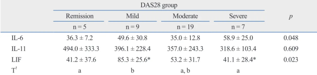
The current study showed that serum concentrations of IL- 36.3 ± 7.2 pg/mL in the remission group, 49.6 ± 30.8 pg/
mL in the mild group, 35.0 ± 12.8 pg/mL in the moderate group, and 58.9 ± 25.0 pg/mL in the severe group. The dif- ferences among the four groups were statistically signifi- cant (p = 0.048), but this was not confirmed by post-hoc analysis. Serum concentrations of IL-11 were 494.0 ± 333.3 pg/mL, 396.1 ± 228.4 pg/mL, 357.0 ± 243.3 pg/mL, and 318.6 ± 103.4 pg/mL in the corresponding group order from mildest to most severe. Serum concentrations of IL-11 were not significantly different among the four groups (p = 0.609). Serum concentrations of LIF were 41.2 ± 37.6 pg/
mL, 85.3 ± 25.6 pg/mL, 53.2 ± 31.7 pg/mL, and 41.1 ± 28.4 pg/mL, in the remission, mild, moderate, and severe groups, respectively. There was a significant difference in the serum concentrations of LIF between the mild and se- vere group (p = 0.023), with serum concentrations of LIF significantly higher in the mild group (Table 3).
Changes in concentrations of IL-6 family cytokines after treatment
To examine whether the serum concentrations of IL-6 fami-
Table 3. A Comparison of the Serum Concentrations of IL-6 Family Cytokines among the Groups Divided Based on DAS28
DAS28 group
p
Remission Mild Moderate Severe
n = 5 n = 9 n = 19 n = 7
IL-6 36.3 ± 7.2 49.6 ± 30.8 35.0 ± 12.8 58.9 ± 25.0 0.048
IL-11 494.0 ± 333.3 396.1 ± 228.4 357.0 ± 243.3 318.6 ± 103.4 0.609
LIF 41.2 ± 37.6 85.3 ± 25.6* 53.2 ± 31.7 41.1 ± 28.4* 0.023
T† a b a, b a
DAS28, disease activity score 28; IL, interleukin; LIF, leukemia inhibitory factor.
Remission group, DAS28 ≤ 2.6; mild disease activity group, 2.6 < DAS28 ≤ 3.2; moderate disease activity group, 3.2 < DAS28 ≤ 5.1;
severe disease activity group, 5.1 < DAS28.
*Tukey’s post-hoc analysis was used.
†The same letters indicate non-significant difference between groups based on Tukey’s multiple comparison test.
Table 4. The Changes in RA Disease Activity and the Concentrations of Cytokines of Five Patients with Severe Disease Activity at Baseline and after Eight-Week Treatment
Initial Follow-up p*
DAS28 6.01 ± 0.48 4.12 ± 1.22 0.013
CRP (mg/L) 41.38 ± 30.90 15.32 ± 17.31 0.233
Pain VAS 8.70 ± 1.20 4.30 ± 2.33 0.022
ESR (mm/hr) 90.60 ± 16.59 58.40 ± 35.42 0.040
IL-6 (pg/mL) 68.13 ± 24.79 16.81 ± 15.21 0.047
IL-11 (pg/mL) 307.0 ± 124.23 56.25 ± 5.39 0.011
LIF (pg/mL) 35.43 ± 34.26 50.63 ± 20.04 0.480
RA, rheumatoid arthritis; DAS28, disease activity score 28; CRP, C-reactive protein; VAS, visual analogue scale; ESR, erythrocyte sedi- mentation rate; IL, interleukin; LIF, leukemia inhibitory factor.
*Paired sample t-test was used.
ed a small number of patients and the types of drugs taken by the patients were not assessed. These factors might re- duce statistical significance. DMARDs can reduce the con- centrations of IL-6 family cytokines;
26therefore, further study is warranted to accurately compare the correlation be- tween disease activity and the concentrations of cytokines, with consideration for the effects of the drugs in a larger pa- tient population.
However, the most interesting finding in the current study was that serum concentrations of IL-6 family cytokines de- creased significantly after treatment in patients with high disease activity. We investigated the changes in cytokine levels associated with changes in disease activity before and after treatment for patients with high disease activity.
We further investigated whether IL-6 family cytokines lev- els can change and found that the concentrations of IL-6 and IL-11 significantly decreased as DAS28 decreased (Table 4). Although cross-sectional analysis did not document a significant correlation between disease activity and cyto- kine concentration, the change in the concentration of cyto- kines correlated well with the clinical course in patients with high disease activity. This finding suggests that the levels of these cytokines could reflect the disease activity.
IL-6 family cytokines have pleiotropic activities in in- flammatory response.
13IL-6 might have dual roles in in- flammation; pro-inflammatiory
27,28or anti-inflammatiory,
29and it is thought to be partly determined by concentration of soluble IL-6 receptors.
30IL-11 also might have pro-in- flammatory
31and anti-inflammatory
32activity, even though the mechanism is obscure. In this study, IL-6 and IL-11 lev- els decreased by treatment, but the level of LIF did not.
This result might be another example of dual effects of IL-6 family cytokines with unique characteristics to LIF which would require further investigation with a large number of subjects.
In summary, we found the serum concentrations of IL-6 family cytokines were significantly increased in patients with RA compared with those of normal controls. The cyto- kine levels were not significantly correlated with RA dis- ease activity in a cross-sectional analysis at baseline. But serum levels of IL-6 correlated with CRP levels, and fol- low-up measurements of cytokine levels in patients with high disease activity showed that the levels of IL-6 and IL- 11 decreased as symptoms improved. These findings sug- gest that IL-6 family cytokines might be involved in the pathogenesis of RA and that levels of IL-6 family cytokines might reflect the activity of the disease.
6, IL-11, and LIF were significantly elevated in patients with RA compared to those in healthy controls. As seen in previous reports, this finding supports the hypothesis that IL-6 family cytokines are involved in the pathogenesis of RA.
13-16IL-6 is a cytokine that causes an acute inflammatory re- sponse, and it is well-documented that IL-6 plays a crucial role in the pathogenesis of various inflammatory diseases including RA.
6,7IL-11, one of the IL-6 family cytokines, is found in high concentrations in the synovial membrane, sy- novial fluid, and serum of patients with RA, and it is also thought to be involved in the pathogenesis of RA.
21Accord- ing to studies where the immunological role of IL-11 in RA was examined, IL-11 was shown to mediate an anti-inflam- matory response by inhibiting the activity of macrophages and thereby diminishing the production of proinflammatory cytokines, such as TNF-α, IL-1β, and IFN-γ.
22However, according to Phase I and II clinical trials where recombi- nant IL-11 was administered to patients with RA, IL-11 did not affect the clinical course.
23There is still a controversy as to the role of IL-11 in the pathogenesis of RA.
It has also been reported that LIF is found in high con- centrations in cartilage and synovial fluid of patients with RA.
16IL-1 and TNF-α mediate the secretion of LIF through chondrocytes, and LIF contributes to cartilaginous destruc- tion by inducing the absorption of proteoglycan in patients with chronic arthritis.
24,25In addition to these previous observations, the significant increase in the IL-6 family cytokines observed in the cur- rent study indicates that these cytokines might play a role in inducing inflammatory responses or mediating anti-inflam- matory responses in the pathogenesis of RA.
However, the only positive correlation was found be- tween serum concentrations of IL-6 and CRP levels at baseline. We could not find any other significant relation- ships between the cytokine levels and the clinical parame- ters that reflect RA disease activity at baseline. This may be explained by the difference of involvement of the IL-6 fam- ily cytokines in the local and systemic reactions; in the synovium and serum. A previous study has shown that the concentrations of IL-6 family cytokines can be measured in both the serum and synovium of patients with RA, but these cytokines are mainly involved in the local reaction.
14In the systemic circulation, this may be the reason that in- creased serum concentrations of cytokines do not signifi- cantly correlate with disease activity.
In addition, the current study was limited; it only includ-
Yonsei Med J http://www.eymj.org Volume 52 Number 1 January 2011 119 leukin-6 type cytokines and soluble interleukin-6 receptor in pa- tients with rheumatoid arthritis. Mediators Inflamm 1998;7:347-53.
16. Waring PM, Carroll GJ, Kandiah DA, Buirski G, Metcalf D. In- creased levels of leukemia inhibitory factor in synovial fluid from patients with rheumatoid arthritis and other inflammatory arthriti- des. Arthritis Rheum 1993;36:911-5.
17. Arnett FC, Edworthy SM, Bloch DA, McShane DJ, Fries JF, Coo- per NS, et al. The American Rheumatism Association 1987 re- vised criteria for the classification of rheumatoid arthritis. Arthritis Rheum 1988;31:315-24.
18. Prevoo ML, van 't Hof MA, Kuper HH, van Leeuwen MA, van de Putte LB, van Riel PL. Modified disease activity scores that in- clude twenty-eight-joint counts. Development and validation in a prospective longitudinal study of patients with rheumatoid arthri- tis. Arthritis Rheum 1995;38:44-8.
19. Smolen JS, Aletaha D. Activity assessments in rheumatoid arthri- tis. Curr Opin Rheumatol 2008;20:306-13.
20. Zatarain E, Strand V. Monitoring disease activity of rheumatoid arthritis in clinical practice: contributions from clinical trials. Nat Clin Pract Rheumatol 2006;2:611-8.
21. Hermann JA, Hall MA, Maini RN, Feldmann M, Brennan FM.
Important immunoregulatory role of interleukin-11 in the inflam- matory process in rheumatoid arthritis. Arthritis Rheum 1998;41:
1388-97.
22. Walmsley M, Butler DM, Marinova-Mutafchieva L, Feldmann M.
An anti-inflammatory role for interleukin-11 in established murine collagen-induced arthritis. Immunology 1998;95:31-7.
23. Moreland L, Gugliotti R, King K, Chase W, Weisman M, Greco T, et al. Results of a phase-I/II randomized, masked, placebo-con- trolled trial of recombinant human interleukin-11 (rhIL-11) in the treatment of subjects with active rheumatoid arthritis. Arthritis Res 2001;3:247-52.
24. Carroll GJ, Bell MC. Leukaemia inhibitory factor stimulates pro- teoglycan resorption in porcine articular cartilage. Rheumatol Int 1993;13:5-8.
25. Campbell IK, Waring P, Novak U, Hamilton JA. Production of leukemia inhibitory factor by human articular chondrocytes and cartilage in response to interleukin-1 and tumor necrosis factor al- pha. Arthritis Rheum 1993;36:790-4.
26. Straub RH, Müller-Ladner U, Lichtinger T, Schölmerich J, Men- ninger H, Lang B. Decrease of interleukin 6 during the first 12 months is a prognostic marker for clinical outcome during 36 months treatment with disease-modifying anti-rheumatic drugs.
Br J Rheumatol 1997;36:1298-303.
27. Kopf M, Baumann H, Freer G, Feudenberg M, Lamers M, Kishi- moto T, et al. Impaired immune and acute-phase responses in in- terleukin-6-deficient mice. Nature 1994;368:339-42.
28. Sasai M, Saeki Y, Ohshima S, Nishioka K, Mima T, Tanaka T, et al.
Delayed onset and reduced severity of collagen-induced arthritis in interleukin-6-deficient mice. Arthritis Rheum 1999;42:1635-43.
29. Xing Z, Gauldie J, Cox G, Baumann H, Jordana M, Lei XF, et al.
IL-6 is an antiinflammatory cytokine required for controlling local or systemic acute inflammatory responses. J Clin Invest 1998;
101:311-20.
30. Cronstein BN. Interleukin-6--a key mediator of systemic and local symptoms in rheumatoid arthritis. Bull NYU Hosp Jt Dis 2007;65 Suppl 1:S11-5.
31. Yin TG, Schendel P, Yang YC. Enhancement of in vitro and in vivo antigen-specific antibody responses by interleukin 11. J Exp Med 1992;175:211-6.
ACKNOWLEDGEMENTS
This work was supported by National Research Foundation of Korea Grant funded by the Korean Government (2009- 0064825).
REFERENCES
1. Hochberg MC, Silman AJ, Smolen JS, Weinblatt ME, Weisman MH. Rheumatology. In: MacGregor AJ, Silman AJ, editors. Classi- fication and epidemiology. 4th ed. Spain: Mosby; 2008. p.755-62.
2. McInnes IB, Schett G. Cytokines in the pathogenesis of rheuma- toid arthritis. Nat Rev Immunol 2007;7:429-42.
3. Brennan F, Beech J. Update on cytokines in rheumatoid arthritis.
Curr Opin Rheumatol 2007;19:296-301.
4. Szekanecz Z, Koch AE. Macrophages and their products in rheu- matoid arthritis. Curr Opin Rheumatol 2007;19:289-95.
5. Huber LC, Distler O, Tarner I, Gay RE, Gay S, Pap T. Synovial fi- broblasts: key players in rheumatoid arthritis. Rheumatology 2006;45:669-75.
6. Nishimoto N, Kishimoto T. Interleukin 6: from bench to bedside.
Nat Clin Pract Rheumatol 2006;2:619-26.
7. Madhok R, Crilly A, Watson J, Capell HA. Serum interleukin 6 levels in rheumatoid arthritis: correlations with clinical and labora- tory indices of disease activity. Ann Rheum Dis 1993;52:232-4.
8. Knudsen LS, Christensen IJ, Lottenburger T, Svendsen MN, Nielsen HJ, Nielsen L, et al. Pre-analytical and biological variabil- ity in circulating interleukin 6 in healthy subjects and patients with rheumatoid arthritis. Biomarkers 2008;13:59-78.
9. Smolen JS, Beaulieu A, Rubbert-Roth A, Ramos-Remus C, Rovensky J, Alecock E, et al. Effect of interleukin-6 receptor inhi- bition with tocilizumab in patients with rheumatoid arthritis (OP- TION study): a double-blind, placebo-controlled, randomised trial.
Lancet 2008;371:987-97.
10. Genovese MC, McKay JD, Nasonov EL, Mysler EF, da Silva NA, Alecock E, et al. Interleukin-6 receptor inhibition with tocilizumab reduces disease activity in rheumatoid arthritis with inadequate re- sponse to disease-modifying antirheumatic drugs: the tocilizumab in combination with traditional disease-modifying antirheumatic drug therapy study. Arthritis Rheum 2008;58:2968-80.
11. Emery P, Keystone E, Tony HP, Cantagrel A, van Vollenhoven R, Sanchez A, et al. IL-6 receptor inhibition with tocilizumab im- proves treatment outcomes in patients with rheumatoid arthritis refractory to anti-tumour necrosis factor biologicals: results from a 24-week multicentre randomised placebo-controlled trial. Ann Rheum Dis 2008;67:1516-23.
12. Kishimoto T, Akira S, Narazaki M, Taga T. Interleukin-6 family of cytokines and gp130. Blood 1995;86:1243-54.
13. Wong PK, Campbell IK, Egan PJ, Ernst M, Wicks IP. The role of the interleukin-6 family of cytokines in inflammatory arthritis and bone turnover. Arthritis Rheum 2003;48:1177-89.
14. Okamoto H, Yamamura M, Morita Y, Harada S, Makino H, Ota Z.
The synovial expression and serum levels of interleukin-6, inter- leukin-11, leukemia inhibitory factor, and oncostatin M in rheu- matoid arthritis. Arthritis Rheum 1997;40:1096-105.
15. Robak T, Gladalska A, Stepień H, Robak E. Serum levels of inter-
regulation of proinflammatory cytokine release and nitric oxide production. J Immunol 1996;157:3627-34.
32. Trepicchio WL, Bozza M, Pedneault G, Dorner AJ. Recombinant human IL-11 attenuates the inflammatory response through down-