Usefulness of Chest Radiographs for Scoliosis Screening:
A Comparison with Thoraco-Lumbar Standing Radiographs
Chang Hyun Oh,
1,2Chan Gyu Kim,
1Myoung Seok Lee,
1Seung Hwan Yoon,
2Hyeong-Chun Park,
2and Chong Oon Park
21Seoul Regional Military Manpower Administration, Seoul;
2Department of Neurosurgery, College of Medicine, Inha University, Incheon, Korea.
Received: October 10, 2011 Revised: December 28, 2011 Accepted: January 5, 2012
Corresponding author: Dr. Seung Hwan Yoon, Department of Neurosurgery,
College of Medicine, Inha University, 27 Inhang-ro, Jung-gu, Incheon 400-711, Korea.
Tel: 82-32-890-2370, Fax: 82-32-890-2374 E-mail: [email protected]
∙ The authors have no financial conflicts of interest.
© Copyright:
Yonsei University College of Medicine 2012 This is an Open Access article distributed under the terms of the Creative Commons Attribution Non- Commercial License (http://creativecommons.org/
licenses/by-nc/3.0) which permits unrestricted non- commercial use, distribution, and reproduction in any medium, provided the original work is properly cited.
Purpose: The purposes of this study were to evaluate the usefulness and limitations of chest radiographs in scoliosis screening and to compare these results with those of thoraco-lumbar standing radiographs (TLSR). Materials and Methods: During Ko- rean conscription, 419 males were retrospectively examined using both chest radio- graphs and TLSR to confirm the scoliosis and Cobb angle at the Regional Military Manpower. We compared the types of spinal curves and Cobb angles as measured from different radiographs. Results: In the pattern of spinal curves, the overall matching rate of chest radiographs using TLSR was about 58.2% (244 of 419 cases).
Cobb angle differences between chest radiographs and TLSR with meaningful dif- ference was observed in 156 cases (37.2%); a relatively high proportion (9.5%) of Cobb angle differences more than 10 degrees was also observed. The matching rate of both spinal curve types and Cobb angle accuracy between chest radiographs and TLSR was 27.9% (117 among 419 cases). Chest radiographs for scoliosis screening were observed with 93.94% of sensitivity and 61.67% of specificity in thoracic curves; however, less than 40% of sensitivity (38.27%, 20.00%, and 25.80%) and more than 95% of specificity (97.34%, 99.69%, and 98.45%) were observed in tho- raco-lumbar, lumbar, and double major curves, respectively. Conclusion: The accu- racy of chest radiographs for scoliosis screening was low. The incidence of thoracic curve scoliosis was overestimated and lumbar curve scoliosis was easily missed by chest radiography. Scoliosis screening using chest radiography has limited values, nevertheless, it is useful method for detecting thoracic curve scoliosis.
Key Words: Scoliosis, screening, chest radiographs, thoraco-lumbar standing ra- diographs
INTRODUCTION
Screening methods for scoliosis include general physical examinations as well as radiological study including chest radiographs. Forward bending test, angle of trunk rotation and Moire’s tomography are the most used first step methods for primary evaluation,1 and the radiography image check is the most common further screening method. Screening with chest radiographs can provide information to
the location and pattern of the curve or curves, such as tho- racic curve, thoraco-lumbar curve, lumbar curve, and double major curve. The Cobb angle was the crossed angle on the perpendicular line from each end vertebrae that are the ver- tebrae at the upper and lower limits of the curve which tilted most severely toward the concavity of the curve.1,2 We con- sidered normal spinal curvature to be a Cobb angle of less than 5 degrees, to compare between two different images although many studies defined as lesser than 10 degrees.
Cobb angles in chest radiographs and TLSR were recorded by a radiologist, an orthopedic surgeon and a neurosurgeon, independently from each other. If the checked Cobb angle was differently depending on different physicians, the Cobb angle was rechecked, and the median angle was selected.
Statistical analysis
To estimate the usefulness of chest radiographs for scoliosis screening, the sensitivity and specificity of chest radio- graphs were calculated. The Cobb angle accuracy was de- fined more than five degrees as a meaningful difference be- tween chest radiographs and TLSR. A statistical analysis was performed using SAS (version 9.1.3, SAS Institute, Inc., Cary, NC, USA). Student t-test was used to compare the tendency of changed curve type of right thoracic curve and left thoracic curve in chest radiographs. Scattered plot and coefficient of correlation were also used to check the distri- bution and relation of Cobb angle between chest radiographs and TLSR. Intraclass correlation was used for interobserver variability.
physicians on the curve of the thoracic spine, and therefore, chest radiographs has been reported to be useful in scoliosis screening.1,2 However, scoliosis screening using chest ra- diograph has inherently been limited due to well-known problems such as ignorance of lumbar curve or chest radio- graph dependence on arbitral posture. Today, the exact use- fulness and limitation of screening programs by chest ra- diograph for early detection of scoliosis have so far not been examined. Therefore, we investigated the usefulness and restricted values of chest radiographs for screening pro- gram of scoliosis.
MATERIALS AND METHODS
Subject selection
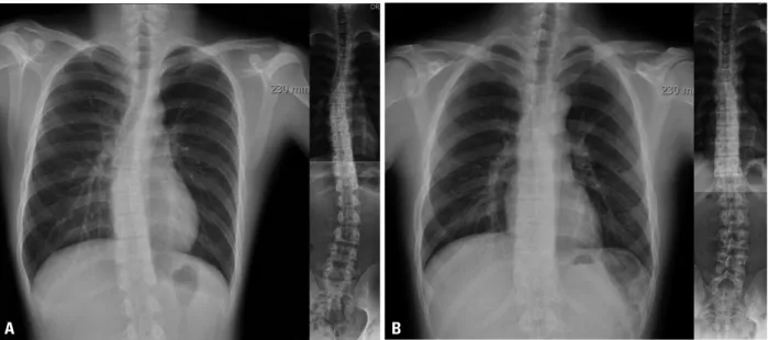
Korea engages in conscription and all men under a medical examination in preparation for this. This survey was conduct- ed at the Regional Military Manpower Administration from April 2008 to May 2010. During this period, 419 men were selected for both chest radiographs and thoraco-lumbar standing radiographs (TLSR) to check the presence of scolio- sis (Fig. 1). All examinees were 19 years old male. Their mean height, mean weight, and mean body mass index were 174.8±5.8 cm, 71.9±13.7 kg, and 23.2±3.9, respectively.
Image studies and analysis
The curvature was recorded using both chest radiograph and TLSR. The type of scoliosis was described with respect to
Fig. 1. Different scoliosis curve pattern according to chest radiographs and thoraco-lumbar standing radiographs (TLSR). (A) Shows screened case as right thoracic curve scoliosis in chest radiographs, but it was confirmed as double major curve convexity right to left by TLSR. (B) Shows a lumbar curve scoliosis case who was screened as normal spinal curvature in chest radiographs.
A B
cases, 6.4%), thoraco-lumbar curve (35 cases, 15.0%), lum- bar curve (37 cases, 15.8%), and double major curve (23 cases, 9.8%). This tendency of chang of curve type was stronger in those with right thoracic curves than those with left thoracic curve on chest radiographs (p-value=0.001).
The curve pattern match rate was 62% in the thoraco-lum- bar curve (50 cases) on chest radiograph, and the others were shown as thoracic curve (2 cases, 4%) and lumbar curve (13 cases, 26%) on TLSR. Additionally, lumbar curves (19 cas- es) on chest radiograph were presented as thoraco-lumbar curve (1 case, 5.2%) on TLSR, and curve patterns were matched in 89.5%.
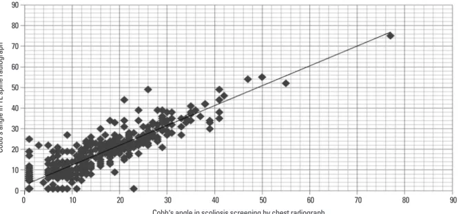
Cobb angle accuracy of chest radiographs and TLSR As for the Cobb angle accuracy, without consideration of curve type match, 263 cases (62.8%) were shown to have less than five degrees Cobb angle difference between chest radiograph and TLSR (Table 2). Conversely, the meaning- ful difference between chest radiographs and TLSR was observed in 156 cases (37.2%). A scatterplot with raw data and a corresponding fitted regression line shows the distri- bution and relation of the Cobb angle between chest radio- graph and TLSR, and the plot is provided in Fig. 2. The mean difference in Cobb angle between chest radiographs and TLSR was 4.02 (0-24) with a coefficient of correlation of 0.903 (p<0.001). However, there was relatively high pro- portion of cases (9.5%) with greater than 10 degree differ- ences in Cobb angles (Table 2).
RESULTS
Interobserver variability
Cobb angles in chest radiographs and TLSR were recorded by a radiologist, an orthopedic surgeon and a neurosurgeon independently from each other. Intraclass correlation be- tween a radiologist and an orthopedic surgeon was found to be 0.910, 0.927 between a radiologist and a neurosurgeon, and 0.925 between an orthopedic surgeon and a neurosur- geon. Cases in which the median Cobb angle was selected, because of more than 5 degrees of different Cobb angle by different physicians, was 23, but no cases exceed 10 de- grees of Cobb angle difference.
Spinal curve pattern matching of chest radiographs and TLSR
A total of 419 examinees were examined using chest radio- graphs and TLSR. The overall matching rate with the focu- son the pattern of spinal curvature of chest radiographs with TLSR was about 58.2% (244 among 419 cases) (Table 1).
Abnormal thoracic curvature on chest radiograph was ob- served in 234 cases; 186 exhibited right thoracic curvature and 48 with left thoracic curvature. The thoracic curve pat- terns observed in chest radiographs were matched only in 122 cases (52.1%) to those in TLSR. Conversely, 110 cases (47.0%) exhibited a change in their final TLSR results with respect to spinal curvature such as normal spinal curve (15
Table 1. Type of Spinal Curve Examined in Chest Radiographs and Thoraco-Lumbar Standing Radiographs (TLSR) in 419 Males
Scoliosis screening
by chest radiograph TLSR
NL RtT RtTL RtL DCRL DCLR LtT LtTL LtL Total
NL 66 3 3 8 0 0 1 8 13 102
RtT 9 92 (67) 13 0 20 0 2 20 30† 186
RtTL 1 0 6 (5) 2 0 0 1 0 3 13
RtL 0 0 1 4 (3) 0 0 0 0 0 5
DCRL 0 1 1 0 8 (5) 0 0 2 1 13
DCLR 0 1 0 0 0 0 0 0 0 1
LtT 6 0 2 3 0 3 30 (17) 0 4 48
LtTL 3 1 0 2 0 0 0 25 (15) 6 37
LtL 0 0 0 1* 0 0 0 0 13 (5) 14
Total 85 98 26 20 28 3 34 55 70 419 (117)
NL, normal spinal curves (Cobb angle of less than 5 degrees); RtT, right thoracic curve; RtTL, right thoraco-lumbar curve; RtL, right lumbar curve; DCRL, double major curve convexity right to left; DCLR, double major curve convexity left to right; LtT, left thoracic curve; LtTL, left thoraco-lumbar curve; LtL, left lumbar curve.
The number in brackets ( ) represents cases with Cobb angle differences of less than five degrees between chest radiographs and TLSR.
*A case of congenital vertebral abnormality with hemivertebrae of the 4th lumbar spine.
†A case of congenital vertebral abnormality with bilateral failure of segmentation from the 2nd to 4th lumbar spine.
curve, 64.7% (66 among 102 cases); right thoracic, 36.0%
(67 among 186 cases); right thoraco-lumbar curve, 38.5%
(5 among 13 cases); right lumbar curve, 80.0% (3 among 5 cases); double major curve convexity right to left, 38.5% (5 among 13 cases); double major curve convexity left to right, 0% (0 among 1 case); left thoracic curve, 35.4% (17 among 48 cases); left thoraco-lumbar curve, 40.5% (15 among 37 cases); and left lumbar curve, 35.7% (5 among 14 cases).
Sensitivity and specificity of chest radiographs
The sensitivity and specificity with chest radiograph and TLSR with resepct to spinal curvature are provided in Table 3.
Regarding thoracic curve, the sensitivity was high (93.94%), but the specificity was low (61.67%). On the contrary, there was a low sensitivity and a high specificity in thoraco-lum- Coincidence of spinal curve type and Cobb angle
accuracy
The coincidence of both types of spinal curves and accura- cy of Cobb angle (a Cobb angle difference of less than five degree between chest radiograph and TLSR) was 27.9%
(117 among 419 cases) (Table 1). Each accuracy according to the type of spinal curve was as follows: normal spinal
Table 2. Differences in Cobb Angles between Chest Radiographs & Thoraco-Lumbar Standing Radiographs (TLSR) Differences in Cobb angle between chest radiograph and TLSR
Total <5 % over total >5 % over total >10 % over total
NL 102 72 70.5% 30 29.5% 8 7.8%
RtT 186 121 65.1% 65 34.9% 15 8.1%
RtTL 13 8 61.5% 5 38.5% 0 0.0%
RtL 5 4 80.0% 1 20.0% 0 0.0%
DCRL 13 8 61.5% 5 38.5% 2 15.4%
DCLR 1 0 0.0% 1 100.0% 1 100.0%
LtT 48 25 52.1% 23 47.9% 5 10.4%
LtTL 37 20 54.1% 17 45.9% 8 21.6%
LtL 14 5 35.7% 9 64.3% 1 7.1%
Total 419 263 62.8% 156 37.2% 40 9.5%
RtT, right thoracic curve; RtTL, right thoraco-lumbar curve; RtL, right lumbar curve; DCRL, double major curve convexity right to left; DCLR, double major curve convexity left to right; LtT, left thoracic curve; LtTL, left thoraco-lumbar curve; LtL, left lumbar curve; NL, normal spinal curve.
Table 3. Sensitivity and Specificity of Spinal Curve as Exam- ined on Chest Radiographs according to Thoraco-Lumbar Standing Radiographs among 419 Males
Spinal curve type Sensitivity (%) Specificity (%)
Thoracic curve 93.94 61.67
Thoraco-lumbar curve 38.27 97.34
Lumbar curve 20.00 99.69
Double major curve 25.80 98.45
Fig. 2. A scatterplot with raw data and corresponding fitted regression line showing the distribution and relation of Cobb angle between chest radiographs and TLSR (r=0.903). TLSR, thoraco-lumbar standing radiographs.
0 10 20 30 40 50 60 70 80 90
Cobb’s angle in TL spine radiograph
0 10 20 30 40 50 60 70 80 90
Cobb’s angle in scoliosis screening by chest radiograph
DISCUSSION
A literature review demonstrated that the prevalence of ado- lescent scoliosis with more than a 10 degree Cobb angle var- ied greatly from 0.3% to 12.6% because of different methods used in the screening of scoliosis as well as variation in the bar, lumbar, and double major curvature. Right thoracic
curvature on chest radiograph showed normal spinal curves (4.8%), right thoraco-lumbar curves (7.0%), double major curve convexity right to left (10.8%), left thoracic curve (1.1%), left thoraco-lumbar curve (10.8%), and left lumbar curve (16.1%) on the TLSR. This tendency of change of right thoracic curve from chest radiographs to TLSR may be categorized with respect to the pattern curve of S shape, with 83.3% of thoracic curves on chest radiograph belong- ing to this pattern (Table 4). Left thoracic curvature in chest radiograph was shown in TLSR as normal spinal curvature (12.5%), double major curve convexity left to right (6.3%), left lumbar curve (8.3%), right thoraco-lumbar curves (4.2%), and right lumbar curve (6.3%). Furthermore, 75.0%
of them belonged to the inverted S shape curve (Table 5).
Congenital vertebral abnormal cases
In this study, two cases of congenital vertebral abnormali- ties were observed, and their curve pattern on chest radio- graphs changed to other pattern on TLSR (Table 1). Right lumbar curve on TLSR was misinterpreted as left lumbar curve on chest radiograph, which was a congenital verte- bral abnormality of the hemivertebra of 4th lumbar spine (Fig. 3). Similarly, a case of congenital vertebral abnormali- ty, which had bilateral failure of segmentation from the 2nd to 4th lumbar spine, was presented as a right thoracic curve on chest radiograph but was finally represented as left lum- bar curve on TLSR.
Table 4. Pattern Curves of Right Thoracic Curves on Chest Radiographs (n=177 Cases, Which Excluded 9 Cases Who Confirmed Normal by Thoraco-Lumbar Standing Radiographs from Total 186 Cases of Right Thoracic Curves on Chest Radiographs)
Pattern curve of S shape No. Pattern curve of inverted S shape No.
RtT 92 LtT 2
RtTL 13 LtTL 20
DCRL 20 DCLR -
LtL 30 RtL -
155 cases (83.3%) 22 cases (11.8%)
RtT, right thoracic curve; RtTL, right thoraco-lumbar curve; RtL, right lumbar curve; DCRL, double major curve convexity right to left; DCLR, double major curve convexity left to right; LtT, left thoracic curve; LtTL, left thoraco-lumbar curve; LtL, left lumbar curve.
Table 5. Pattern Curves of Left Thoracic Curves on Chest Radiographs (n=42 Cases, Which Excluded 6 Cases Who Were Con- firmed Normal by Thoraco-Lumbar Standing Radiographs from Total 48 Cases of Left Thoracic Curves on Chest Radiographs)
Pattern curve of inverted S shape No. Pattern curve of S shape No.
LtT 30 RtT -
LtTL - RtTL 2
DCLR 3 DCRL -
RtL 3 LtL 4
36 cases (75.0%) 6 cases (12.5%)
RtT, right thoracic curve; RtTL, right thoraco-lumbar curve; RtL, right lumbar curve; DCRL, double major curve convexity right to left; DCLR, double major curve convexity left to right; LtT, left thoracic curve; LtTL, left thoraco-lumbar curve; LtL, left lumbar curve.
Fig. 3. Congenital vertebral abnormalities with hemivertebra of the 4th lum- bar spine. The right lumbar curve on TLSR was misinterpreted as left lum- bar curve on chest radiographs due to restricted field of sight. TLSR, thora- co-lumbar standing radiographs.
Chest X-ray TL spine standing
radiographs have been very useful in detecting not only lung parenchyma disease but also scoliosis in the thoracic spine. Furthermore, the spinal alignment on chest radio- graph could be flexible depending on the position, given the fact that most curves we observed on chest radiograph lacked consistency. In this study, a total of 19 cases were examined for normal spinal curvature in TLSR, although they were observed to have scoliosis on chest radiographs.
A further limitation of chest radiographs is that the lumbar spinal curve is hidden, therefore, the observer cannot detect lumbar scoliosis. Furthermore, thoracic or thoraco-lumbar curves have been misunderstood as the S or inverted S shaped patterns (Table 4 and 5). The S curve patterns (222 cases; right thoracic curve, right thoraco-lumbar curve, dou- ble major curve convexity right to left, left lumbar curve of TLSR in Table 1) were more common than inverted S pat- terns (114 cases; left thoracic curve, left thoraco-lumbar curve, double major curve convexity left to right, right lum- bar curve of TLSR in Table 1), and it made stronger tenden- cy to change the curve type of right thoracic curves in chest radiographs than left thoracic curves (p<0.001). And, anoth- er reason for different result by chest radiographs and TLSR could be due to different position, inspiration/expira- tion difference, and posterior-anterior/posterior-anterior im- age difference. Although both chest radiographs and TLSR were taken by standing position, careful correction of radio- graphic position was carried out by TLSR. Chest radio- graphs can differently be checkable by inspiration or expi- ration, and it could contribute to the Cobb angle difference geography. Physical examinations are the most frequently
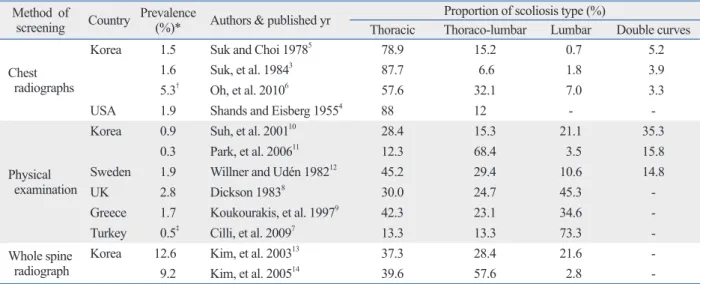
used first step methods for primary evaluation, and the radi- ography image check is used for further screening. Howev- er, this step has often been neglected.2-6 Table 6 provides a summary of the prevalence of scoliosis and the proportion of scoliosis type according to the method used for scoliosis screening. The prevalence of scoliosis was relatively higher in studies screening using chest radiographs as opposed to those using physical examination. A higher proportion of thoracic and thoraco-lumbar curvature was prominent when screening with chest radiographs as compared to oth- er types and was comparable to curve distribution observed when screening by physical examination. This tendency is not different from the current data. Our data showed a high proportion of thoracic and thoraco-lumbar curve scoliosis (a Cobb angle greater than 5 degrees) in screening by chest radiograph with thoracic in 55.8% of cases, thoraco-lumbar in 11.9%, lumbar in 4.5%, and double major curve in 3.3%.
This proportion is similar to that of other studies using chest radiograph to screen for scoliosis.3-6 However, this distribu- tion changed to thoracic 31.5%, thoraco-lumbar 19.3%, lumbar 21.5%, and double major curve 7.4% on TLSR, the finding that is more similar to the proportions in studies us- ing physical examination or whole spine radiographs.7-14
The different proportions were arbitrarily created by the restricted field of sight on chest radiograph. On chest radio- graph, the whole thoracic spine and upper lumbar spine can be included in the radiographic field, but the area below the mid lumbar area is easily excluded (Figs. 1 and 3). Chest
Table 6. Prevalence and Proportion of Scoliosis Type according to Screening Method Method of
screening Country Prevalence(%)* Authors & published yr Proportion of scoliosis type (%)
Thoracic Thoraco-lumbar Lumbar Double curves Chest
radiographs
Korea 1.5 Suk and Choi 19785 78.9 15.2 0.7 5.2
1.6 Suk, et al. 19843 87.7 6.6 1.8 3.9
5.3† Oh, et al. 20106 57.6 32.1 7.0 3.3
USA 1.9 Shands and Eisberg 19554 88 12 - -
Physical examination
Korea 0.9 Suh, et al. 200110 28.4 15.3 21.1 35.3
0.3 Park, et al. 200611 12.3 68.4 3.5 15.8
Sweden 1.9 Willner and Udén 198212 45.2 29.4 10.6 14.8
UK 2.8 Dickson 19838 30.0 24.7 45.3 -
Greece 1.7 Koukourakis, et al. 19979 42.3 23.1 34.6 -
Turkey 0.5‡ Cilli, et al. 20097 13.3 13.3 73.3 -
Whole spine
radiograph Korea 12.6 Kim, et al. 200313 37.3 28.4 21.6 -
9.2 Kim, et al. 200514 39.6 57.6 2.8 -
*Criteria of scoliosis was a Cobb angle of more than 10 degrees.
†Only male prevalence.
‡Criteria of scoliosis was a Cobb angle of more than 5 degrees.
REFERENCES
1. Sugita K. [Epidemiological study on idiopathic scoliosis in high school students. Prevalence and relation to physique, physical strength and motor ability]. Nihon Koshu Eisei Zasshi 2000;47:
320-5.
2. Sugita K, Ihara Y, Hamazaki H, Kasamatsu T, Hashimoto T. [Ap- plication of tuberculosis medical examination radiographs to sco- liosis screening in high schools]. Nihon Koshu Eisei Zasshi 1997;44:167-73.
3. Suk SI, Jo JH, Choi JS, Cho HO, Lee YG. The prevalence of sco- liosis junior and senior high school students, Pusan, Korea. J Ko- rean Orthop Assoc 1984;19:431-5.
4. Shands AR Jr, Eisberg HB. The incidence of scoliosis in the state of Delaware; a study of 50,000 minifilms of the chest made during a survey for tuberculosis. J Bone Joint Surg Am 1955;37-A:1243-9.
5. Suk SI, Choi IH. The incidence of scoliosis in Korea, Part II: the incidence of scoliosis in the middle and high school male students.
J Korean Orthop Assoc 1978;13:317-23.
6. Oh CH, Jahng YJ, Lee JH, Yoon SH, Park HC, Park CO. Scoliosis in a nineteen years old male: prevalence study. Korean J Spine 2010;7:161-6.
7. Cilli K, Tezeren G, Taş T, Bulut O, Oztürk H, Oztemur Z, et al.
[School screening for scoliosis in Sivas, Turkey]. Acta Orthop Traumatol Turc 2009;43:426-30.
8. Dickson RA. Scoliosis in the community. Br Med J (Clin Res Ed) 1983;286:615-8.
9. Koukourakis I, Giaourakis G, Kouvidis G, Kivernitakis E, Blazos J, Koukourakis M. Screening school children for scoliosis on the island of Crete. J Spinal Disord 1997;10:527-31.
10. Suh SW, Hur CY, Chae IJ, Hong JS, Yoo JC, Kang CS, et al. Idio- pathic scoliosis in Korean middle school students: prevalence study. J Korean Orthop Assoc 2001;36:33-8.
11. Park MS, Lee CS, Kim YT, Ko SH, Eo J, Cho SD. Idiopathic sco- liosis in the eleven years old: prevalence study. J Korean Orthop Assoc 2006;41:263-7.
12. Willner S, Udén A. A prospective prevalence study of scoliosis in Southern Sweden. Acta Orthop Scand 1982;53:233-7.
13. Kim MJ, Alamin TF, Lee GH, Choi KS, Park SB, Oh JH, et al.
Prevalence of adolescent scoliosis in a Korean urban middle and high school students. Spine J 2003;3:98.
14. Kim KB, Jung HR. A survey study of the juvenile idiopathic sco- liosis using radiation indirect examination. J Radiol Sci Technol 2005;28:327-32.
between chest radiographs and TLSR. Also, cinematogra- phy view could contribute to the difference as chest radio- graphs by posterior-anterior view and TLSR by anterior- posterior view.
In this study, the use of chest radiograph in scoliosis screen- ing exhibited a high sensitivity and low specificity for tho- racic curves, and very low sensitivity and high specificity for thoraco-lumbar, lumbar and double major curves (Table 3). This result indicates that chest radiographs are excellent in detecting thoracic type scoliosis, and poor in the detec- tion of thoraco-lumbar, lumbar, and double major curves.
Sugita, et al.2 suggested that tuberculosis examination ra- diographs may be useful for scoliosis screening in high schools. They examined 2068 first year high school students who had chest radiographs taken, and found 24 cases with scoliosis involved a Cobb angle of more than 10 degrees. The correlation coefficient between the Cobb angle measured in the tuberculosis examination radiographs and in the total spi- nal radiographs taken by the hospital was 0.815 (p<0.001). In a recent study, the correlation coefficient between the Cobb angle measured in chest radiograph and TLSR was 0.903 (p<0.001). However, 37.2% of cases exhibited a greater than five degree difference in Cobb angle between chest radio- graph and TLSR, and 9.5% exhibited a Cobb angle differ- ence of more than five degrees (Table 2). Moreover, the coin- cidence of both types of spinal curve and accuracy of Cobb angle, with a difference of less than five degrees between chest radiograph and TLSR, was only 27.9%.
In conclusion, the coincidence of spinal curve type was 58.2% and the consentaneity of Cobb angle was 62.8% be- tween chest radiographs and TLSR. The accuracy for using chest radiographs as scoliosis screening was only 27.9%.
Furthermore, thoracic curve scoliosis was overestimated, and lumbar curve scoliosis was easily missed on chest ra- diographs. Scoliosis screening using chest radiography has limited values, nevertheless, it is useful method for detect- ing thoracic curve scoliosis.