The Role of Sonography in Patients with Breast Cancer Presenting as an Axillary Mass
Objective: To compare sonography and mammography in terms of their diag- nostic value in breast cancer cases which initially presented as an axillary mass without a palpable mass or other clinical symptoms.
Materials and Methods: Seven patients with enlarged axillary lymph nodes who first presented with no evidence of palpable breast lesions and who under- went both mammography and sonography were enrolled in this study. In six of the seven, the presence of metastatic adenocarcinoma was confirmed preopera- tively by axillary needle aspiration biopsy; in four, subsequent sonographically- guided breast core biopsy performed after careful examination of the primary site indicated that primary breast cancer was present. In each case, the radiologic findings were evaluated by both breast sonography and mammography.
Results: Breast lesions were detected mammographically in four of seven cases (57%); in three of the four, the lesion presented as a mass, and in one as microcalcification. In three of these four detected cases, fatty or scattered fibrog- landular breast parenchyma was present; in one, the parenchyma was dense. In the three cases in which lesions were not detected, mammography revealed the presence of heterogeneously dense parenchyma. Breast sonography showed that lesions were present in six of seven cases (86%); in the remaining patient, malignant microcalcification was detected at mammography. Final pathologic examination indicated that all breast lesions except one, which was a ductal car- cinoma in situ, with microinvasion, were infiltrating ductal carcinomas whose size ranged from microscopic to greater than 3 cm. At the time of this study, all seven patients were alive and well, having been disease free for up to 61 months after surgery.
Conclusion: In women with a palpable axillary mass confirmed as metastatic adenocarcinoma, breast sonography may be a valuable adjunct to mammogra- phy.
xillary lymph node metastases without clinical or diagnostic evidence of a primary site have been seen as a manifestation of ipsilateral breast can- cer, defined as ‘occult’. Since Halstead reported three cases of occult breast cancer presenting as an axillary mass (1), the overall incidence of the condition has ranged from 0.3 to 0.8% (2). While the treatment for occult breast cancer is con- troversial, most studies have recommended mastectomy (3).
Efforts to detect occult breast lesions have frequently relied on the use of mammog- raphy. The results have been disappointing, however: primary tumors were discov- ered in more than 40% of patients whose mammograms were negative (4). Several re- cent reports have shown that otherwise occult primary breast cancers can be detected by MR imaging (5). In addition, however, the modality can assist in the selection of pa- Sun Young Park, MD
1Eun-Kyung Kim, MD
1Ki Keun Oh, MD
1Kyong Sik Lee, MD
2Byeong-Woo Park, MD
2Index terms :
Breast neoplasms, diagnosis Breast neoplasms, radiography Breast neoplasms, US
Korean J Radiol 2002; 3: 189-193 Received December 13, 2001; accepted after revision May 4, 2002.
Departments of 1Diagnostic Radiology and 2General Surgery, Yonsei University College of Medicine
Address reprint requests to :
Eun-Kyung Kim, MD, Department of Diagnostic Radiology, Research Institute of Radiological Science, Yonsei University College of Medicine, Shinchon-dong 134, Seodaemun-gu, Seoul 120-752, South Korea.
Telephone: (822) 361-5750 Fax: (822) 393-3035
e-mail: [email protected]
A
tients who are most likely to benefit from breast-conserva- tion therapy, including initial chemoradiation. MR imaging is, however, inherently expensive, and, in any case, an MR image-guided localization system is not commercially available. For proper identification and biopsy of a lesion detected by MR imaging, directed post-MRI sonographic examination of the likely location of such a lesion must, therefore, be performed (6).
Few studies have compared the usefulness of fundamen- tal modalities such as breast sonography and mammogra- phy for detecting and diagnosing occult breast lesions ac- companied by axillary lymph node metastasis. In this study, breast cancers initially presenting as an axillary mass without other clinical symptoms were reviewed according to the radiologic findings, and breast sonography and mammography were compared in terms of their useful- ness.
MATERIALS AND METHODS
Between 1996 and 2000, the presence of breast cancer was pathologically proven in 1,445 patients who under- went biopsy or curative surgery at this institute. Using a ra- diologic database, ten patients (0.7%) were found to have enlarged axillary lymph nodes at initial clinical presenta- tion, without evidence of palpable breast lesions, and in seven of these, who had undergone both mammography and sonography, the findings were jointly analyzed by two radiologists. The remaining three were excluded because their imaging studies were unavailable at the time of this investigation. All patients were female and aged between 43 and 64 (mean, 53.4) years.
Preoperatively, six of the seven patients involved under- went sonographically-guided needle aspiration biopsy of the axillary lymph nodes, and the presence of metastatic
adenocarcinoma was demonstrated. The patient in whom biopsy was not performed underwent curative surgery.
After the primary site was carefully determined by means of mammography and breast sonography, the breast nod- ules discovered were also biopsied. Pathological examina- tion of all biopsies but one revealed the presence of infil- trating ductal cancer. All seven patients underwent cura- tive resection of the primary lesions and three also re- ceived conjoined neoadjuvant chemotherapy. Surgery in- volved modified radical mastectomy in four patients, and partial mastectomy and axillary dissection in three.
The sonographic and mammographic findings were eval- uated in each case, and clinical findings such as the pathol- ogy and size of a breast lesion, the duration and size of an axillary mass, and period of survival, were also deter- mined. No patient involved in this study underwent MR imaging for the detection of occult breast lesions.
RESULTS
After initial clinical evaluation, palpable axillary masses persisted for approximately 10 days to 6 months. At both mammography and breast sonography, all clinically mani- fested axillary lesions were clearly visualized. They were either single (n=1) or conglomerated (n=6), measuring 1-3 cm in three patients and more than 3 cm in the other four.
As mentioned above, six of seven cases were confirmed at needle aspiration biopsy as metastatic adenocarcinoma.
At subsequent sonographically-guided core biopsy of five patients with symptoms other than microcalcification, infil- trating ductal carcinoma was confirmed in four and benign fibrosis in one; in that patient the presence of infiltrating ductal carcinoma was proven by later curative surgery.
Even though this had not been demonstrated at mammog- raphy, it was clearly visualized at sonography, measuring
Table 1. Mammographic and Sonographic Findings in Seven Patients with Breast Cancer Presenting as an Axillary Mass Patient No. Axilla LN Breast Lesion Mammographic Finding
Pathologic /Age Number Size (cm) (cm) Breast Pattern
Findings BIRADS Sonographic Finding
Finding
(Grade) Category
1 /45 1 2.5 1.5 1.5 1.0 3 ( ) 1 Mass (irregular, IDCa
microlobulated and hypoechoic)
2 /51 3 4 3.0 3.0 1.2 0.9 3 Mass 5 Mass (ill-defined and hypoechoic) IDCa
3 /63 3 3.0 3.0 1.4 1.0 2 Mass 4 Mass (ill-defined and hypoechoic) IDCa
4 /48 3 4 2.4 1.7 4.5 3.0 1 Microcalcification 4 ( ) IDCa
5 /43 4 5 2.5 1.4 3.0 3.0 3 ( ) 1 Mass (ill-defined and hypoechoic) IDCa
6 /60 2 3 >4 0.5 0.5 3 ( ) 1 Mass (ill-defined and hypoechoic) DCIS with
microinvasion
7 /64 2 3 3 0.9 0.8 2 Mass 5 Mass (ill-defined and hypoechoic) IDCa
Note. IDCa = infiltrating ductal cancer, DCIS = ductal carcinoma in situ, BIRADS = breast imaging-report and data system
about 1.5 1.0 cm. Final pathologic examination of all sev- en ipsilateral cancer patients who underwent surgery showed that other than in one with a ductal carcinoma in situ, with microinvasion, all lesions were infiltrating ductal carcinomas.
Three of the seven primary tumors identified were less than 1 (mean, 0.6) cm in size, two measured 1 2 (mean, 1.4) cm, and two were larger than 2 (mean, 3.8) cm.
After surgery, all patients were followed up by means of periodic clinical and radiologic examinations. At the time of this study, all seven were alive and well, showing a good prognosis after surgery and with a postoperative disease- free survival time of up to 61 months.
The mammographic and sonographic findings are sum- marized in Table 1. The mammograms optained were sus- picious in four cases (57%) [BIRADS category 4 (n=2) and 5 (n=2)] and negative in three. The suspicious mammo- grams demonstrated a mass in three patients (Fig. 1A) and microcalcification in one (Fig. 2A); fatty or scattered fi- broglandular breast parenchyma was present in three, and dense parenchyma in one. In the three non-suspicious cas- es, mammography demonstrated heterogenously dense breast parenchyma (Fig. 3A).
At sonography, enlarged axillary lymph nodes were clearly visualized (Fig. 1B), and breast lesions in the form of a malignant nodule were observed in six (86%) of the seven cases (Figs. 1C, 3B). However, one patient in whom
mammography revealed a malignant microcalcification showed a false negative finding at sonography.
DISCUSSION
In the absence of an obvious inflammatory lesion, persis- tent discrete axillary adenopathy among adult females is frequently the first sign of breast cancer. Fieuerman et al.
(7) reported 14 cases of metastatic adenocarcinoma in an axillary lymph node, ten of which had metastasized from the breast. The sites of primary origin of axillary node metastasis, other than breast lesions, are known to include melanomas and carcinomas of the lung, thyroid, gastroin- testinal tract, ovary, or other organs.
Breast cancer, while still in the intraductal stage and clin- ically obscure, appears to be able to metastasize to regional lymph nodes because of its ability to penetrate basement membrane (14). Indeed, a small primary breast tumor and a relatively larger metastatic mass exhibit differential growth potential, as shown in our cases.
It has been recommended that a carcinoma found in an axillary node should be treated as breast cancer, even if this is not clinically obvious (1, 8). In previously reported cases, the prognosis after radical mastectomy was better than if palpable breast cancer and axillary metastases re- mained (4), and at biopsy of a suspicious axillary node, a radiologist or clinician must therefore bear in mind the likelihood of breast cancer.
It is generally agreed that mammography is efficacious in detecting an ‘occult’ primary tumor. However, the report-
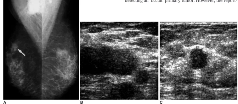
Fig. 1. A 64-year-old woman with axillary node metastasis.
A. Mammography reveals not only enlarged nodes but also scattered fibroglandular parenchyma, with a 1-cm spiculated nodule in the right upper outer quadrant (arrow).
B. At sonography, multiple enlarged axillary lymph nodes are seen, the largest of which has a diameter of approximately 3.5 cm.
C. At the corresponding site of mammographic abnormality, sonography depicts a 1cm-sized hypoechoic nodule, confirmed to be an in- filtrating ductal carcinoma.
A B C
ed frequency of detection of such tumors in this way has been disappointingly low (6). Positive mammographic find- ings appear to be of value in the detection of primary breast tumors, but where these findings are negative, the breast as the source of a primary tumor cannot be exclud- ed.
Recently, with the development of MR technology, it has been demonstrated that the sensitivity of MR imaging in the detection of breast cancer is extremely high, ranging
from 86% to 100% (9). In the future, because it can be of help in breast-sparing approaches to treatment, the use of MR imaging may more often be considered valuable.
There are, however, there are several pitfalls. First, its specificity, at 37 86%, is lower and more variable than that of other modalities (10), and a MR imaging-guided lo- calization system is not commercially available. In addi- tion, the cost of breast MR imaging is high. Morris et al. re- ported a case in which a lesion was successfully identified
Fig. 2. A 48-year-old-woman with axil- lary node metastasis.
A. Mammography reveals the presence of clustered microcalcifications (arrows), without mass, in the right inner central area. At sonography, however, neither a breast nodule nor the above-mentioned microcalcifications are seen. Surgical excision after needle localization con- firmed the presence of ductal carcinoma in situ, with microinvasion.
B. Axillary view depicts multiple hyper- dense enlarged lymph nodes.
A B
Fig. 3. A 60-year-old woman with axil- lary node metastasis.
A. Mammography demonstrates hetero- geneous fibroglandular densities, with no sign of malignancy. The right axillary lymph nodes are, however, enlarged.
B. Careful examination of this sonogram shows that the right upper central area contains a malignant microlobulated nodule, 1cm in diameter (arrow).
Sonograpahically-guided core biopsy confirmed the presence of an infiltrating ductal carcinoma.
A B