DOI 10.3349/ymj.2008.49.4.537
Purpose: Polymerase chain reaction (PCR) assay, introduced as a fast and sensitive diagnostic method, is useful in detecting Mycobacterium tuberculosis. The purpose of this study was to evaluate the usefulness of in-house PCR assay in the detection of Mycobacterium tuberculosis by comparing PCR results with conventional diagnostic techniques and Cobas Amplicor M.
tuberculosisTMkit. Materials and Methods: We retrospectively assessed the diagnostic yield of in-house PCR method employed for the amplification IS6110 sequences in 2,973 specimens. We also compared in-house PCR with Cobas Amplicor M. tuberculosisTM kit in 120 specimens collected from June to July 2006. Routine acid-fast stain (AFS) and culture assay were also performed and analyzed. Results: Of 2,973 cases, 2,832 cases (95.3%) showed consistent results between in house PCR, AFS and culture methods, whereas 141 (4.7%) displayed inconsistent results. The sensitivities, specificities, and positive and negative predictive values of each method were as follows: 77.5%, 99.7%, 95.5%, and 98.0%, respectively for PCR; 49.2%, 100%, 100%, and 95.7%, respectively, for AFS method; and 80.7%, 100%, 100%, and 98.3%, respectively, for culture assay. Consistent results between PCR and Cobas Amplicor M. tuberculosisTMkit were shown in 109 cases (90.8%). The sensitivities, specificities, and positive and negative predictive values of each method were as follows: 81.3%, 98.9%, 96.3%, and 93.5% respectively for PCR and 71.9%, 100%, 100%, and 90.7%, respectively, for Cobas AmplicorTM kit. Conclusion: In-house PCR and Cobas AmplicorTM kit show high sensitivity and specificity, and are reliable tests in the diagnosis of tuberculosis.
Key Words: Mycobacterium tuberculosis, in-house polymerase chain reaction assay, Cobas Amplicor M. tuberculosisTM kit
INTRODUCTION
Tuberculosis is a pandemic and highly con- tagious disease. Of 6.1 billion world population, 2 billion people, which are approximately one-third of the entire population, have been infected with Mycobactericum tuberculosis, and 1% of the population is being introduced as carriers every year. Of these carriers, 7 - 8 million people develop tuberculosis and 2 million die of the disease.1 In Korea, the incidence of tuberculosis was 72.1 in 100,000 in the year 2001, decreased to 67.2 in 2002, 64.0 in 2003, and then slightly increased to 65.4 in 2004.2 Death from tuberculosis in Korea was 6.7 in 100,000 in the year 2001, 7.0 in 2002, and 6.9 in 2003.3
Due to the world wide increase in the incidences of immune-related diseases such as acquired immunodeficiency syndrome (AIDS), the incidence of tuberculosis is also being increased. In that context, Korea is no exception, therefore more prompt and accurate diagnosis of tuberculosis is required.4
Diagnosis of tuberculosis consists of signs and symptoms, X-ray findings, and detection of M.
tuberculosis. Currently, bacterial culture, AFS, and PCR are employed to isolate M. tuberculosis.
Bacterial culture method is feasible only if > 100 M. tuberculosis are present in 1 mL of specimen.
Although the specificity of the culture method is close to 100% so as to be used for final diagnosis, 3 - 8 weeks are required to cultivate the bacteria.
The culture method is also not cost efficient.5-8 AFS is a relatively fast simple procedure and cost efficient. Nevertheless, it needs 5,000 - 10,000 bacteria
Comparison of In-house PCR with Conventional Techniques and Cobas Amplicor M. tuberculosis
TMKit for Detection of Mycobacterium tuberculosis
Myeong-Hee Kim, Hee-Young Yang, Jin-Tae Suh, and Hee Joo Lee
Department of Laboratory Medicine, Kyung Hee University College of Medicine, Seoul, Korea.
Received August 23, 2007 Accepted January 2, 2008
Reprint address: requests to Dr. Hee Joo Lee, Department of Laboratory Medicine, Kyung Hee University College of Medicine, 1 Hoegi-dong, Dongdaemun-gu, Seoul 130-702, Korea. Tel: 82-2- 958-8672, Fax: 82-2-958-8609, E-mail: [email protected]
present in 1 mL of sample and does not discrimi- nate M. tuberculosis against other non-tuberculosis mycobacteria, leading to low sensitivity (22 - 78%).5-8 PCR detects M. tuberculosis directly in the specimen and does not require weeks-long in- cubation time so as to be fit for early diagnosis.
The disadvantage of PCR method is that it detects not only viable M. tuberculosis but non-viable M.
tuberculosis. Thus, the method is not used for definitive diagnosis.5,9-11 Moreover, variations in diagnostic procedures and specimens lead to different rate of specificity between different labs.12-20
Given the above mentioned information, this study was designed to evaluate in-house PCR method by comparing it with conventional AFS, bacterial culture method, and commonly used Cobas Amplicor M. tuberculosisTM kit (Roche Molecular System, Inc., Branchburg, NJ, USA).
MATERIALS AND METHODS Patients
In this study, we retrospectively analyzed the results of in-house PCR, AFS, and culture for tuberculosis diagnosis in 2,973 patients who visited Kyung Hee Medical Center between July 2003 and July 2006. We also compared 29 in-house PCR positive and 91 in-house PCR negative cases (total 120 cases) between April 2006 and July 2006 with commercially available Cobas Amplicor M.
tuberculosisTM kit, AFS, and culture.
Specimens
Sputum, bronchial aspirate, pleural fluid, urine, cerebrospinal fluid, tissue, and other body fluid
were analyzed. Sputum, bronchial aspirate, urine, and pus were incubated for 15 minutes at room temperature in 4% NaOH and then centrifuged for 15 - 20 minutes at 3000 g. Tissues were minced with scissors, to which 1 mL of K buffer and 300
g/mL
μ proteinase K were added and incubated for 3 hours at 55°C. After heating for 10 minutes at 95°C and centrifugation for 5 minutes at 7000g, 2 L of supernatant were collected and analyzed.μ Cerebrospinal and other body fluid were centrifuged without any pretreatmet.
AFS
Fluorochrome-stain positive specimens were Ziehl-Neelsen stained and the results were then determined under 1,000 × magnification with > 300 field according to classification of the Center for Disease Control.21
Culture of Mycobacterium tuberculosis
Specimens were inoculated onto 3% Ogawa media and then incubated for at least 8 weeks at 37°C. Specimens were observed once every week.
In-house PCR
Samples were collected in Tris ethylenedia- minetetraacetic acid (EDTA) [10 mM Tris-HCl ([H 8.0], 1 mM EDTA), centrifuged twice at 7000 rpm for 5 minutes. After discarding supernatant, samples were heated for 10 minutes in 5% Chelex 50 - 200 L and Tris EDTA buffer and centrifugedμ at 12,000 rpm for 5 minutes. Two L of supernatantμ was added to 20 L of reaction mixture [14μ μL sterilized water, 2 L PCR buffer, 3.0μ mM MgCl2, 0.8 L 2.5μ mM deoxynucleoside triphosphate (Core Biosystem, Seoul, Korea), loading dye 1 L, Taqμ
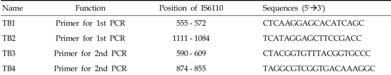
Table 1. Primer Sequences of In-house PCR for Detection of Mycobacterium tuberculosis
Name Function Position of IS6110 Sequences (5' 3')à
TB1 Primer for 1st PCR 555 - 572 CTCAAGGAGCACATCAGC
TB2 Primer for 1st PCR 1111 - 1084 TCATAGGAGCTTCCGACC
TB3 Primer for 2nd PCR 590 - 609 CTACGGTGTTTACGGTGCCC
TB4 Primer for 2nd PCR 874 - 855 TAGGCGTCGGTGACAAAGGC
PCR, polymerase chain reaction.
DNA polymerase (Core Biosystem, Seoul, Korea) 0.1 L 1.0μ μL primers (Bioneer, Daejeon, Korea)]
and amplified (GeneAmp PCR system 9600;
Perkin-Elmer Medical Instruments, CT, USA) with 30 cycles at 94°C for 20 seconds, 60°C for 20 seconds, and 72°C for 30 seconds. Primer sequences are shown in Table 1. Two L of 10μ times-diluted PCR product was again amplified with the same condition at the first PCR. PCR product (285 bp) was checked for size by com- paring positive M. tuberculosis using 2% agarose gel electrophoresis. To minimize cross-contamina- tion, DNA extraction and amplification were performed in separate rooms.
Cobas Amplicor M. tuberculosisTM
DNA extraction and amplification were per- formed according to the manufacturer's instruc- tions.
Diagnosis of tuberculosis
Tuberculosis was diagnosed when culture was positive. Culture negative tuberculosis was also diagnosed based on positive results of other diagnostic methods such as AFS and PCR. In addition, clinical signs, X-ray and hemological findings, and responses to anti-tuberculosis drugs were also considered.7,8,22 Culture-positive non- tuberculosis Mycobacterium was excluded.
RESULTS
Analysis of in-house PCR, AFS, and culture Of 2,973 samples, pulmonary samples were 1,134 (38.1%) [sputum 864 (29.0%), bronchial aspirates 271 (9.1%)] non-pulmonary samples were 1,839 (61.9%) of which pleural fluid was 834 (28.1%) (Table 2). Of pulmonary samples, 212 Table 2. Diagnostic Results of In-house PCR, AFB Stain, and Culture
Type of sample Total no. of
samples (%)
Positive samples (n)
AFB stain Culture In-house PCR
Pulmonary specimens 1,134 (38.1)
Sputum 863 (29.0) 70 111 93
Bronchial aspirate 271 (9.1) 10 18 26
Extrapulmonary specimens 1,839 (61.9)
Pleural fluid 834 (28.1) 14 30 27
Cerebrospinal fluid 313 (10.5) 0 0 1
Urine 248 (8.3) 6 12 15
Tissue 147 (4.9) 6 8 8
Pus 109 (3.7) 13 16 25
Peritoneal fluid 59 (2.0) 0 1 0
Blood 34 (1.1) 0 0 0
Gastric aspirate 12 (0.4) 0 0 0
Pericardial fluid 9 (0.3) 0 0 0
Bone marrow 7 (0.2) 0 0 0
Other 67 (2.3) 1 0 3
Total 2,973 (100.0) 120 197 198
PCR, polymerase chain reaction; AFB, acid fast bacilli.
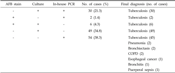
(18.7%) were positive for in-house PCR, 80 (7.1%) for AFS, and 129 (11.4%) for culture. Of non- pulmonary samples, 79 (4.3%) were positive for in-house PCR, 40 (2.2%) for AFS, and 67 (3.6%) for culture (Table 2). Of 2,973, 244 cases (8.2%) were diagnosed as tuberculosis, 112 of which were consistently positive for AFS, culture, and PCR (Table 3). Of 132 cases that displayed inconsistent results in AFS, culture, and PCR, 123 were finally diagnosed as tuberculosis and the remaining 9 were diagnosed as pneumonia (2), bronchiectasia (2), chronic obstructive pulmonary diseases (2), esophageal cancer (1), peripartum infection (1), and bronchitis (1). The 9 cases were all positive for in-house PCR (Table 4). The sensitivities for in-house PCR, AFS, and culture were 77.5%, 49.2%, and 80.7%, respectively, and specificities were 99.7%, 100%, and 100%, respectively (Table 5).
Analysis of in-house PCR and Cobas Amplicor M.
tuberculosisTM kit
Of 120 samples, pulmonary samples were 81 (67.5%) and non-pulmonary samples were 39 (32.5%). Of pulmonary samples, 17 (21.0%) were positive for in-house PCR and 20 (24.7%) for Cobas AmplicorTM. Of non-pulmonary samples, 10 (25.6%) were positive for in-house PCR and 3 (7.7%) for Cobas AmplicorTM (Table 6). Nineteen cases were consistently positive in both in-house PCR and Cobas AmplicorTM, and 3 were con- sistently negative. Inconsistent results were found in 10 cases, of which 9 (3 sputum samples posi- tive for Cobas AmplicorTM and 2 pleural fluid, 2 urine, and 2 tissue samples positive for in-house PCR) were diagnosed as tuberculosis and 1 as chronic obstructive pulmonary disease (1 sputum sample weakly positive for in-house PCR). The Table 4. Final Diagnosis
AFB stain Culture In-house PCR No. of cases (%) Final diagnosis (no. of cases)
- + + 30 (21.3) Tuberculosis (30)
+ - + 2 (1.4) Tuberculosis (2)
+ + - 6 (4.3) Tuberculosis (6)
- + - 49 (34.8) Tuberculosis (49)
- - + 54 (38.3) Tuberculosis (45)
Pneumonia (2) Bronchiectasis (2) COPD (2)
Esophageal cancer (1) Bronchitis (1)
Puerperal sepsis (1) AFB, acid fast bacilli; PCR, polymerase chain reaction; COPD, chronic obstructive pulmonary disease.
Table 5. Sensitivities, Specificities, and Positive and Negative Predictabilities of In-house PCR, AFB Stain, and Culture
Sensitivity (%) Specificity (%) PPV (%) NPV (%)
AFB stain 49.2 100.0 100.0 95.7
Culture 80.7 100.0 100.0 98.3
In-house PCR 77.5 99.7 95.5 98.0
PCR, polymerase chain reaction; AFB, acid fast bacilli; PPV, positive predictability; NPV, negative predictability.
sensitivities of in-house PCR and Cobas AmplicorTM were 81.3% and 71.9%, respectively (pulmonary samples, 77.3% and 90.9%. non-pulmonary samples, 90.0% and 30.0%, respectively) (Table 7).
DISCUSSION
Diagnosis of tuberculosis usually includes clinical symptoms, X-ray findings, and detection
of M. tuberculosis. To detect M. tuberculosis in clinical samples, laboratory employs AFS, culture of M. tuberculosis, and PCR. AFS is fast but the sensitivity is low whereas culture of M. tuberculosis needs long turnover time but sensitivity and specificity are high. PCR assay, the sensitivity and specificity of which are high and widely used in many clinical laboratories, requires only very small amounts of M. tuberculosis to diagnose tuberculosis.
Table 7.Sensitivities, Specificities, and Positive and Negative Predictabilities of In-house PCR, Cobas Amplicor M. tuberculosisTM Kit, AFB Stain, and Culture in Pulmonary and Non-Pulmonary Samples
Sensitivity (%) Specificity (%) PPV (%) NPV (%)
AFB stain 35.5 100.0 100.0 80.6
Culture 74.2 100.0 100.0 91.7
In-house PCR 81.3 98.9 96.3 93.5
Pulmonary samples 77.3 100.0 100.0 92.2
Non-pulmonary samples 90.0 96.6 90.0 96.6
Cobas AmplicorTM 71.9 100.0 100.0 90.7
Pulmonary samples 90.9 100.0 100.0 96.7
Non-pulmonary samples 30.0 100.0 100.0 80.6
PCR, polymerase chain reaction; AFB, acid fast bacilli; PPV, positive predictability; NPV, negative predictability.
Table 6. Diagnostic Results of In-house PCR, Cobas AmplicorTM, AFB Stain, and Culture in Pulmonary and Non-pulmonary Samples
Type of sample Total no. of samples (%)
Positive samples (n)
AFB stain Culture In-house PCR Cobas AmplicorTM Pulmonary specimens 81 (67.5)
Sputum 65 (54.2) 10 15 14 17
Bronchial aspirate 15 (12.5) 0 3 2 2
Lung 1 (0.8) 0 1 1 1
Extrapulmonary specimens 39 (32.5)
Pleural fluid 25 (20.8) 0 1 3 0
Urine 8 (6.7) 0 3 4 2
Tissue 1 (0.8) - - 1 0
Gastric aspirate 1 (0.8) 0 0 0 0
Stool 1 (0.8) 1 0 1 1
Other 3 (2.5) 0 0 1 0
Total 120 (100.0) 11 23 27 23
PCR, polymerase chain reaction; AFB, acid fast bacilli.
In a study by 6 laboratories AFS, culture, and PCR were analyzed, and the pooled sensitivities were found to be 55.5%, 89.3%, 85.2%, respec- tively, and pooled specificity was 99.7% for all 3 diagnostic methods.22 Our study showed that the sensitivities for AFS, culture, and PCR were 49.2%, 80.7%, 77.5%, respectively, and specificities were 100%, 100%, and 99.7%, respectively, in- dicating lower sensitivity and higher specificity.
Another study demonstrated that the sensitivities for AFS, culture, and PCR were 41.3%, 65.7%, and 59%, respectively, which are lower than those in our study and the specificities > 97%, and also demonstrated that PCR in combination with AFS increased sensitivity up to 65%, which is similar to that of culture method, suggesting the possi- bility for standard diagnostic procedure for tuber- culosis.7 Given the high sensitivity and specificity of PCR method, we suggest that PCR is very useful for the diagnosis of tuberculosis, except when drug sensitivity and detection of non- tuberculosis mycobacteria are required. PCR may be used in AFB smear-positive samples for final diagnosis of tuberculosis.8
Nine false-positive results were found with in-house PCR in this study. Two were pneumonia, 2 bronchiectasia, 2 chronic obstructive pulmonary diseases, 1 esophageal cancer, 1 peripartum infec- tion and 1 bronchitis. False positive may be caused by the contamination of amplicons.9,23-25 The contamination is even higher in in-house PCR, which requires multiple steps of specimen processing procedures than in commercial kit.23 False positive may also be caused by the fact that PCR can not distinguish between viable and non-viable M. tuberculosis.9,23-25
Fifty-five cases in this study were found to be false negative. False negative may be caused by the presence of inhibitors, loss of M. tuberculosis during DNA extraction procedures, and amplifi- cation methods.9,25 Since this is a retrospective study, determination of the causes of false- positive and false-negative results is limited.
The sensitivity and specificity estimates for in-house PCR vary widely, ranging from 63 - 100%
for the sensitivity and 62 - 100% for the specificity.
5,26-34 The difference in the sensitivity of PCR may be due to analytical sensitivity of PCR, PCR assay shows difference in sensitivity depending on the
target of PCR and whether it is nested PCR or not.
In particular, high sensitivity was obtained when IS6110 was the target. In addition, the distribution of positive samples in the specimen, especially in weak positive samples,5 affects the sensitivity of PCR, particularly contamination by amplicon.34
In this study, the sensitivity and specificity were 81.3% and 98.9% in in-house PCR and 71.9%
and 100% in Cobas Amplicor M. tuberculosisTM kit, indicating higher sensitivity and lower specificity in in-house PCR. Intriguingly, when samples were subdivided into pulmonary and non-pulmonary, non-pulmonary samples showed lower sensitivity (30%) than pulmonary samples (Table 7). This result is not in line with a study in which diagnostic results of in-house PCR and Cobas Amplicor M. tuberculosisTM kit in pulmonary and non-pulmonary samples were analyzed.20 The reason of why inconsistent results were observed in our study could be due to the fact that the amount of specimens was inadequate or specimens were weakly positive for M. tuberculosis.35
Another study in which diagnostic results of in- house PCR and Cobas Amplicor M. tuberculosisTM were analyzed demonstrated sensitivities of 91.08% and 66.33% for in-house PCR and Cobas Amplicor M. tuberculosisTM kit and specificities of 99.85% and 99.71%,35 which are similar to our results. Lower sensitivity in Cobas Amplicor M.
tuberculosisTM kit may be attributed to the use of single copy and ELISA or smaller amount of sample (0.1 mL in Cobas AmplicorTM and 1.0 mL in in-house PCR). The possibility that lower sensitivitiy in Cobas Amplicor M. tuberculosisTM kit may be due to the presence of inhibitors has also been suggested.36
Our laboratory employed nested PCR that requires 2 rounds of amplification procedure and confirmed PCR products not by Southern blotting but agarose gel electrophoresis. Thus, different primers, the amount of samples, and amplification method might have affected sensitivity. As demon- strated by the manufacturer, Cobas AmplicorTM displayed significantly lower sensitivity in non- pulmonary samples and higher sensitivity in pulmonary samples than in-house PCR (p < 0.01).
More samples and further followup are required for in-depth study, nevertheless we suggest that Cobas AmplicorTM is a useful and time-efficient
diagnostic method.
In summary, despite the high false-positive diagnostic results, we demonstrated that in-house PCR at Kyung Hee Medical Center can be used in place of AFS and culture for the early diagnosis of tuberculosis. Furthermore, in-house PCR showed higher sensitivity in non-pulmonary samples than Cobas AmplicorTM kit. Cobas AmplicorTM kit is time efficient and has the advantage of lower false-positive rates in the diagnosis of tuber- culosis.
REFERENCES
1. Dye C, Scheele S, Dolin P, Pathania V, Raviglione MC.
Global burden of tuberculosis: estimated incidence, prevalence, and mortality by country. WHO Global Surveillance and Monitoring Project. JAMA 1999;282:
677-86.
2. Korea Center for Disease Control and Prevention, Korean National Tuberculosis Association. Report on the tuberculosis prevalence survey in Korea. 2004.
Available from: URL: http://www.knta.or.kr/korea/
img/study/DP2-2.JPG.
3. Korea National Statistical Office. Annual report on the caues of death statistics 2003. 2004. Available from:
URL: http://www.knta.or.kr/korea/study05_4.asp.
4. Kim SJ, Im JG, Seong CK, Song JW, Yeon KM, Han MC.
Pulmonary infection in AIDS. J Korean Radiol Soc 1998;
39:933-9.
5. Yeam YS, Jeong OY, Jang SJ, Moon DS, Park YJ.
Comparison of culture, acid-fast stain and polymerase chain reaction assay for detection of Mycobacterium Tuberculosis. Korean J Lab Med 1995;15:594-603.
6. Kivihya-Ndugga L, Cleeff M, Juma E, Kimwomi J, Githui W, Oskam L, et al. Comparision of PCR with the routine procedure for diagnosis of tuberculosis in a population with high prevalences of tuberculosis and human immunodeficiency virus. J Clin Microbiol 2004;
42:1012-5.
7. Almeda J, García A, González J, Quintó L, Ventura PJ, Vidal R, et al. Clinical evaluation of an in-house IS6110 polymerase chain reaction for diagnosis of tuberculosis.
Eur J Clin Microbiol Infect Dis 2000;19:859-67.
8. American Thoracic Society. Diagnostic standards and classification of tuberculosis in adults and children. Am J Respir Crit Care Med 2000;161:1376-95.
9. Kim JH, Jang SJ, Moon DS, Park YJ. Evaluation of two PCR-hybridization methods for the detection of Mycobacterium tuberculosis. Korean J Lab Med 2003;23:
32-8.
10. Lee KE, Cho JH, Moon YH. Comparison of stain methods with PCR and culture for the detection of Mycobacterium tuberculosisin the sputum. Korean J Lab
Med 1998;18:201-7.
11. Shin WS. Diagnosis of tuberculosis; serodiagnosis and molecular biologic approach [in Korean]. Tuberc Respir Dis 1992;39:1-6.
12. Bodmer T, Gurtner A, Scholkmann M, Matter L.
Evaluation of the COBAS AMPLICOR MTB system. J Clin Microbiol 1997;35:1604-5.
13. D'Amato RF, Wallman AA, Hochstein LH, Colaninno PM, Scardamaglia M, Ardila E, et al. Rapid diagnosis of pulmonary tuberculosis by using Roche AMPLICOR Mycobacterium tuberculosis PCR test. J Clin Microbiol 1995;33:1832-4.
14. Moore DF, Curry JI. Detection and identification of Mycobacterium tuberculosis directly from sputum sediments by Amplicor PCR. J Clin Microbiol 1995;33:
2686-91.
15. Piersimoni C, Callegaro A, Nista D, Bornigia S, De Conti F, Santini G, et al. Comparative evaluation of two commercial amplification assays for direct detection of Mycobacterium tuberculosis complex in respiratory specimens. J Clin Microbiol 1997;35:193-6.
16. Rolfs A, Beige J, Finckh U, Köhler B, Schaberg T, Lokies J, et al. Amplification of Mycobacterium tuberculosis from peripheral blood. J Clin Microbiol 1995;33:3312-4.
17. Schirm J, Oostendorp LA, Mulder JG. Comparison of Amplicor, in-house PCR, and conventional culture for detection of Mycobacterium tuberculosis in clinical samples. J Clin Microbiol 1995;33:3221-4.
18. Stauffer F, Mutschlechner R, Hasenberger P, Stadlbauer S, Schinko H. Detection of Mycobacterium tuberculosis complex in clinical specimens by a commercial polymerase chain reaction kit. Eur J Clin Microbiol Infect Dis 1995;14:1046-51.
19. Wobeser WL, Krajden M, Conly J, Simpson H, Yim B, D'costa M, et al. Evaluation of Roche Amplicor PCR assay for Mycobacterium tuberculosis. J Clin Microbiol 1996;34:134-9.
20. Reischl U, Lehn N, Wolf H, Naumann L. Clinical evaluation of the automated COBAS AMPLICOR MTB assay for testing respiratory and nonrespiratory specimens. J Clin Microbiol 1998;36:2853-60.
21. Forbes BA, Sahm DF, Weissfeld AS. Bailey and Scott's diagnostic microbiology. 11th ed. St. Louis: Mosby;
2002.
22. Bogard M, Vincelette J, Antinozzi R, Alonso R, Fenner T, Schirm J, et al. Multicenter study of a commercial, automated polymerase chain reaction system for the rapid detection of Mycobacterium tuberculosis in respira- tory specimens in routine clinical practice. Eur J Clin Microbiol Infect Dis 2001;20:724-31.
23. Cohen RA, Muzaffar S, Schwartz D, Bashir S, Luke S, McGartland LP, et al. Diagnosis of pulmonary tuberculosis using PCR assays on sputum collected within 24 hours of hospital admission. Am J Respir Crit Care Med 1998;157:156-61.
24. Beige J, Lokies J, Schaberg T, Finckh U, Fischer M, Mauch H, et al. Clinical evaluation of a Mycobacterium tuberculosis PCR assay. J Clin Microbiol 1995;33:90-5.
25. Beavis KG, Linchty MB, Jungkind DL, Giger O. Evalua- tion of Amplicor PCR for direct detection of Mycobacterium tuberculosis from sputum specimens. J Clin Microbiol 1995;33:2582-6.
26. Abe C, Hirano K, Wada M, Kazumi Y, Takahashi M, Fukasawa Y, et al. Detection of Mycobacterium tuberculosis in clinical specimens by polymerase chain reaction and Gen-Probe Aamplified Mycobacterium tuberculosisDirect Test. J Clin Microbiol 1993;31:3270-4.
27. Miller N, Hernandez SG, Cleary TJ. Evaluation of Gen-Probe Amplified Mycobacterium Tuberculosis Direct Test and PCR for direct detection of Mycobacterium tuberculosisin clinical specimens. J Clin Microbiol 1994;
32:393-7.
28. Shawar RM, el-Zaatari FA, Nataraj A, Clarridge JE.
Detection of Mycobacterium tuberculosis in clinical samples by two-step polymerase chain reaction and nonisotopic hybridization methods. J Clin Microbiol 1993;31:61-5.
29. Pierre C, Lecossier D, Boussougant Y, Bocart D, Joly Y, Yeni P, et al. Use of a reamplification protocol improves sensitivity of detection of Micobacterium tuberculosis in clinical samples by amplification of DNA. J Clin Microbiol 1991;29:712-7.
30. Cousins DV, Wilton S, Francis BR, Gow BL. Use of polymerase chain reaction for rapid diagnosis of tuberculosis. J Clin Microbiol 1992;30:255-8.
31. Bodmer T, Gurtner A, Schopfer K, Matter L. Screening of respiratory tract specimens for the presence of Mycobacterium tuberculosis by using the Gen-Probe Amplified Mycobacterium tuberculosis Direct Test. J Clin Microbiol 1994;32:1483-7.
32. Pfyffer GE, Kissling P, Wirth R, Weber R. Direct detection of Mycobacterium tuberculosis complex in respiratory specimens by a target-amplified test system.
J Clin Microbiol 1994;32:918-23.
33. Hermans PW, Schuitema AR, Van Soolingen D, Verstynen CP, Bik EM, Thole JE, et al. Specific detec- tion of Mycobacterium tuberculosis complex strains by polymerase chain reaction. J Clin Microbiol 1990;28:
1204-13.
34. Noordhoek GT, Kolk AH, Bjune G, Catty D, Dale JW, Fine PE, et al. Sensitivity and specificity of PCR for detection of Mycobacterium tuberculosis: a blind comparison study among seven laboratories. J Clin Microbiol 1994;32:277-84.
35. Eing BR, Becker A, Sohns A, Ringelmann R. Comparison of Roche Cobas Amplicor Mycobacterium tuberculosis assay with in-house PCR and culture for detection of M. tuberculosis. J Clin Microbiol 1998;36:2023-9.
36. Schirm J, Oostendorp LA, Mulder JG. Comparison of Amplicor, in-house PCR, and conventional culture for detection of Mycobacterium tuberculosis in clinical samples. J Clin Microbiol 1995;33:3221-4.