J. Exp. Biomed. Sci. 2010, 16(2): 127~131
Detection of Mycobacterium leprae by Real-time PCR Targeting
Mycobacterium leprae-Specific Repetitive Element Sequence
Hyunwoo Jin
1, Hye-Young Wang
1, Jong-Pill Kim
2, Sang-Nae Cho
3and Hyeyoung Lee
1,† 1Department of Biomedical Laboratory Science, College of Health Sciences, Yonsei University234 Maeji-ri, Heungup-myun, Wonju-si, Kangwon-do 220-710;
2Affiliated Hospital, Korean Leprosy Control Association, Euiwang-si, Kyunggi-do 437-823;
3Department of Microbiology, Yonsei University College of Medicine, 134 Shinchon-dong, Seoul 120-752, Korea
Mycobacterium leprae detection is difficult even with molecular biological techniques due to the low sensitivity of current methodologies. In this report, real-time PCR targeting the M. leprae-specific repetitive element (RLEP) sequence was developed as a new diagnostic tool and evaluated using clinical specimens. For this, M. leprae DNAs were extracted from skin biopsy specimens from 80 patients and analyzed by real-time PCR using TaqMan probe. Then, the detection efficiency of the real-time PCR was compared with that of standard PCR. In brief, the rate of positive detection by the standard PCR and real-time PCR was 32.50% and 66.25%, respectively. The results seemed to clearly show that the TaqMan real-time PCR developed in this study may be a useful tool for sensitive detection of M. leprae from clinical specimens.
Key Words: Mycobacterium leprae, Real-time PCR, Repetitive element sequence (RLEP)
서 론
나병은 나균인 Mycobacterium leprae가 피부와 말초신 경에 주로 침해하여 발생하는 전염성 만성면역질환으로 질병의 발병은 줄어들고 있지만, 아직도 세계적으로 문 제가 되는 질환이다 (Chae et al., 2002). WHO의 보고에 의하면, 2005년 한 해 전세계에서 발생한 신규나병환자 는 296,499명이었고 그 중 68%인 201,635명은 동남아시 아에서 발생하였다 (WHO, 2006). 나균은 인공배지에서 배양이 되지 않기 때문에 나병 진단을 위해서는 피부도말을 항산성 염색을 하여 확인 하고 피부병변 및 신경손상 등의 임상소견을 기초로 확 진한다 (Kampirapap et al., 1998). 하지만 항산성 염색 결 과는 모든 Mycobacterium spp.가 양성으로 나타나기 때문 에 나균만 감별할 수 없고, 피부조직에서 균수가 최소 104개 이상이 존재해야 양성으로 나타나기 때문에 민감 도와 특이도가 낮은 검사법이라 할 수 있다 (Kurabachew et al., 1998 Donoghue et al., 2001; Kang et al., 2003).
따라서 최근에는 분자생물학적 방법인 중합효소 연쇄 반응 (polymerase chain reaction; PCR)을 이용하여 나균의 특정한 DNA 염기서열을 증폭시켜 조직 내의 나균을 검 출하는 방법이 많이 사용되고 있다 (De wit et al., 1991; Yoon et al., 1993; Donoghue et al., 2001; Kang et al., 2003; Patrocinio et al., 2005).
최근에는 real-time PCR 기술이 많은 병원체의 진단에 서 검증되어 (Rondini et al., 2003; Kesanopoulos et al., 2005) 결핵균을 포함한 다른 마이코박테리아의 진단에도 응 용되고 있다. Real-time PCR 기술은 PCR 방법의 최대의 단점인 실험실내 오염사고를 예방할 수 있다는 장점과 더불어 프로브를 사용한다는 장점에 의해 기존의 PCR 에 비해 민감도와 특이도가 높다는 평가를 받고 있다 (Kramme et al., 2004; Martinez et al., 2006; Shamsi et al., 2007). 따라서 본 연구에서는 나균 검출을 목적으로 한 real-time PCR을 개발하고 이어 이 방법의 나균 검출에서의 민감도 및 특이도를 평가하여 이를 기존의 PCR법에 의 한 결과와 비교함으로써 real-time PCR의 나병 진단에서 *접수일: 2010년 3월 19일 / 수정일: 2010년 6월 18일 채택일: 2010년 6월 21일
†Corresponding author: Hyeyoung Lee. Department of Biomedical Laboratory Science, College of Health Sciences, Yonsei University 234 Maeji-ri, Heungup-myun, Wonju-si, Kangwon-do 220-710, Korea. Tel: 82-33-760-2740, Fax: 82-33-760-2561
e-mail: [email protected]
의 유용성을 평가하였다.
재료 및 방법
나균 및 생검조직에서 DNA의 추출
표준균주로 사용된 M. leprae strain 4264는 Colorado State University에서 정제된 100 mg (평균 2.9×10⁹ bacilli/
mg 포함)을 제공받아 QIAamp DNA Mini Kit (Tissue protocol; Qiagen GmbH, Hilden, Germany)를 이용하여 DNA를 분리하고, 최종농도를 50 ng/μl로 희석하여 사용 하였다. 임상검체 평가에 이용된 DNA는 한센병연구원 에서 나병 환자들로부터 채취한 피부생검조직으로부터 QIAamp DNA Mini Kit (Qiagen GmbH, Hilden, Germany)를 이용하여 추출한 DNA 80개를 제공받아 사용하였다.
항산균에서 DNA의 추출
Real-time PCR에 사용되는 프라이머와 프로브의 특이 도를 검사하기 위해 다양한 마이코박테리아 균주로부터 DNA를 분리하여 사용하였다. 실험에 사용된 마이코박 테리아 균종은 M. tuberculosis H37Rv, M. abscessus (ATCC 19977), M. africanum (KCTC 9504), M. aichiens (ATCC 27280), M. aurum (ATCC 23366), M. avium (ATCC 25291), M. avium (ATCC 35719), M. celatum (ATCC 51130), M. celatum (ATCC 51131), M. celatum sub. chelonae (ATCC 35749), M. fortuitum (ATCC 49403), M. fortuitum (ATCC 49404), M. fortuitum sub. fortuitum (KCTC 9510), M. gallinarum (KCTC 9511), M. gastri (ATCC 15754), M. genevence (ATCC 51233), M. gordonae (ATCC 14470), M. gordonae (KCTC 9513), M. hassiacum (ATCC 700660), M. intracellulare (ATCC 13950), M. intracellulare (KCTC 9514), M. kansasii (ATCC 12478), M. kansasii (KCTC 9515), M. kubicae (ATCC 700732), M. malmonense (ATCC 29571), M. marinum (ATCC 927), M. microti (ATCC 19422), M. moriokaense (KCTC 9516), M. neoaurum (ATCC 25795), M. nonchromogenicum (ATCC 19530), M. peregrinum (ATCC 14467), M. phlei (ATCC 11758), M. porcinum (KCTC 9517), M. scrofulacium (ATCC 19981), M. senegalense (ATCC 35796), M. septicum (ATCC 700731), M. smegmatis (KCTC 9108), M. szulgai (ATCC 35799), M. terrae (ATCC 15755), M. thermoreisistable (ATCC 19527), M. trivial (ATCC 23292), M. vaceae (ATCC 15483), M. xenopi (ATCC 19250)이었다.
DNA를 분리하기 위해 배양한 균체에 lysozyme을 5
mg/ml로 가하여 37℃에서 1시간, 1 mg/ml의 proteinase K 및 1% SDS를 가하여 55℃에서 24시간 반응시켰다. Cetyl Trimethyl Ammonium Bromide (CTAB)를 첨가하여 65℃ 에서 10분간 반응시키고 페놀 처리 및 에탄올 침전하여 DNA를 얻었다.
Real-time PCR
National Center for Biotechnology information (NCBI)에서 제공하는 Blast search (http://www.ncbi.nlm.nih.gov/BLAST/) 를 이용하여 나균 DNA의 repetitive sequence (GenBank accession No. AL583917)의 유사염기서열을 추출한 후 MultAlin program을 이용하여 분석하였다.
이를 이용하여 예상되는 증폭산물이 107-bp의 크기 가 되도록 MLF (5'-GTGTCGGCGTGGTCAATGTG-3')와 MLR (5'-CGATACCAGCGGCAGAAATGG-3') 한 쌍의 primer를 제작하고 5' 끝에 5-carboxyfluoroscein (FAM)과 3' 끝에 N,N,N',N'-tetrametyl-6-carboxyrhodamine (TAMRA) 의 형광염료를 부착한 MLSP (5'-FAM-CCGCACCTGAA- CAGGCACGTCCC-TAMRA-3') TaqMan probe를 제작하였 다 (Bioneer, 대전, 한국). Real-time PCR 반응액은 총 20 μl 내에 iQTM Supermix (Bio-Rad, USA) 10 μl, 각각 10 pmol
의 primer 1 μl와 probe 0.5 μl, template DNA 5 μl를 첨가하 여 사용하였다. PCR 반응조건은 predenaturation을 94℃에 서 3분간 1회 수행한 후에 94℃에서 20초 denaturation, 56℃에서 40초 annealing 및 extension 후, 45회 실시하였 고 형광측정을 사용하였다. iCycler iQTM Multicolor
Real-time PCR Detection System (Bio-Rad, USA)을 사용하였고 iCycler iQTM Optical System Software Version 3.1 (Bio-Rad,
USA)을 사용하여 결과를 분석하였다.
비교실험을 위해 이용된 PCR은 touch-down (TD) PCR로, 예상되는 증폭산물은 129-bp가 되도록 F (5'-TGCATGTCATGGCCTTGAGG-3')와 R (5'-CACCGATAC- CAGCGGCAGAA-3')의 primer를 이용하였다 (Kang et al., 2003). 반응조건으로는 먼저 pre-denaturation을 94℃에서 5분간 1회 수행한 후에, 94℃에서 45초 denaturation, 64℃ 에서 58℃까지 1℃씩 감소시키면서 7회 동안 반복 시행 한 후에 다시 94℃에서 45초 denaturation, 58℃에서 45초 annealing, 72℃에서 90초 extension으로 35회 실시하였다.
결과 및 고찰
나균은 시험관 내에서 배양할 수 없기 때문에 감염병의 진단에 필수적인 실험실 내에서 균 배양과 동정을 거 치지 못하는 것이 나병 진단의 가장 큰 취약점이다. 따라 서 현재까지 나병의 진단은 임상소견과 항산성균 염색에 주로 의존하여 왔다 (Talhari, 1996). 항산성균 염색은 적 어도 균체가 104개 이상 존재하여야 양성으로 판정되어 민감도가 낮고 다른 mycobacteria와 구별이 되지 않기 때문에 특이도가 낮다는 단점이 있다 (Kurabachew et al., 1998). 최근에는 분자생물학적인 방법을 이용한 나균의 DNA 나 RNA를 직접 증폭시켜 검출하는 방법이 널리 사용 되고 있다 (Karamme et al., 2003). DNA를 증폭하는 방법 에 사용되는 표적 유전자는 65 kDa 항원 유전자 gene encoding 65 kDa antigen (Plikaytis et al., 1990), 18 kDa 항원 유전자 (Kim et al., 1996; Williams et al., 1990), 36 kDa 항원 유전자 (Parkash et al., 2004), repetitive sequences (Wood and
Cole, 1989; Yoon et al., 1993) 등이 널리 사용되고 있다. RNA를 증폭하는 방법에 사용되는 표적은 16S rRNA 부 위에 나균에 특이적인 부분을 검출하는 방법이 사용되고 있다 (Kurabachew et al., 1998; Phetsuksiri et al., 2006).
본 연구에 사용된 repetitive sequence (RLEP)는 다른 세 균이나 항산성균에는 존재하지 않는 나균에만 존재하는 유전자로 알려져 있으며, 약 28 copy가 존재하기 때문에 다른 유전자보다 검출 민감도가 훨씬 높다는 장점이 있 다 (Donoghue et al., 2001; Kang et al., 2003).
나균 검출을 위한 real-time PCR법의 결과판정을 위해 측정된 threshold cycle (Ct) 값을 분석하여 36 이하를 M. leprae 양성, 36초과하는 값에 대하여 음성으로 판단하도 록 cut off value를 결정하였다.
나균을 진단하기 위하여 제작된 MLF, MLR primer 쌍 이 M. leprae에만 특이적으로 반응하는지를 알아보기 위
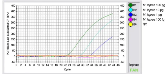
A
Fig. 1. The sensitivity of the (A) real-time PCR and (B) standard PCR for detecting M. leprae genomic DNA. Using the diluted DNA of the M. leprae reference strain 4264, the sensitivity of the two PCR assays were compared. (A), real-time PCR with 100 pg (B1), 10 pg (B2), 1 pg (B3), 100 fg (B4), and no DNA (B8). (B) PCR with from 100 pg to 100 ag DNA. M; 100 bp DNA ladder (Bioneer, Daejeon, Korea), N; no DNA added negative control.
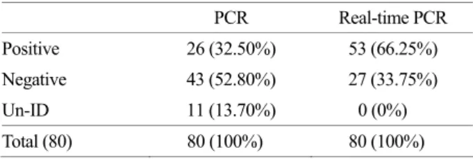
해 M. leprae strain 4264와 35종의 Mycobacterium spp. DNA를 가지고 real-time PCR을 수행하였다. 측정된 Ct 값을 분석한 결과 M. leprae strain 4264만 양성으로 판정 되고 35종의 Mycobacterium spp. DNA는 모두 음성으로 측정됨을 확인하였다. 이런 결과로 보아 RLEP 부위를 검출하는 real-time PCR법은 나균에 대한 특이성이 높다 는 것을 확인하였다. 또한, 나균 검출을 위한 real-time PCR법의 민감도를 비교 분석하기 위해 M. leprae strain 4264 DNA를 연속적 으로 10배씩 희석한 후 real-time PCR의 template DNA로 각각 사용하였다. 그 결과 1 pg 이상에서 증폭되는 것을 확인할 수 있었고 측정된 Ct값으로 1 pg에서 양성으로 측정됨을 확인하였다 (Fig. 1). 따라서, 민감도 부분에서는 비교실험에 이용된 single PCR과 real-time PCR이 비슷한 민감도를 가지고 있었으나, 나병으로 의심되는 환자의 피부조직에서 추출된 DNA로부터 RLEP의 특이 유전자 검출을 실시하였다. 그 결과 총 80개의 시료 중 PCR을 이용한 방법으로는 26개 (32.5%)의 검체에서 양성 결과 가 나온 반면 real-time PCR을 이용한 방법으로는 53개 (66.25%)의 검체에서 양성 결과가 나왔다 (Table 1). 따라 서 real-time PCR을 이용한 방법이 single PCR을 이용한 진단법보다 높은 민감도를 나타내어 높은 진단 효율성 을 나타내었다. Real-time PCR은 형광물질이 부착된 probe를 사용하여 실시간으로 증폭산물의 유무를 확인하는 방법으로 전기 영동을 하지 않기 때문에 일반 PCR보다 빠른 결과를 얻 을 수 있고 PCR 전기영동 상에서 눈으로 확인하는 것보 다 정확한 형광의 신호를 측정하여 정량측정도 가능하 다. 그리고 PCR에서 교차오염으로 인한 위양성 결과가 real-time PCR은 뚜껑을 한번 닫으면 열지 않기 때문에 교차오염의 위험이 없다는 장점이 있다. 따라서, RLEP를 검출하는 real-time PCR법은 피부 생검 조직에서 직접 균을 검출할 때에 PCR을 대신하여 나균 진단에 유용할 것으로 생각된다.
REFERENCES
Chae GT, Kim MJ, Kang TJ, Lee SB, Shin HK, Kim JP, Ko YH, Kim SH, Kim NH. DNA-PCR and RT-PCR for the 18-kDa gene of Mycobacterium lepraeto assess the efficacy of multidrug therapy for leprosy. J Med Microbiol. 2002. 51: 417-422.
De wit MYL, Feber WR, Krieg SR. Application of a polymerase chain reaction for the detection of Mycobacterium leprae in skin tissues. J Clin Microbiol. 1991. 29: 906-910.
Donoghue HD, Holton J, Spigelman M. PCR primers that can detect low levels of Mycobacterium leprae DNA. J Med Microbiol. 2001. 50: 177-182.
Kampirapap K, Singtham N, Klatser PR, Wiriyawipart S. DNA amplification for detection of leprosy and assessment of efficacy of leprosy chemotherapy. Int J Lepr Other Mycobact Dis. 1998. 66: 16-21.
Kang TJ, Kim SJ, Lee SB, Chae GT, Kim JP. Comparison of Two different PCR amplification products (the 18-kDa protein gene vs. RLEP repetitive sequence) in the diagnosis of
Mycobacterium leprae. Clin Exp Dermatol. 2003. 28: 420
-424.
Kesanopoulos K, Tzanakaki G, Levidiotou S, Blackwell C, Kremastinou J. Evaluation of touch-down real-time PCR based on SYBR Green I fluorescent dye for the detection of Neisseria meningitidis in clinical samples. FEMS Immunol Med Microbiol. 2005. 43: 419-424.
Kramme S, Bretzel G, Panning M, Kawuma J, Drosten C. Detection and quantification of Mycobacterium lepraein tissue samples by real-time PCR. Med Microbiol Immunol. 2004. 193: 189 -193.
Kurabachew M, Wondimu A, Ryon JJ. Reverse transcription-PCR detection of Mycobacterium leprae in clinical specimens. J Clin Microbiol. 1998. 36: 1352-1356.
Martinez AN, Britto CF, Nery JA, Sampaio EP, Jardim MR, Sarno EN, Moraes MO. Evaluation of real-time and conventional PCR targeting complex 85 genes for detection of
Mycobacterium leprae DNA in skin biopsy samples from
patients diagnosed with leprosy. J Clin Microbiol. 2006. 44: 3154-3159.
Patrocinio LG, Goulart IM, Goulart LR, Patrocinio JA, Ferreira FR, Fleury RN. Detection of Mycobacterium leprae in nasal mucosa biopsies by the polymerase chain reaction. FEMS Immunol Med Microbiol. 2005. 44: 311-316.
Table 1. Comparison of sensitivity between the real-time PCR and the standard PCR using clinical samples
PCR Real-time PCR
Positive 26 (32.50%) 53 (66.25%)
Negative 43 (52.80%) 27 (33.75%)
Un-ID 11 (13.70%) 0 (0%)
Phetsuksiri B, Rudeeaneksin J, Supapkul P, Wachapong S, Mahotarn K, Brennan PJ. A simplified reverse transcriptase PCR for rapid detection of Mycobacterium leprae in skin specimens. FEMS Immunol Med Microbiol. 2006. 48: 319 -328.
Rondini S, Mensah-Quainoo E, Troll H, Bodmer T, Pluschke G. Development and application of real-time PCR assay for quantification of Mycobacterium ulcerans DNA. J Clin Microbiol. 2003. 41: 4231-4237.
Shamsi FA, Chaudhry IA, Moraes MO, Martinez AN, Riley FC. Detection of Mycobacterium lepraein ocular tissues by histopathology and real-time polymerase chain reaction. Ophthalmic Res. 2007. 39: 63-68.
Talhari S. Leprosy diagnosis, classification and prognosis. Int J
Lepr Other Mycobact Dis. 1996. 64: S13-S15.
World Health Organization. New case detection trends in leprosy. 2006. Available from: <http://www.who.int/lep/situation/ NCDetection2006.pdf>
Yoon KH, Cho SN, Lee MK, Abalos RM, Cellona RV, Fajardo Jr TT, Guido LS, Dela Cruz EC, Walsh GP, Kim JD. Evaluation of polymerase chain reaction amplification of Mycobacterium
leprae-specific repetitive sequence in biopsy specimens from
leprosy patients. J Clin Microbiol. 1993. 31: 895-899. Yoon KH, Cho SN, Gerald PW, Lee JB, Kim JD. Detection of
Mycobacterium leprae in Skin Biopsy Specimens From
Leprosy Patients by Polymerase Chain Reaction. Kor J Dermatol. 1994. 32: 409-415.