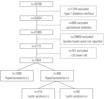
• 검색 결과가 없습니다.

Association between Metformin Use and Risk of Lactic Acidosis or Elevated Lactate Concentration in Type 2 Diabetes

N/A
N/A
Protected

Academic year: 2022

Share "Association between Metformin Use and Risk of Lactic Acidosis or Elevated Lactate Concentration in Type 2 Diabetes"

Copied!
7
0
0

로드 중.... (전체 텍스트 보기)

전체 글

(1)

INTRODUCTION

Metformin is an effective anti-hyperglycemic agent and the drug of choice in patients with type 2 diabetes.

1,2

It is well known that metformin has many beneficial effects on body weight, serum lipids, fibrinolysis, blood pressure, and endothe- lial function.

3-5

Furthermore, metformin can reduce diabetes- related risks up to 32%, diabetes-related death up to 42%, and all-cause mortality up to 36%.

3

However, metformin use is limited because of its potential adverse effects associated with lactic acidosis (LA), particularly in patients with reduced renal

Association between Metformin Use and Risk

of Lactic Acidosis or Elevated Lactate Concentration in Type 2 Diabetes

Eun Young Lee

1

, Sena Hwang

2

, Yong-ho Lee

3

, Seo Hee Lee

3

, Young Mi Lee

4

, Hua Pyong Kang

3

, Eugene Han

3

, Woonhyoung Lee

5

, Byung-Wan Lee

3

, Eun Seok Kang

3

, Bong Soo Cha

3

, and Hyun Chul Lee

3

1

Division of Endocrinology and Metabolism, Department of Internal Medicine, Seoul St. Mary’s Hospital, College of Medicine, The Catholic University of Korea, Seoul;

2

Chaum Life Center, CHA University School of Medicine, Seoul;

3

Division of Endocrinology and Metabolism, Department of Internal Medicine, Yonsei University College of Medicine, Seoul;

4

Department of Internal Medicine, Dongtan Jeil Women’s Hospital, Hwasung;

5

Department of Laboratory Medicine, Kosin University College of Medicine, Busan, Korea.

Purpose: Metformin can reduce diabetes-related complications and mortality. However, its use is limited because of potential lactic acidosis-associated adverse effects, particularly in renal impairment patients. We aimed to investigate the association of metformin use with lactic acidosis and hyperlactatemia in patients with type 2 diabetes.

Materials and Methods: This was a cross-sectional study from a tertiary university-affiliated medical center. A total of 1954 type 2 diabetes patients were recruited in 2007–2011, and stratified according to the estimated glomerular filtration rate of 60 mL/

min/1.73 m

2

. Lactic acidosis was defined as plasma lactate levels >5 mmol/L and arterial pH <7.35.

Results: Metformin was used in 61.4% of the patients with type 2 diabetes mellitus. Plasma lactate levels were not different in the patients with and without metformin use. There was no difference in prevalence of hyperlactatemia and lactic acidosis between the patients with and without metformin use (18.9% vs. 18.7%, p=0.905 for hyperlactatemia and 2.8% vs. 3.3%, p=0.544 for lactic acidosis). Similar results were observed in the patients with estimated glomerular filtration rate <60 mL/min/1.73 m

2

. Most pa- tients with lactic acidosis had at least one condition related to hypoxia or poor tissue perfusion. Multiple regression analysis indi- cated no association between metformin use and lactic acidosis, whereas tissue hypoxia was an independent risk factor for lactic acidosis [odds ratio 4.603 (95% confidence interval, 1.327–15.965)].

Conclusion: Metformin use was not associated with hyperlactatemia or lactic acidosis in patients with type 2 diabetes.

Key Words: Metformin, lactic acidosis, diabetes mellitus, lactate Yonsei Med J 2017 Mar;58(2):312-318

https://doi.org/10.3349/ymj.2017.58.2.312 pISSN: 0513-5796 · eISSN: 1976-2437

Received: December 22, 2015 Revised: July 5, 2016 Accepted: August 23, 2016

Corresponding author: Dr. Hyun Chul Lee, Division of Endocrinology and Metabo- lism, Department of Internal Medicine, Yonsei University College of Medicine, 50-1 Yonsei-ro, Seodaemun-gu, Seoul 03722, Korea.

Tel: 82-2-2228-1943, Fax: 82-2-393-6884, E-mail: [email protected]

•The authors have no financial conflicts of interest.

© Copyright: Yonsei University College of Medicine 2017

This is an Open Access article distributed under the terms of the Creative Com- mons Attribution Non-Commercial License (http://creativecommons.org/licenses/

by-nc/3.0) which permits unrestricted non-commercial use, distribution, and repro-

duction in any medium, provided the original work is properly cited.

(2)

function.

6

The adverse effects of biguanides associated with LA have become a concern in clinical practice with the use of the first line biguanide, phenformin. Phenformin inhibits lactate oxi- dation and increases lactate production.

7,8

It has been reported that the incidence of phenformin-associated LA ranges between 40 and 129 cases per 100000 patient-years.

9,10

However, despite being a biguanide, metformin does not affect anaerobic lactate production.

11

In fact, the incidence of LA is 10 to 20 times lower with the use of metformin than with that of phenformin.

12

Fur- thermore, several recent studies have suggested that the oc- currence of LA with the use of metformin might be a coinci- dence rather than a consequence. Scale and Harvey

13

reported that LA was more common in patients with diabetes but was not more frequent in patients who had taken metformin. An- other study has shown that the prevalence of LA is lower in pa- tients with metformin therapy than those with sulfonylurea therapy (3.3 and 4.8 cases per 100000 patient-years, respective- ly).

14

Therefore, we aimed to investigate whether metformin use was associated with LA and hyperlactatemia in patients with type 2 diabetes.

MATERIALS AND METHODS

Patients and study design

This was a cross-sectional study. In the present study, patients from the diabetes registry of the Severance Diabetes Center between January 2007 and December 2011 were retrospec- tively evaluated. Of total 33758 diabetic patients, patients with type 1 diabetes, gestational diabetes, and patients whose lac- tate levels were not reported were excluded. A total of 1954 pa- tients aged ≥20 years and with type 2 diabetes were included in this study (Fig. 1). Medical history including medication use, history of operation, and underlying diseases was collected.

Hyperlactatemia was defined as plasma lactate levels above the normal upper limit of 2.2 mmol/L. LA was defined as plas- ma lactate levels of >5 mmol/L and arterial pH <7.35. All patients were stratified according to the use of metformin or the estimat- ed glomerular filtration rate (eGFR) of 60 mL/min/1.73 m

2

. The metformin therapy group comprised patients who used met- formin during the study period, and the non-metformin thera- py group comprised subjects who did not use metformin in the year prior to data collection. This study was approved by the research ethics committee of the Severance Hospital, un- der protocol No. 4-2014-0420.

Laboratory measurements

Fasting and postprandial plasma glucose levels were mea- sured using the glucose oxidase method on a Hitachi 747 au- tomatic analyzer (Hitachi Instruments Service, Tokyo, Japan).

Glycated hemoglobin (HbA1c) levels were measured using high- performance liquid chromatography and a Variant II Turbo

hemoglobin testing system (Bio-Rad Laboratories, Hercules, CA, USA). Plasma lactate levels were measured using the am- perimetric electrode method. Arterial blood pH was analyzed in patients with increased lactate levels. Serum creatinine lev- els were measured using the Jaffe method on a Hitachi 7600 analyzer. The eGFR was calculated using the 4-variable Modi- fication of Diet in Renal Disease study equation: eGFR=186.3×

(creatinine)

-1.154

×age

-0.203

×0.742 (for women).

15

Serum aspartate aminotransferase (AST) and alanine aminotransferase (ALT) levels were measured using the Internal Federation of Clinical Chemistry standardization without pyridoxal phosphate using a Hitachi 7600 analyzer. Among participants, we measured metformin concentration in those taking metformin and with reduced renal function (creatinine ≥1.5 mg/dL for men and

≥1.4 mg/dL for women) (n=31). None of them had severe ill- ness such as liver cirrhosis, acute coronary syndrome, malig- nancy, acute infectious disease, and acute or severe renal fail- ure (renal failure occurred within 7 days or serum creatinine

≥3 mg/dL). Metformin concentration was measured by high- performance liquid chromatography-tandem mass spectrom- etry using a 1200 Series HPLC system (Agilent Technologies, Santa Clara, CA, USA) and API 4000

TM

mass spectrometer (AB Sciex, Framingham, MA, USA). The coefficient of variance ranged between 4.7% and 7.7%.

Statistical analysis

All continuous variables are shown as mean±standard devia- tion or median (interquartile range) for data not normally dis- tributed. Data were analyzed after stratification considering a threshold eGFR value of 60 mL/min/1.73 m

2

. Variable compari- son between two groups was performed using Student’s t-test or chi-square test, as appropriate. The Mann-Whitney U test

Fig. 1. Flow diagram of the study population.

n=1586 Hyperlactatemia (-)

n=368 Hyperlactatemia (+)

n=1104 excluded

; type 1 diabetes mellitus

n=689 excluded

; gestational diabetes

n=29850 excluded

; lactate levels were not reported

n=161 excluded

; <20 years old

n=310 Lactic acidosis (-)

n=58 Lactic acidosis (+) n=33758

n=32654

n=31965

n=2115

n=1954

(3)

Table 1. Baseline Characteristics of Subjects According to the Metformin Use and Renal Function Variables

Total (n=1954) eGFR 60 mL/min/1.73 m

2

(n=1438) eGFR <60 mL/min/1.73 m

2

(n=516) No metformin use (n=754) Metformin use (n=1200) p value No metformin use (n=584) Metformin use (n=854) p value No metformin use (n=170) Metformin use (n=346) p value Age (yr) 62.3 ± 11.7 64.6 ± 11.0 <0.001 62.4 ± 11.5 64.0 ± 11.0 0.008 62.2 ± 12.4 65.9 ± 11.0 <0.001 Sex (women, %) 315 (41.8) 480 (40.0) 0.232 238 (40.8) 328 (38.4) 0.201 77 (45.3) 152 (43.9) 0.421 BMI (kg/m

2

) 24.3 ± 3.6 24.1 ± 3.8 0.208 24.2 ± 3.5 24.0 ± 3.7 0.545 24.8 ± 3.8 24.3 ± 3.9 0.182 Metformin dose (mg/day) − 1000 (500–1000) − 1000 (500–1000) − 1000 (500–1000) Lactate (mmol/L) 1.2 (0.8–1.9) 1.1 (0.8–1.9) 0.050 1.1 (0.8–1.8) 1.1 (0.8–1.8) 0.095 1.4 (0.9–2.6) 1.2 (0.8–2.5) 0.087 Cr (mg/dL) 1.3 ± 1.6 1.2 ± 1.1 0.531 0.8 ± 0.2 0.8 ± 0.2 0.508 2.9 ± 2.8 2.3 ± 1.7 0.009 eGFR (mL/min/1.73 m

2

) 81.8 ± 34.0 79.0 ± 36.5 0.097 95.4 ± 24.1 95.8 ± 28.2 0.745 35.1 ± 17.6 37.6 ± 15.5 0.117 Albuminuria (mg/day) 26.3 (11.9–101.2) 14.0 (0–88.6) <0.001 18.8 (9.8–60.8) 9.2 (0–35.0) <0.001 71.8 (19.9–411.7) 68.3 (13.2–442.2) 0.313 CO

2

(mmol/L) 21.8 ± 4.6 21.8 ± 5.7 0.720 22.5 ± 4.2 22.6 ± 5.7 0.792 19.4 ± 5.3 19.4 ± 5.1 0.952 AST (IU/L) 22 (16–36) 23 (16–40) 0.186 23 (17–36) 23 (16–39) 0.437 21 (16–41) 24 (16–44) 0.243 AL T (IU/L) 20 (12–33) 19 (13–34) 0.554 20 (13–34) 19 (13–33) 0.255 17 (12–31) 19 (12–36) 0.451 FPG (mg/dL) 154.6 ± 67.4 148.4 ± 65.2 0.053 152.9 ± 62.0 148.1 ± 61.3 0.151 160.1 ± 83.4 149.4 ± 75.1 0.161 PPG (mg/dL) 220.7 ± 97.4 208.7 ± 78.0 0.041 221.7 ± 99.3 206.0 ± 77.3 0.022 217.3 ± 91.3 216.1 ± 79.7 0.913 HbA1c (%) 7.6 ± 4.3 7.4 ± 1.6 0.116 7.7 ± 4.9 7.4 ± 1.5 0.100 7.4 ± 1.6 7.4 ± 1.8 0.943 Total cholesterol (mg/dL) 156.0 ± 49.9 152.9 ± 48.7 0.196 157.4 ± 49.2 155.6 ± 49.7 0.510 151.1 ± 52.2 145.6 ± 45.2 0.234 Triglyceride (mg/dL) 126 (89–171) 118 (85–164) 0.147 126 (88–167) 118 (84–161) 0.306 127 (93–201) 119 (88–169) 0.268 HDL-cholesterol (mg/dL) 41.8 ± 14.6 42.0 ± 14.0 0.814 42.8 ± 13.9 43.1 ± 14.0 0.792 39.3 ± 16.0 39.5 ± 13.6 0.894 LDL-cholesterol (mg/dL) 95.5 ± 37.1 90.3 ± 45.8 0.081 98.3 ± 37.8 93.6 ± 50.1 0.207 87.9 ± 34.2 82.1 ± 31.2 0.161 AL T, alanine aminotransferase; AST , aspartate aminotransferase; BMI, body mass index; CO

2

, bicarbonate; Cr , creatinine; eGFR, estimated glomerular filtration rate; FPG, fasting plasma glucose; HbA1c, glycated

hemoglobin, HDL, high density cholesterol; LDL, low density cholesterol; PPG, postprandial plasma glucose. Data are mean

± SD, median (interquartile range), or n (%).

(4)

was used to compare two groups if variables were not normally distributed. Pearson’s correlation coefficient was used to as- sess the association between metformin and lactate levels.

Multiple logistic regression analysis was used to investigate the association between metformin use and LA after adjustment for confounding factors. Statistical analyses were performed using SPSS 18.0 software (SPSS Inc., Chicago, IL, USA), and a p value<0.05 was considered significant.

RESULTS

Baseline characteristic of the study subjects

Overall, 61.4% of the patients with diabetes were treated with metformin. Metformin therapy was more common in patients with eGFR level of <60 mL/min/1.73 m

2

than in those with eGFR level ≥60 mL/min/1.73 m

2

(67.1% vs. 59.4%, p=0.002).

Table 1 shows the baseline characteristics of patients with and

without metformin therapy. Among total participants, patients with metformin therapy were older but had lower albumin- uria and postprandial glucose levels than those without metfor- min therapy. There were no significant differences either in plasma lactate levels or in total bicarbonate (CO

2

) levels be- tween the patients with and without metformin therapy [1.2 (in- terquartile range, 0.8–1.9) mmol/L vs. 1.1 (interquartile range, 0.8–1.9) mmol/L, p=0.050 for plasma lactate; 21.8±5.7 mmol/L vs. 21.8±4.6 mmol/L, p=0.720 for total CO

2

]. This was consistent- ly observed in both eGFR subgroups. In addition, no significant correlation was found between metformin dose and plasma lactate levels (r=0.051, p=0.350) regardless of the eGFR levels.

In 31 patients with reduced renal function, metformin con- centration was measured. Median (interquartile range) met- formin concentration was 0.97 (0.30–1.90) μg/mL and it in- creased as metformin dose increased. However, there was no significant correlation between metformin concentration and plasma lactate levels (r=-0.018, p=0.924) or total CO

2

(r=-0.042, p=0.833) levels (Supplementary Fig. 1 and 2, only online).

Incidence of hyperlactatemia and lactic acidosis Among the 1954 patients with diabetes, 18.8% of them pre- sented with hyperlactatemia and 3.0% of them with LA (Table 2). There was no significant difference in the prevalence of hy- perlactatemia (18.9% vs. 18.7%, p=0.905) or LA (2.8% vs. 3.3%, p=0.544) between the patients with and without metformin therapy. Regardless of eGFR levels, there was no significant difference in the prevalence of hyperlactatemia or LA. Al- though not significant, the prevalence of LA in patients with metformin therapy was lower than that in patients without metformin therapy, even in the patient subgroup with eGFR

<60 ml/min/1.73 m

2

.

Comparison of subjects with hyperlactatemia according to the development of lactic acidosis

We further evaluated patients with hyperlactatemia according to the occurrence of LA (Table 3). LA occurred in 16.0% of the patients with hyperlactatemia. The mortality rates of patients with hyperlactatemia and LA were 15.2% and 39.0%, respec- tively. Compared with patients with hyperlactatemia but with- Table 2. Prevalence of Hyperlactatemia and Lactic Acidosis in Patients with Type 2 Diabetes According to Metformin Use

Stratification by eGFR Total, n (%) No metformin use, n (%) Metformin use, n (%) p value

Total 1954 754 1200

Hyperlactatemia 368 (18.8) 141 (18.7) 227 (18.9) 0.905

Lactic acidosis 58 (3.0) 25 (3.3) 33 (2.8) 0.544

eGFR ≥60 mL/min/1.73 m

2

1438 584 854

Hyperlactatemia 225 (15.6) 93 (15.9) 132 (15.5) 0.810

Lactic acidosis 22 (1.5) 12 (2.1) 10 (1.2) 0.180

eGFR <60 mL/min/1.73 m

2

516 170 346

Hyperlactatemia 143 (27.7) 48 (28.2) 95 (27.5) 0.853

Lactic acidosis 36 (7.0) 13 (7.6) 23 (6.9) 0.769

eGFR, estimated glomerular filtration rate.

Fig. 2. Odds ratio for the development of lactic acidosis in subgroup with hyperlactatemia. The odds ratio of eGFR was expressed as an in- creased risk per 10 mL/min/1.73 m

2

increase of eGFR. ALT, alanine ami- notransferase; BMI, body mass index; CI, confidence interval; DM, dia- betes mellitus; eGFR, estimated glomerular filtration rate; HbA1c, glycated hemoglobin.

0.978 (0.948–1.009) Age

Odds ratio (95% CI) Variables

0.450 (0.192–1.057) Sex

1.010 (0.906–1.125) BMI

0.999 (0.952–1.049) DM duration

0.803 (0.695–0.929) eGFR

1.004 (1.002–1.006) ALT

1.107 (0.886–1.385) HbA1c

2.648 (1.019–6.885) Insulin

1.617 (0.581–4.498) Sulfonylurea

0.957 (0.415–2.210) Metformin

4.603 (1.327–15.965)

1 2 3 4 5 16

Underlying causes

(5)

out LA, patients with hyperlactatemia and LA had higher plas- ma lactate levels [10.4 (interquartile range, 6.4–13.3) mmol/L vs. 3.3 (interquartile range, 2.7–4.3) mmol/L, p<0.001], but lower arterial pH (7.2±0.1 vs. 7.4±0.1, p<0.001) and total CO

2

levels (14.2±5.3 mmol/L vs. 20.5±4.3 mmol/L, p<0.001). The eGFR level significantly decreased in patients with LA in com- parison with those without LA (53.5±37.3 mL/min/1.73 m

2

vs.

71.9±32.6 mL/min/1.73 m

2

, p<0.001). Liver enzymes signifi- cantly increased in patients with LA compared with those with- out LA [92 (interquartile range, 28–238) IU/L vs. 25 (interquar- tile range, 18–45), p<0.001 for AST and 49 (interquartile range, 20–137) IU/L vs. 22 (interquartile range, 15–39) IU/L, p<0.001 for ALT]. There was no significant difference in the number of pa- tients with metformin therapy or in the metformin dose be-

Table 3. Biochemical Profile of Subjects with Hyperlactatemia According to the Occurrence of Lactic Acidosis

Variables Hyperlactatemia (n=368)

p value No lactic acidosis (n=310) Lactic acidosis (n=58)

Age (yr) 64.5±11.2 63.5±13.2 0.556

Sex (women, %) 142 (45.8) 18 (31.0) 0.023

BMI (kg/m

2

) 24.0±4.0 23.9±3.0 0.777

Duration of diabetes (yr) 2 (0–9) 3 (1–8) 0.193

Metformin dose (mg/day) 500 (0–1000) 500 (0–1000) 0.750

Lactate (mmol/L) 3.3 (2.7–4.3) 10.4 (6.4–13.3) <0.001

Cr (mg/dL) 1.3±1.1 2.1±1.6 0.001

eGFR (mL/min/1.73 m

2

) 71.9±32.6 53.5±37.3 <0.001

Albuminuria (mg/day) 17.4 (0–96.0) 33.0 (8.1–227.4) 0.065

Arterial pH 7.4±0.1 7.2±0.1 <0.001

CO

2

(mmol/L) 20.5±4.3 14.2±5.3 <0.001

AST (IU/L) 25 (18–45) 92 (28–238) <0.001

ALT (IU/L) 22 (15–39) 49 (20–137) <0.001

FPG (mg/dL) 168.7±95.4 171.5±76.5 0.836

PPG (mg/dL) 224.7±88.4 259.5±94.7 0.065

HbA1c (%) 7.3±1.6 7.5±1.8 0.404

Total cholesterol (mg/dL) 157.5±52.2 134.4±56.9 0.003

Triglyceride (mg/dL) 130 (94–169) 123 (62–169) 0.345

HDL-cholesterol (mg/dL) 42.0±14.4 36.0±13.3 0.026

LDL-cholesterol (mg/dL) 95.7±37.3 85.3±39.8 0.146

Insulin (n, %) 105 (33.9) 26 (44.8) 0.110

Metformin use (n, %) 194 (62.6) 33 (56.9) 0.414

Sulfonylurea (n, %) 81 (26.1) 16 (27.6) 0.817

Other OHA (n, %) 37 (11.9) 9 (15.5) 0.449

ALT, alanine aminotransferase; AST, aspartate aminotransferase; BMI, body mass index; CO

2

, bicarbonate; Cr, creatinine; eGFR, estimated glomerular filtration rate; FPG, fasting plasma glucose; HbA1c, glycated hemoglobin, HDL, high density lipoprotein; LDL, low density lipoprotein; OHA, oral hypoglycemic agent; PPG, postprandial plasma glucose.

Data are mean±SD, median (interquartile range), or n (%).

Table 4. Underlying Causes According to the Lactic Acidosis

Underlying causes No lactic acidosis, n (%) Lactic acidosis, n (%) p value

Sepsis 43 (13.9) 18 (30.5) 0.002

Infection 138 (44.7) 26 (44.1) 0.933

Myocardial infarction* 6 (1.9) 2 (3.4) 0.376

Heart failure* 14 (4.5) 2 (3.4) 0.512

Hypoxia 39 (12.6) 6 (10.3) 0.598

Shock 56 (18.1) 21 (35.6) 0.003

Liver cirrhosis 15 (4.9) 12 (20.3) <0.001

Grand mal seizure* 1 (0.3) 2 (3.4) 0.068

Bleeding 36 (11.7) 17 (28.8) 0.001

*Fisher’s exact test.

(6)

tration with lactate levels in patients with chronic kidney dis- ease but without LA.

22

In accordance with our results, this study demonstrated no correlation of lactate levels with metformin concentration. In Asian population with type 2 diabetes, an ob- servational study showed that the mean plasma lactate level was not associated with total daily dose of metformin.

25

In the present study, most patients with LA had at least one condition that could result in tissue hypoxia. In accordance with our results, previous case series of metformin-associated LA have demonstrated that all patients except one case had clinical shock or tissue hypoxia.

24,26

In the largest study of met- formin-treated patients with LA (n=49), all patients had at least one disease that could lead to tissue hypoxia, such as cardio- pulmonary failure, sepsis, or hemorrhage.

20

Among the patients treated with other hypoglycemic agents except metformin, se- vere medical condition causing tissue hypoxia has been con- sistently observed in all patients with LA.

27

Taken together, previous studies suggest that underlying disease associated with tissue hypoxia rather than metformin use is linked to LA in diabetic patients. In addition to confirming the previous ob- servation, we also found that this condition had an indepen- dent association with LA, even after adjustment for possible confounders.

It is one of strengths of our study that we measured plasma lactate and bicarbonate levels in a relatively large sample (n=1954). Previous studies with a large sample size usually used the diagnosis of acidosis or LA from the medical record. We also adjusted possible confounding factors to evaluate the as- sociation of metformin use and the presence of LA. Although a small number of participants (n=31), measurement of metfor- min concentration is an another strength of this study. To the best of our knowledge, this is the largest study of metformin- treated patients with LA in Asian population. However, there are several limitations. The retrospective and cross-sectional nature of the study did not allow us to accurately determine the causal relationship between metformin use and LA. Second, we could not adjust the severity of underlying diseases. Lastly, our results could not be generalized to other population.

In conclusion, we demonstrated herein that there is no asso- ciation between metformin use and the risk of LA or elevated lactate concentration in type 2 diabetes, suggesting that metfor- min might be a bystander rather than the cause of LA. Further prospective studies are needed to elucidate the direct role of metformin on the development of LA in subjects with type 2 diabetes.

REFERENCES

1. Inzucchi SE, Bergenstal RM, Buse JB, Diamant M, Ferrannini E, Nauck M, et al. Management of hyperglycemia in type 2 diabetes, 2015: a patient-centered approach: update to a position statement of the American Diabetes Association and the European Associa- tion for the Study of Diabetes. Diabetes Care 2015;38:140-9.

2. Kim DL. Metformin based dual-combination therapies in drug tween the patients with and without LA.

Multiple regression analysis for lactic acidosis

More than 60% of the patients with hyperlactatemia and 84.7%

of the patients with LA had at least one underlying disease that could cause tissue hypoxia, such as sepsis, heart failure, hypox- emia, shock, liver cirrhosis, or bleeding (Table 4). Sepsis, shock, liver cirrhosis, and bleeding were more prevalent in patients with LA compared to those without LA. Multiple logistic re- gression analysis indicated that metformin therapy was not as- sociated with the development of LA (Fig. 2). In contrast, the presence of underlying disease leading to tissue hypoxia showed a significant association with LA (odds ratio, 4.603;

95% confidence interval, 1.327–15.965).

DISCUSSION

In the present study, we demonstrated that there was no sig- nificant difference in the prevalence of hyperlactatemia and LA between patients with and without metformin therapy. De- spite the concerns for LA in patients with reduced renal func- tion, the prevalence of these conditions was not increased in the metformin-treated patients with GFR <60 mL/min/1.73 m

2

or <45 mL/min/1.73 m

2

(data not shown). In consistent with our results, a recent meta-analysis from 194 prospective studies found that neither fatal nor nonfatal LA occurred in 36893 pa- tients-years in patients with metformin therapy.

16

The estimat- ed incidence of LA in patients with and without metformin therapy was 8.1 and 9.9 cases per 100000 patients-years, respec- tively. This meta-analysis also included elderly patients and those with renal impairment. Other studies involving elderly pa- tients with renal insufficiency did not find any case of LA.

12,17,18

There have been growing evidences that metformin does not increase plasma lactate levels. Patients with diabetes showed similar lactate turnover and lactate oxidation rates re- gardless of the metformin therapy.

19

Previous studies including a randomized controlled trial showed that lactate levels did not change in patients with metformin therapy or were not correlated with the use of metformin.

5,12,20-22

The switch from phenformin to metformin in patients with type 2 diabetes re- sulted in decreased plasma lactate levels from 28 mg/dL to 15 mg/dL (the normal range is 9–18 mg/dL).

17

Although we mea- sured metformin concentration in a small number of partici- pants (n=31), serum lactate levels were not correlated with plasma metformin concentration in patients with reduced re- nal function (mean eGFR 41.7 mL/min/1.73 m

2

). Previously, few studies have measured plasma metformin concentrations.

A review of case reports for LA found that there was no quan-

titative association between plasma metformin and lactic acid

concentration (n=19).

23

Another study performed in 14 patients

with LA showed similar results.

24

As far as we are aware of, only

one study has directly compared plasma metformin concen-

(7)

naïve type 2 diabetic patients. Diabetes Metab J 2013;37:429-32.

3. UK Prospective Diabetes Study (UKPDS) Group. Effect of inten- sive blood-glucose control with metformin on complications in overweight patients with type 2 diabetes (UKPDS 34). Lancet 1998;352:854-65.

4. Bailey CJ, Turner RC. Metformin. N Engl J Med 1996;334:574-9.

5. DeFronzo RA, Goodman AM. Efficacy of metformin in patients with non-insulin-dependent diabetes mellitus. The Multicenter Metformin Study Group. N Engl J Med 1995;333:541-9.

6. Suh S. Metformin-associated lactic acidosis. Endocrinol Metab 2015;30:45-6.

7. Kreisberg RA, Pennington LF, Boshell BR. Lactate turnover and gluconeogenesis in obesity. Effect of phenformin. Diabetes 1970;

19:64-9.

8. Searle GL, Siperstein MD. Lactic acidosis associated with phen- formin therapy. Evidence that inhibited lactate oxidation is the causative factor. Diabetes 1975;24:741-5.

9. Gan SC, Barr J, Arieff AI, Pearl RG. Biguanide-associated lactic ac- idosis. Case report and review of the literature. Arch Intern Med 1992;152:2333-6.

10. Bergman U, Boman G, Wiholm BE. Epidemiology of adverse drug reactions to phenformin and metformin. Br Med J 1978;2:464-6.

11. Stumvoll M, Nurjhan N, Perriello G, Dailey G, Gerich JE. Metabol- ic effects of metformin in non-insulin-dependent diabetes melli- tus. N Engl J Med 1995;333:550-4.

12. Stang M, Wysowski DK, Butler-Jones D. Incidence of lactic acido- sis in metformin users. Diabetes Care 1999;22:925-7.

13. Scale T, Harvey JN. Diabetes, metformin and lactic acidosis. Clin Endocrinol (Oxf) 2011;74:191-6.

14. Bodmer M, Meier C, Krähenbühl S, Jick SS, Meier CR. Metformin, sulfonylureas, or other antidiabetes drugs and the risk of lactic acidosis or hypoglycemia: a nested case-control analysis. Diabe- tes Care 2008;31:2086-91.

15. Levey AS, Bosch JP, Lewis JB, Greene T, Rogers N, Roth D. A more accurate method to estimate glomerular filtration rate from se- rum creatinine: a new prediction equation. Modification of Diet in Renal Disease Study Group. Ann Intern Med 1999;130:461-70.

16. Salpeter SR, Greyber E, Pasternak GA, Salpeter EE. Risk of fatal

and nonfatal lactic acidosis with metformin use in type 2 diabetes mellitus. Cochrane Database Syst Rev 2010;(4):CD002967.

17. Velussi M, Cernigoi AM, Viezzoli L, Caffau C. [Median-term (4 months) treatment with glibenclamide + metformin substituting for glibenclamide + fenformin lowers the lacticemia levels in type-2 diabetics (NIDDM)]. Clin Ter 1992;141:483-92.

18. Pilger E, Schmid P, Goebel R. [Effect of biguanide therapy on lac- tate metabolism during graded submaximal ergometric testing].

Acta Med Austriaca 1978;5:91-5.

19. Cusi K, Consoli A, DeFronzo RA. Metabolic effects of metformin on glucose and lactate metabolism in noninsulin-dependent dia- betes mellitus. J Clin Endocrinol Metab 1996;81:4059-67.

20. Lalau JD, Race JM. Lactic acidosis in metformin-treated patients.

Prognostic value of arterial lactate levels and plasma metformin concentrations. Drug Saf 1999;20:377-84.

21. Dell’Aglio DM, Perino LJ, Kazzi Z, Abramson J, Schwartz MD, Mor- gan BW. Acute metformin overdose: examining serum pH, lactate level, and metformin concentrations in survivors versus nonsurvi- vors: a systematic review of the literature. Ann Emerg Med 2009;

54:818-23.

22. Duong JK, Roberts DM, Furlong TJ, Kumar SS, Greenfield JR, Kirkpatrick CM, et al. Metformin therapy in patients with chronic kidney disease. Diabetes Obes Metab 2012;14:963-5.

23. Stades AM, Heikens JT, Erkelens DW, Holleman F, Hoekstra JB.

Metformin and lactic acidosis: cause or coincidence? A review of case reports. J Intern Med 2004;255:179-87.

24. Lalau JD, Lacroix C, Compagnon P, de Cagny B, Rigaud JP, Bleich- ner G, et al. Role of metformin accumulation in metformin-asso- ciated lactic acidosis. Diabetes Care 1995;18:779-84.

25. Lim VC, Sum CF, Chan ES, Yeoh LY, Lee YM, Lim SC. Lactate levels in Asian patients with type 2 diabetes mellitus on metformin and its association with dose of metformin and renal function. Int J Clin Pract 2007;61:1829-33.

26. Kim MJ, Han JY, Shin JY, Kim SI, Lee JM, Hong S, et al. Metformin- associated lactic acidosis: predisposing factors and outcome. En- docrinol Metab (Seoul) 2015;30:78-83.

27. Brown JB, Pedula K, Barzilay J, Herson MK, Latare P. Lactic acido-

sis rates in type 2 diabetes. Diabetes Care 1998;21:1659-63.

수치

Fig. 1. Flow diagram of the study population.
Table 1. Baseline Characteristics of Subjects According to the Metformin Use and Renal Function Variables
Fig. 2. Odds ratio for the development of lactic acidosis in subgroup  with hyperlactatemia
Table 3. Biochemical Profile of Subjects with Hyperlactatemia According to the Occurrence of Lactic Acidosis

참조

관련 문서

27 Insulin itself is a growth promoting hormone with mito- genic (but not mutagenic) effects and diabetes associated cancer cells express insulin and IGF-1 receptors which play a

Association between Vitamin C and Type 2 Diabetes Mellitus in Korea: Korean National Health and Nutrition Examination Survey 2012-2015.. Jin Young Kim, Hee Jeong Kim, Ji Yun Jeong,

Association between Vitamin A, E and Type 2 Diabetes Mellitus in Korea: Korean National Health and Nutrition Examination Survey 2016–2018.. Kyungpyo Kim 1 , Junho Choi 2, *, Serng

The solid line represents the point estimates of relative risk for the association between the biomarker score and the risk of incident type 2 diabetes mellitus using

Kim for expressing interest and giving com- ments on our article entitled “Association of thigh muscle mass with insulin resistance and incident type 2 diabetes mellitus in

Fasting plasma levels of ghrelin (pmol/L); comparison be- tween (A) all study subjects (type 2 diabetes mellitus [T2DM] sub- jects and controls) with or without insulin resistance

W, Dietz R, Gawaz M: Platelet degranulation is associated with progres- sion of intima-media thickness of the common carotid artery in patients with diabetes mellitus type

ORIGINAL INVESTIGATION Association between sodium glucose co-transporter 2 inhibitors and a reduced risk of heart failure in patients with type 2 diabetes mellitus: a real-world