Introduction Case Report
The most common malignancy of the extrahepatic bile duct is adenocarcinoma. Neuroendocrine tumors of the extrahepatic bile duct are extremely rare.
1LCNEC has been reported in lung by Travis
2et al., but it is also found in a variety of extra-pulmonary locations. Here we report a rare case of a coexisting large neuroendocrine carcinoma and adenocarcinoma of the ampulla of Vater.
A 60-year-old male patient experienced jaundice, fever and body chills for 3 days. Physical examination revealed icteric sclerae and right upper quadrant tenderness.
Laboratory findings revealed elevated liver enzymes and bilirubin levels: γ-GTP 644 IU/L, AST 55 IU/L, ALT 127 IU/L, alkaline phosphatase 473 IU/L, total bilirubin 6.5 mg/dL and direct bilirubin 3.6 mg/dL. The levels of carcinoembryonic antigen and CA19-9 were within
Composite Large Cell Neuroendocrine Carcinoma and Adenocarcinoma of the Ampulla of Vater
Large Cell Neuroendocrine Tumors (LCNEC) in the ampulla of Vater are extremely rare. This report addresses a case of concurrent LCNEC and adenocarcinoma in the ampulla of Vater. A 60-year-old male patient experienced fever, body chills and jaundice. He had a periampullary ulcerative lesion and underwent radical pancreaticoduodenectomy. Histopathologically, the tumor consisted of an LCNEC component and an adenocarcinoma component. Simultaneous LCNEC and adenocarcinoma has been reported in a few cases. Our patient had a coexisting LCNEC and an adenocarcinoma of the ampulla of Vater. We also present a review of the literature.
Composite Large Cell Neuroendocrine Carcinoma and Adenocarcinoma of the Ampulla of Vater
Key Words : Large Cell Neuroendocrine Carcinoma, Adenocarcinoma, Ampulla of Vater
카 톨 릭 대 학 교 성 가 병 원 외 과 학 교 실 1 , 내과학교실2, 병리학교실3
손보성1, 남유희1, 이진아1, 박일영1, 한남익2, 이희정3
Department of Surgery1, Internal medicine2 and Pathology3, Holy Family Hospital, The Catholic University of Korea, Seoul, Korea Bo Sung Sohn, M.D.1, Yu Hee Nam, M.D.1, Jin A Lee, M.D.1, Il Young Park, M.D.1, Nam Ik Han, M.D.2, Hee Jeong Lee. M.D.3
책임저자
박 일 영 우 420-717
주소: 경기도 부천시 원미구 소사동 카톨릭대학교 성가병원 외과학교실 Tel : 032-340-7021
Fax : 032-340-7021 E-mail : [email protected]
Received: 2009. 6. 5.
Accepted: 2009. 6. 22.
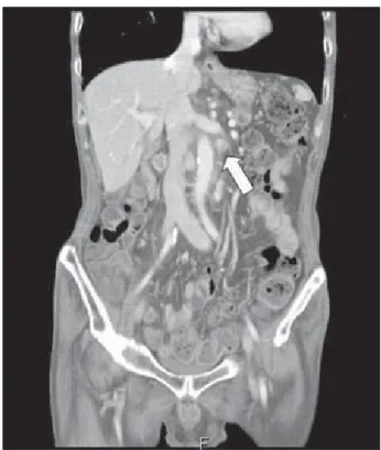
normal limits. A CT scan of the abdomen demonstrated a 2.5cm enhancing ampullary mass associated with retropancreatic nodules (Fig. 1). The liver was normal
except for dilatation of the biliary duct and narrowing of the ampulla. The patient underwent percutaneous transhepatic biliary drainage (PTBD). Imaging showed
Fig. 1. CT scan of abdomen demonstrated 2.5cm sized enhancing ampullary mass(white arrow) associated with retropancreatic nodularity.
Fig. 2. Endoscopy showed a 3cm sized ulceroinfiltrative mucosal lesion in periampullary area. We perfomed biopsy in the ulcerating lesion for two times.
Fig. 3. Specimen showed 3.5cm ulceroinfiltrative mass located in the ampullary region.(A, B black arrow). In cut section, distal common
bile duct(CBD) was dilated, the tumor invade the pancreatic tissue.(B, white arrow)
Fig. 4. (A) In Hematoxylin & eosin stain(H&E), tumor cell revealed combined LCNEC and adenocarcinoma pattern.(H&E, ×100) (B) LCNEC component in H&E stain showed organoid growth pattern, peripheral nuclear palisading, rosette-like structures(H&E, ×400).
(C) In immunohistochemical staining, Large cell neuroendocrine cells were positive for chromogranin and other neuroendocrine markers, such as synaptophysin, neuron-specific endolase and CD56 showed same pattern. (D) LCNEC component showed negative for CEA.
dilatation of the peripheral ducts and complete obstruction of the distal common bile duct.
Endoscopy indicated a 3cm sized ulceroinfiltrative mucosal lesion in the periampullary area (Fig. 2). The first biopsy indicated a high grade neuroendocrine tumor.
A re-biopsy was conducted and the pathologic examination showed a poorly differentiated adenocarcinoma. The patient underwent radical pancreaticoduodenectomy. Macroscopic examination showed a 3.5 cm ulcero-infiltrative mass located in the ampullary region (Fig. 3A). The tumor had invaded the pancreas (Fig. 3B). Histopathologically, the tumor was ulcerated and showed expansion and infiltration. It
exhibited necrotic lesions as well as lymphatic invasion.
Metastases were observed in 4 of 18 lymph nodes.
Microscopic findings showed a composite LCNEC and a
poorly differentiated adenocarcinoma (Fig. 4A). The
neuroendocrine component showed an organoid growth
pattern, peripheral nuclear palisading, and rosette-like
structures in a hematoxylin-eosin stain (Fig. 4B). On
immunohistochemical staining, LCNEC component cells
were positive for neuroendocrine markers such as
chromogranin (Fig. 4C), synaptophysin, neuron-specific
endolase and CD56, but negative for carcinoembryonic
antigen (CEA)(Fig. 4D). Adenocarcinoma cells were
reactive only for CEA.
At five months post surgery, a PET-CT scan showed multiple metastatic lymphadenopathies in the left posterior cervical space (Fig. 5A), aortocaval space and retrocaval space as well as a superior mesenteric vein lesion (Fig. 5B). The patient had received radiation therapy to the supraclavicular lymph nodes (5040 cGy) and on the para-aortic lymph nodes (7020 cGy) for two months. After radiation therapy, lesions on the supraclavicular lymph nodes disappeared, but lesions on para aortic and mesenteric lymph nodes had progressed (see CT scan in Fig. 6). The patient then received a fourth round of treatment with 5-FU chemotherapy from the Department of Oncology, and survived for a period of 18 months post operation.
Fig. 5. PET-CT shows metastatic lymphadenopathy in the left posterior cervical space(A, white arrow) and paraaortic space(B, black arrow) in 5 months after operation.
The most common periampullary tumors are adenocarcinomas, which account for more than 90% of periampullary malignancies. Neuroendocrine neoplasms are rare in this location.
1The WHO has c l a s s i f i e d n e u r o e n d o c r i n e t u m o r s o f t h e g a s t r o i n t e s t i n a l t r a c t i n t o t h e f o l l o w i n g:
1)well-differentiated endocrine tumor (carcinoid).
2)Well-differentiated endocrine carcinoma (atypical carcinoid). 3)Poorly-differentiated endocrine carcinomas (small cell carcinoma and large cell neuroendocrine carcinoma).
3Travis
2et al. described pulmonary neuroendocrine tumors as having three grades, namely, low grade (typical carcinoid), intermediate grade (atypical carcinoid), high grade LCNEC, and small cell carcinoma. Our case was classified as high grade LCNEC. Histologically, LCNEC has a neuroendocrine morphology including an
Discussion
Fig. 6. CT scan of 11months after surgery, we noted progression of multiple LN recurrent metastasis of the paraaortic and mesenteric nodes.(white arrow).
organoid growth pattern, trabeculae and rosette formation. Clinically, it presents typically as a tumor with aggressive behavior and prognosis is poor.
4A correct diagnosis of LCNEC is based on typical morphology with hematoxylin-eosin staining and must
be confirmed by positive immunohistochemical staining for neuroendocrine markers.
4-5There are two types of coexisting neuroendocrine carcinoma and adenocarcinoma. One is a composite case and the other is a collision case.
6In our study, there was a gradual transitional zone between LCNEC and adenocarcinoma components: neuroendocrine cells were found in some adenocarcinoma areas. Therefore, this is a composite tumor arising from multipotent stem cells. Biopsies were performed twice due to pathological results. It is difficult to diagnose tumors of the ampulla of Vater using preoperative endoscopic forceps biopsies. Deep biopsy after sphincterotomy and appropriate use of immunohistochemical staining is
necessary for improving diagnostic accuracy.
5.7According to several case reports, involvement of lymph nodes is common and the liver is the predominant site for metastasis of large cell neuroendocrine tumors.
4-5,8-12We believe the treatment of choice is radical pancreaticoduodenectomy.
Although the role of chemotherapy and radiotherapy is uncertain, tumor targeted radiation therapy reflects a new treatment option.
13We experienced a very rare case. Our case is only the third one of a coexisting large cell neuroendocrine carcinoma and an adenocarcinoma. We know very little about this tumor due to the small number of cases.
Further investigation is required to understand the pathogenesis and clinical features of this disease.
References
1. Carriaga MT, Henson DE. Liver, gallbladder, extrahepatic bile ducts, and pancreas. Cancer. 1995;75:171-190.
2. Travis WD, Linnoila RI, Tsokos MG, et al. Neuroendocrine tumors of the lung with proposed criteria for large-cell neuroendocrine carcinoma. An ultrastructural, immunohistochemical, and flow cytometric study of 35 cases. Am J Surg Pathol. 1991;15:529-553.
3. Solcia E, Kloppel G, Sobin LH. Histological typing of endocrine tumours, 2nd ed. Heidelberg:Springer;2000.
4. Cavazza A, Gallo M, Valcavi R, De Marco L, Gardini G. Large cell neuroendocrine carcinoma of the ampulla of Vater. Arch Pathol Lab Med. 2003;127:221-223.
5. Selvakumar E, Vimalraj V, Rajendran S, et al. Large cell neuroendocrine carcinoma of the ampulla of Vater. Hepatobiliary Pancreat Dis Int. 2006;5:465-467.
6. Lewin K. Carcinoid tumors and the mixed (composite) glandular- endocrine cell carcinomas. Am J Surg Pathol. 1987;11:71-86.
7. Menzel J, Poremba C, Dietl KH, Bocker W, Domschke W.
Tumors of the papilla of Vater - inadequate diagnostic impact of endoscopic forceps biopsies taken prior to and following sphincterotomy.
Ann Oncol. 1999;10:1227-1231.
8. Cheng SP, Yang TL, Chang KM, Liu CL. Large cell neuroendocrine carcinoma of the ampulla of Vater with glandular differentiation. J Clin Pathol. 2004;57:1098-1100.
9. Hartel M, Wente MN, Bergmann F, Schmidt J, Buchler MW, Friess H. Large-cell neuroendocrine carcinoma of the major duodenal papilla: case report. Gastrointest Endosc. 2004;60:838-841.
10. Nassar H, Albores-Saavedra J, Klimstra DS. High-grade neuroendocrine carcinoma of the ampulla of Vater: a clinicopathologic
..
..